
Article Figures & Data
Figures
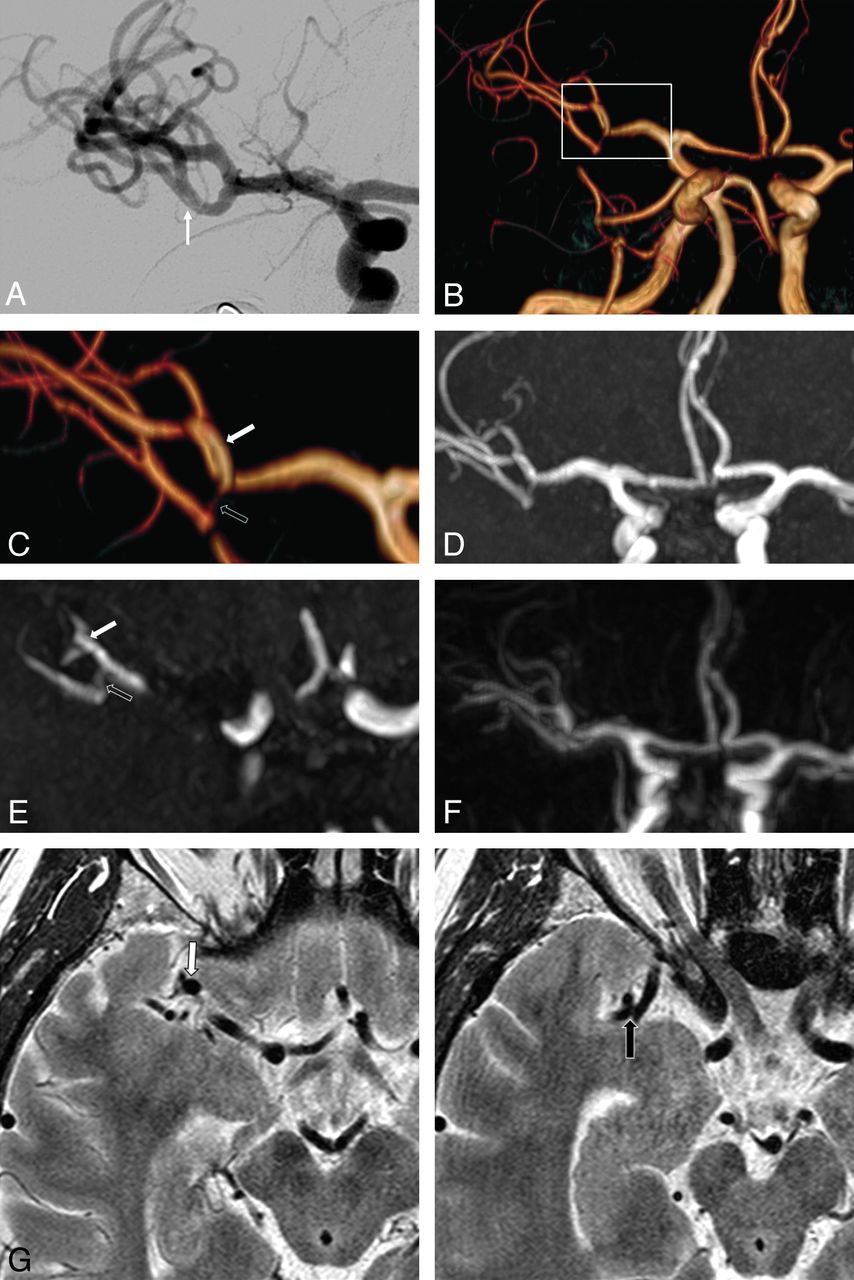
- Fig 1.
Eighteen-month MR imaging follow-up of de novo arterial stenosis and dilation (patient 2). Final post-MET angiographic run (A) of a 53-year-old patient treated for a right M1 middle cerebral artery occlusion, with 5 Solitaire passes in the superior and inferior M2 MCA branches, shows a flap in the inferior stenosis suggestive of dissection (arrow). TOF volume-rendering (B and C), MIP (D), native image (E), gadolinium-enhanced MRA (F), and high-resolution T2 (G) reveal an asymptomatic dilation (white arrow) of the superior M2 MCA branch and >50% stenosis (black arrow) of the inferior M2 MCA branch of the target vessel. Wall analysis at the stenosis is difficult on high-resolution T2 due to the vertical orientation and the size of the stenosed arterial segment.
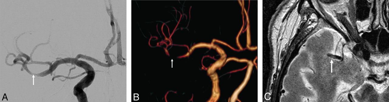
- Fig 2.
Seventeen-month MR imaging follow-up of pre-existing arterial stenosis (patient 5). Final post-MET angiographic run (A) of a 77-year-old patient treated for a right M1 middle cerebral artery occlusion with 2 Solitaire passes, 1 in the superior and 1 in the inferior M2 MCA branch, shows >50% stenosis in the inferior M2 MCA division (arrow). TOF volume-rendering (B) shows persistent stenosis (worsening aspect was considered due to TOF stenosis overestimation). High-resolution T2 MRA (C) shows M2 posterior wall thickening. Atrial fibrillation was established as the etiology of the stroke.
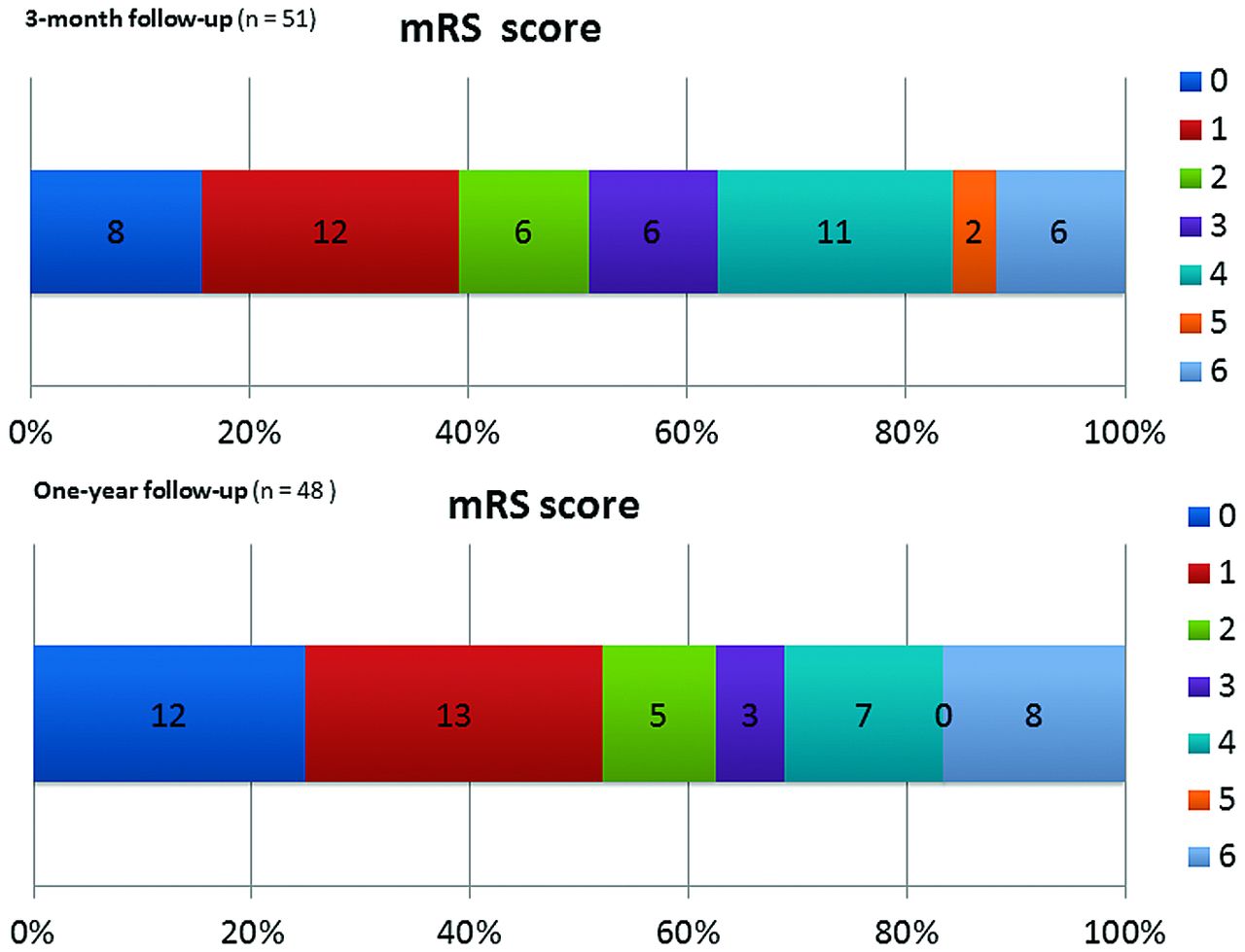
- Fig 3.
Clinical follow-up. Distribution of 3-month and 1-year mRS (0–6). Values on the bar graphs indicate the number of patients.


Tables
- Table 1:
Baseline characteristics of patients (40 survivors at 1-year follow-up) and recanalization therapy
Population (n = 40) ACO (n = 30) PCO (n = 10) Basic data Age (yr), mean (SD; range) 57.4 (15.2; 27–79) 60 (14.9; 35–79) 49.6 (14.1; 27–71) Female sex (%) 12 (30%) 10 2 Pretreatment NIHSS score, mean (SD) 15.9 (5.5) 16.1 (4.9) 15.5 (7.3) Premorbid status Charlson Index ≥3 (%) 4 (10) 3 1 Etiology TOAST I—large-artery atherosclerosis 10 5 5 TOAST II—cardiac embolism 16 14 2 TOAST IV—other determined etiology 4 3 1 TOAST V—undetermined etiology 9 7 2 Initial imaging CT 8 8 3 MRI 32 25 7 DWI-ASPECTS <5 5 5 Target-vessel territories M1 segment of the MCA 29 29 M2 segment of the MCA 1 1 Basilar artery 10 10 Recanalization strategy Stand-alone 21 14 7 Bridging 10 8 2 Rescue 9 8 1 Endovascular therapy Solitaire passes, mean (range) 2.0 (1–7) 1.9 (1–4) 2.4 (1–7) Procedural time (min), mean (SD, range) 75 (40, 30–189) 69 (30, 30–150) 96 (54, 30–189) Time to recanalization (min), mean (SD, range) 322 (127, 127–793) 288 (90, 67–430) 421 (170, 150–793) Note:—ACO indicates anterior circulation occlusion; PCO, posterior circulation occlusion.
Population (n = 48) ACO PCO mRS 0–1 (%) 25 (52.1) 17/34 (50.0) 8/14 (57.1) mRS 0–2 (%) 30 (62.5) 22/34 (64.7) 8/14 (57.1) mRS 3 (%) 3 (6.2) 2/34 (5.9) 1/14 (7.1) mRS 4–5 (%) 7 (14.6) 6/34 (17.6) 1/14 (7.1) mRS 6 (mortality) (%) 8/48 (16.7) 4/34 (11.8) 4/14 (28.6) SF-36 PCS (mean) (median, range) 51.2 (52.8, 5.6–100) 49.9 (51.9, 5.6–100) 56.3 (60.6, 32.9–71.0) SF-36 MCS (mean) (median, range) 51.4 (47.1, 4.0–98.8) 50.4 (47.1, 4.0–98.8) 53.9 (48.6, 32.3–81.1)


Jump to section
Related Articles
Cited By...
- Stroke thrombectomy complication management
- MRI Vessel Wall Imaging after Intra-Arterial Treatment for Acute Ischemic Stroke
- Delayed mid-basilar artery stenosis following paediatric acute mechanical thrombectomy: a rare complication from a rare case
- Thrombectomy using the EmboTrap device: core laboratory-assessed results in 201 consecutive patients in a real-world setting
- Mechanical thrombectomy for pediatric acute ischemic stroke: review of the literature
- Mechanical thrombectomy with the ERIC retrieval device: initial experience