Advertisement

Main menu

  • Home
  • Content
    • Current Issue
    • Publication Preview--Ahead of Print
    • Past Issue Archive
    • Case of the Week Archive
    • Classic Case Archive
    • Case of the Month Archive
  • For Authors
  • About Us
    • About AJNR
    • Editors
    • American Society of Neuroradiology
  • Submit a Manuscript
  • Podcasts
    • Subscribe on iTunes
    • Subscribe on Stitcher
  • More
    • Subscribers
    • Permissions
    • Advertisers
    • Alerts
    • Feedback
  • Other Publications
    • ajnr

User menu

  • Subscribe
  • Alerts
  • Log in

Search

  • Advanced search
American Journal of Neuroradiology

American Journal of Neuroradiology

  • Subscribe
  • Alerts
  • Log in

Advanced Search

  • Home
  • Content
    • Current Issue
    • Publication Preview--Ahead of Print
    • Past Issue Archive
    • Case of the Week Archive
    • Classic Case Archive
    • Case of the Month Archive
  • For Authors
  • About Us
    • About AJNR
    • Editors
    • American Society of Neuroradiology
  • Submit a Manuscript
  • Podcasts
    • Subscribe on iTunes
    • Subscribe on Stitcher
  • More
    • Subscribers
    • Permissions
    • Advertisers
    • Alerts
    • Feedback
  • Follow AJNR on Twitter
  • Visit AJNR on Facebook
  • Follow AJNR on Instagram
  • Join AJNR on LinkedIn
  • RSS Feeds
Letter

Carotid Web: Appearance at MR Angiography

J.E. Lantos, J.L. Chazen and A. Gupta
American Journal of Neuroradiology January 2016, 37 (1) E5-E6; DOI: https://doi.org/10.3174/ajnr.A4598
J.E. Lantos
aDepartment of Radiology Weill Cornell Medical College New York, New York
  • Find this author on Google Scholar
  • Find this author on PubMed
  • Search for this author on this site
  • ORCID record for J.E. Lantos
J.L. Chazen
aDepartment of Radiology Weill Cornell Medical College New York, New York
  • Find this author on Google Scholar
  • Find this author on PubMed
  • Search for this author on this site
  • ORCID record for J.L. Chazen
A. Gupta
aDepartment of Radiology Weill Cornell Medical College New York, New York
  • Find this author on Google Scholar
  • Find this author on PubMed
  • Search for this author on this site
  • ORCID record for A. Gupta
  • Article
  • Figures & Data
  • Info & Metrics
  • References
  • PDF
Loading

In their article entitled “Carotid Webs and Recurrent Ischemic Strokes in the Era of CT Angiography,” Choi et al1 described the prevalence, demographics, clinical presentation, imaging features, histopathology, and stroke risk associated with carotid webs. We read this thorough review with great interest, and it has already helped us in our clinical practice.

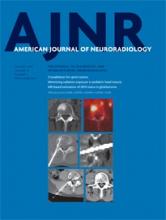
Recently, an otherwise healthy 49-year-old woman presented to our hospital with neck pain and left-sided numbness, prompting an imaging work-up, including CTA of the head and neck. On sagittal reformatted images (Fig 1A), we noticed a thin intraluminal filling defect along the posterior wall of the carotid bulb, which appeared as a septum on axial images (Fig 2A). When we used the definition set forth by Choi et al,1 our patient had findings consistent with a carotid web. No other abnormality was identified on her CTA. The carotid web was ipsilateral to the patient's clinically diagnosed TIA. Work by Choi et al and and Morgenlander and Goldstein2 has demonstrated an association between TIA and ipsilateral carotid webs and has provided evidence to support the diagnosis in this patient.

Fig 1.
  • Download figure
  • Open in new tab
  • Download powerpoint
Fig 1.

Sagittal reformat (A) from a CT angiogram demonstrates a thin intraluminal filling defect (arrow) along the posterior wall of the carotid bulb just beyond the carotid bifurcation. Axial source image (B) demonstrates a thin posterior septum (arrow) projecting into the lumen of the proximal right internal carotid artery. This finding is consistent with a carotid web using the definition set forth by Choi et al.1

Fig 2.
  • Download figure
  • Open in new tab
  • Download powerpoint
Fig 2.

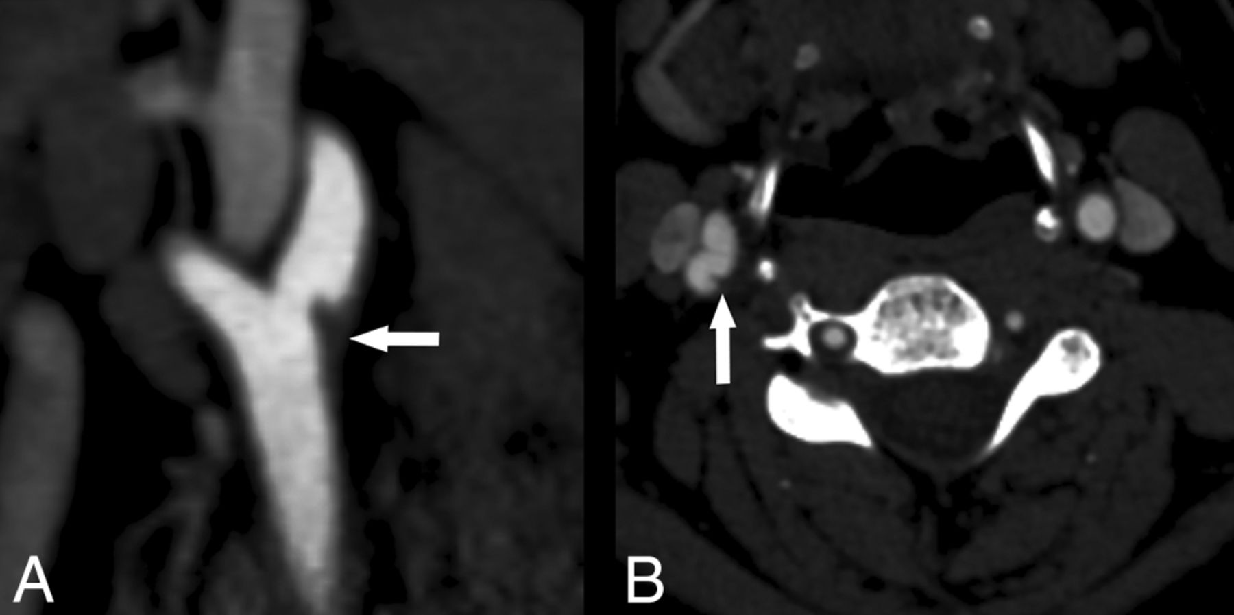
Axial source image from a 2D TOF MR angiogram (A) demonstrates some loss of flow-related enhancement (arrow) associated with the web, indicative of diminished or absence of flow. Axial T1-weighted fat-suppressed image (B) through the level of the carotid bulb demonstrates T1 shortening (arrow) consistent with hemorrhage associated with the carotid web seen on the CTA.

Choi et al1 mentioned that the MR imaging appearance of carotid webs has not been reported in the literature. Our patient also underwent MRA of the neck during her hospitalization, which included 2D time-of-flight and a black-blood, axial T1-weighted fat-suppressed sequence for the evaluation of vessel wall pathology. On the T1-weighted fat-saturated images, we observed a crescentic hyperintense signal (Fig 2A) consistent with hemorrhage.3,4 The finding of Choi et al1 of hemorrhage in 2 of the 4 patients for which carotid endarterectomy specimens were available is consistent with our findings on T1-weighted black-blood imaging in this patient. Axial source images from the 2D TOF image (Fig 2B) demonstrated abnormal flow-related enhancement in the area of the known web, possibly due to diminished or turbulent flow. The significance of abnormal flow in carotid webs in the pathogenesis of ipsilateral brain ischemia is uncertain but may be explained by the hypothesis of Choi et al1 that “the existence of turbulence and stasis in a cul-de-sac upstream to the web…could potentially create a thrombogenic milieu.”

Our case also demonstrates that when evaluated without CTA, a carotid web containing hemorrhage can be indistinguishable from hemorrhage in a nonstenosing atherosclerotic plaque. Whether carotid webs containing plaque hemorrhage confer a greater risk of ipsilateral ischemic events than webs without hemorrhage is unclear and requires future investigation.

In conclusion, we thank the authors for bringing the perhaps underdiagnosed entity of carotid web to our attention.

References

  1. 1.↵
    1. Choi PM,
    2. Singh D,
    3. Trivedi A, et al
    . Carotid webs and recurrent ischemic strokes in the era of CT angiography. AJNR Am J Neuroradiol 2015 Jul 30. [Epub ahead of print] doi:10.3174/ajnr.A4431 pmid:26228877
    Abstract/FREE Full Text
  2. 2.↵
    1. Morgenlander JC,
    2. Goldstein LB
    . Recurrent transient ischemic attacks and stroke in association with an internal carotid artery web. Stroke 1991;22:94–98 doi:10.1161/01.STR.22.1.94 pmid:1987677
    Abstract/FREE Full Text
  3. 3.↵
    1. Gupta A,
    2. Baradaran H,
    3. Schweitzer AD, et al
    . Carotid plaque MRI and stroke risk: a systematic review and meta-analysis. Stroke 2013;44:3071–77 doi:10.1161/STROKEAHA.113.002551 pmid:23988640
    Abstract/FREE Full Text
  4. 4.↵
    1. Gupta A,
    2. Baradaran H,
    3. Kamel H, et al
    . Intraplaque high-intensity signal on 3D time-of-flight MR angiography is strongly associated with symptomatic carotid artery stenosis. AJNR Am J Neuroradiol 2014;35:557–61 doi:10.3174/ajnr.A3732 pmid:24008170
    Abstract/FREE Full Text
  • © 2016 by American Journal of Neuroradiology
PreviousNext
Back to top

In this issue

American Journal of Neuroradiology: 37 (1)
American Journal of Neuroradiology
Vol. 37, Issue 1
1 Jan 2016
  • Table of Contents
  • Index by author
  • Complete Issue (PDF)
Advertisement
Print
Download PDF
Email Article

Thank you for your interest in spreading the word on American Journal of Neuroradiology.

NOTE: We only request your email address so that the person you are recommending the page to knows that you wanted them to see it, and that it is not junk mail. We do not capture any email address.

Enter multiple addresses on separate lines or separate them with commas.
Carotid Web: Appearance at MR Angiography
(Your Name) has sent you a message from American Journal of Neuroradiology
(Your Name) thought you would like to see the American Journal of Neuroradiology web site.
CAPTCHA
This question is for testing whether or not you are a human visitor and to prevent automated spam submissions.
Citation Tools
Carotid Web: Appearance at MR Angiography
J.E. Lantos, J.L. Chazen, A. Gupta
American Journal of Neuroradiology Jan 2016, 37 (1) E5-E6; DOI: 10.3174/ajnr.A4598

Citation Manager Formats

  • BibTeX
  • Bookends
  • EasyBib
  • EndNote (tagged)
  • EndNote 8 (xml)
  • Medlars
  • Mendeley
  • Papers
  • RefWorks Tagged
  • Ref Manager
  • RIS
  • Zotero
Share
Carotid Web: Appearance at MR Angiography
J.E. Lantos, J.L. Chazen, A. Gupta
American Journal of Neuroradiology Jan 2016, 37 (1) E5-E6; DOI: 10.3174/ajnr.A4598
del.icio.us logo Digg logo Reddit logo Twitter logo Facebook logo Google logo Mendeley logo
  • Tweet Widget
  • Facebook Like
  • Google Plus One
Purchase

Jump to section

  • Article
    • References
  • Figures & Data
  • Info & Metrics
  • References
  • PDF

Related Articles

  • No related articles found.
  • PubMed
  • Google Scholar

Cited By...

  • Carotid Bulb Webs as a Cause of "Cryptogenic" Ischemic Stroke
  • Crossref
  • Google Scholar

This article has not yet been cited by articles in journals that are participating in Crossref Cited-by Linking.

Similar Articles

Advertisement

News and Updates

  • Lucien Levy Best Research Article Award
  • Thanks to our 2021 Distinguished Reviewers
  • Press Releases

Resources

  • Evidence-Based Medicine Level Guide
  • How to Participate in a Tweet Chat
  • AJNR Podcast Archive
  • Ideas for Publicizing Your Research
  • Librarian Resources
  • Terms and Conditions

Opportunities

  • Share Your Art in Perspectives
  • Get Peer Review Credit from Publons
  • Moderate a Tweet Chat

American Society of Neuroradiology

  • Neurographics
  • ASNR Annual Meeting
  • Fellowship Portal
  • Position Statements

© 2022 by the American Society of Neuroradiology | Print ISSN: 0195-6108 Online ISSN: 1936-959X

Powered by HighWire