Advertisement

Main menu

  • Home
  • Content
    • Current Issue
    • Publication Preview--Ahead of Print
    • Past Issue Archive
    • Case of the Week Archive
    • Classic Case Archive
    • Case of the Month Archive
  • For Authors
  • About Us
    • About AJNR
    • Editors
    • American Society of Neuroradiology
  • Submit a Manuscript
  • Podcasts
    • Subscribe on iTunes
    • Subscribe on Stitcher
  • More
    • Subscribers
    • Permissions
    • Advertisers
    • Alerts
    • Feedback
  • Other Publications
    • ajnr

User menu

  • Subscribe
  • Alerts
  • Log in
  • Log out

Search

  • Advanced search
American Journal of Neuroradiology

American Journal of Neuroradiology

  • Subscribe
  • Alerts
  • Log in
  • Log out

Advanced Search

  • Home
  • Content
    • Current Issue
    • Publication Preview--Ahead of Print
    • Past Issue Archive
    • Case of the Week Archive
    • Classic Case Archive
    • Case of the Month Archive
  • For Authors
  • About Us
    • About AJNR
    • Editors
    • American Society of Neuroradiology
  • Submit a Manuscript
  • Podcasts
    • Subscribe on iTunes
    • Subscribe on Stitcher
  • More
    • Subscribers
    • Permissions
    • Advertisers
    • Alerts
    • Feedback
  • Follow AJNR on Twitter
  • Visit AJNR on Facebook
  • Follow AJNR on Instagram
  • Join AJNR on LinkedIn
  • RSS Feeds
Research ArticleHEAD & NECK

MR Imaging in Sudden Sensorineural Hearing Loss. Time to Talk

G. Conte, F. Di Berardino, C. Sina, D. Zanetti, E. Scola, C. Gavagna, L. Gaini, G. Palumbo, P. Capaccio and F. Triulzi
American Journal of Neuroradiology August 2017, 38 (8) 1475-1479; DOI: https://doi.org/10.3174/ajnr.A5230
G. Conte
aFrom the Postgraduation School of Radiodiagnostics (G.C.)
  • Find this author on Google Scholar
  • Find this author on PubMed
  • Search for this author on this site
  • ORCID record for G. Conte
F. Di Berardino
bAudiology (F.D.B., D.Z.)
  • Find this author on Google Scholar
  • Find this author on PubMed
  • Search for this author on this site
  • ORCID record for F. Di Berardino
C. Sina
cNeuroradiology (C.S., E.S., C.G., F.T.)
  • Find this author on Google Scholar
  • Find this author on PubMed
  • Search for this author on this site
  • ORCID record for C. Sina
D. Zanetti
bAudiology (F.D.B., D.Z.)
  • Find this author on Google Scholar
  • Find this author on PubMed
  • Search for this author on this site
  • ORCID record for D. Zanetti
E. Scola
cNeuroradiology (C.S., E.S., C.G., F.T.)
  • Find this author on Google Scholar
  • Find this author on PubMed
  • Search for this author on this site
  • ORCID record for E. Scola
C. Gavagna
cNeuroradiology (C.S., E.S., C.G., F.T.)
  • Find this author on Google Scholar
  • Find this author on PubMed
  • Search for this author on this site
  • ORCID record for C. Gavagna
L. Gaini
dOtolaryngology (L.G., P.C.) Units, Department of Clinical Sciences and Community Health, Fondazione IRCCS Ca'Granda Ospedale Maggiore Policlinico
  • Find this author on Google Scholar
  • Find this author on PubMed
  • Search for this author on this site
  • ORCID record for L. Gaini
G. Palumbo
eSchool of Medicine (G.P.)
  • Find this author on Google Scholar
  • Find this author on PubMed
  • Search for this author on this site
  • ORCID record for G. Palumbo
P. Capaccio
dOtolaryngology (L.G., P.C.) Units, Department of Clinical Sciences and Community Health, Fondazione IRCCS Ca'Granda Ospedale Maggiore Policlinico
  • Find this author on Google Scholar
  • Find this author on PubMed
  • Search for this author on this site
  • ORCID record for P. Capaccio
F. Triulzi
cNeuroradiology (C.S., E.S., C.G., F.T.)
fDepartment of Pathophysiology and Transplantation (F.T.), Università degli Studi di Milano, Milan, Italy.
  • Find this author on Google Scholar
  • Find this author on PubMed
  • Search for this author on this site
  • ORCID record for F. Triulzi
  • Article
  • Figures & Data
  • Supplemental
  • Info & Metrics
  • References
  • PDF
Loading

Abstract

SUMMARY: Sudden sensorineural hearing loss is defined as acute hearing loss of the sensorineural type of at least 30 dB over 3 contiguous frequencies that occurs within a 72-hour period. Although many different causative factors have been proposed, sudden sensorineural hearing loss is still considered “idiopathic” in 71%–85% of cases, and treatments are empiric, not based on etiology. MR imaging implemented with a 3D FLAIR sequence has provided new insights into the etiology of sudden sensorineural hearing loss. Herein, we review the current management trends for patients with sudden sensorineural hearing loss, from the initial clinical diagnosis to therapeutic strategies and diagnostic work-up. We focused primarily on MR imaging assessment and discuss the relevance that MR imaging findings might have for patient management, pointing out different perspectives for future clinical research.

ABBREVIATION:

SSHL
sudden sensorineural hearing loss

According to the guidelines of the Committee on Hearing and Equilibrium of the American Academy of Otolaryngology, sudden sensorineural hearing loss (SSHL) is defined as acute hearing loss of the sensorineural type of at least 30 dB over 3 contiguous frequencies occurring within a 72-hour period.1 SSHL occurs most often in the fourth decade of life, with an annual incidence that varies between 5 and 20 cases among 100,000 people. The severity of hearing loss is variable and is usually unilateral, though bilateral involvement has been reported in up to 4% of patients.2

Although SSHL has been attributed to many different causative factors,3 there are still no audiologic means of accurately assessing its etiology,4 so it is still considered “idiopathic” in 71%–85% of cases.5 Consequently, there is not a treatment that targets the etiology, which partly explains the inconsistent results of the various empiric drug protocols proposed, which include systemic steroids, antiviral medications, vasodilators, and carbogen or hyperbaric oxygen alone or in combination, none of which has been proved to be superior to the others.6⇓–8

Over the past decade, MR imaging has provided new insights about SSHL etiology because of high-resolution sequences that are able to detect subtle changes in the inner ear.9⇓⇓⇓⇓⇓–15 However, for many reasons, the use of this imaging technique for SSHL is still not widespread. First, many radiologists are not yet aware of the possibilities offered by advanced MR imaging studies of the inner ear and/or of the possible MR imaging findings in SSHL. Second, it is still unclear and is not yet addressed in the literature how MR imaging could change therapeutic strategies.

In this paper, we review the current management trends of patients with SSHL, from the initial clinical diagnosis to the therapeutic strategies, through the diagnostic work-up. Our work focuses particularly on MR imaging assessment. We discussed how MR imaging should be performed and the relevance that the possible MR imaging findings in the inner ear might have. Finally, we addressed the perspectives of possible changes in the therapeutic management of SSHL based on MR imaging findings, which could impact the patient's prognosis.

Current Diagnostic Work-Up of SSHL

SSHL is suspected in patients with a sudden onset of generally unilateral decrease or loss of hearing, occurring instantaneously or rapidly developing over a period of hours or days.3 In other instances, primary care or emergency department physicians refer patients to an audiologist.16,17

The evaluation usually begins with a careful history and physical examination to look for potential causes such as infections, systemic diseases, and exposure to known ototoxic medications. Otomicroscopy is generally negative for external and middle ear pathologies. Pure-tone audiometry is the main diagnostic tool used to differentiate between conductive and sensorineural hearing loss, which have very different management strategies. The physician can differentiate SSHL (symptoms occur within 3 days) from progressive or fluctuating SSHL. The presence of bilateral sudden hearing loss, recurrent episodes of sudden hearing loss, or focal neurologic findings suggests systemic disorders, autoimmune or metabolic disorders, bilateral Menière disease, or primary neurologic disorders.

If SSHL is diagnosed, an empiric treatment is started while the diagnostic work-up continues. A complete audiovestibular evaluation, including speech audiometry, speech in noise test, tympanometry, acoustic reflexes, and otoacoustics emissions, is performed in an attempt to investigate whether the SSHL is cochlear or retrocochlear.8

Brain MR imaging with and without gadolinium is often used to exclude a vestibular schwannoma (reported in up to 10%–20% of patients with SSHL) as well as rarer causes of retrocochlear hearing loss such as other cerebellopontine tumors, brain stem infarctions, and demyelinating disease.3,8,18⇓⇓–21 A number of studies have advocated the use of MR imaging without gadolinium as the more appropriate means of screening patients with asymmetric SSHL suspected to be retrocochlear.22

In cases of suspected cochlear SSHL, different studies have proposed a tailored temporal bone MR imaging with 3D-FLAIR sequence to exclude abnormalities in the inner ear structures.9⇓⇓⇓⇓⇓–15 MR imaging of the temporal bone can be negative, and the SSHL is therefore defined as idiopathic, or the MR imaging can show abnormalities in the inner ear structures, suggesting a specific etiopathogenesis (vascular or inflammatory).14 In both cases, because no trial has yet investigated different medical protocols guided by MR imaging findings, physicians continue with empiric therapy, so temporal bone MR imaging with a 3D-FLAIR sequence has not had any effect on the therapeutic management of cochlear SSHL in clinical practice. Nevertheless, in our opinion, it is important that radiologists perform temporal bone MR imaging with a 3D-FLAIR sequence to clarify the cochlear origin of SSHL, suggesting the probable pathogenesis, and provide prognostic information to physicians.

How We Perform MR Imaging in Patients with SSHL

MR imaging of the temporal bone is challenging because of the complexity and small dimensions of the anatomic structures. Thus, MR imaging should preferably be performed on a 3T scanner, which provides high-resolution images with a higher signal-to-noise ratio compared with a 1.5T scanner. The basic MR imaging protocol should include an axial precontrast 3D steady-state free procession sequence, a pre- and postcontrast T1-weighted FSE sequence, and a pre- and postcontrast 3D-FLAIR sequence.4⇓⇓⇓⇓⇓⇓⇓⇓⇓⇓–15 The On-line Table summarizes the MR imaging scan parameters used in our department. It is recommended to use both postcontrast 3D-FLAIR and postcontrast T1-weighted FSE sequences because the former are more sensitive than a T1-weighted sequence in detecting intralabyrinthine contrast enhancement,11,14,23 and the latter can clarify the presence of a small schwannoma.

Postcontrast sequences should be acquired approximately 10 minutes after administration of the contrast agent.13,15 To cover part of the 10 minutes needed before the acquisition of postcontrast sequences, it is advantageous to inject a contrast agent before the acquisition of the 3D steady-state free procession sequence. Although the contrast enhancement of some structures can be seen on the 3D steady-state free procession sequence,24 the assessment of the inner ear anatomic structures and internal auditory canal on this sequence is usually not hindered by the presence of the contrast agent.

What Can MR Imaging Detect?

Neuroradiologists should review MRIs and look for asymmetry of the signal between the affected and unaffected sides. In the literature, abnormalities on MR imaging are reported in 27%–53% of SSHL cases.10,14

Two patterns can be recognized based on the MR imaging signal of the inner ear on precontrast T1-weighted and 3D-FLAIR images:

  1. The vascular pattern shows hyperintensity on precontrast T1-weighted and 3D-FLAIR images because of the presence of methemoglobin in the inner ear (Fig 1); and

  2. The inflammatory pattern shows hyperintensity only on 3D-FLAIR images because of the presence of proteinaceous exudate in the inner ear (Fig 2).

Fig 1.
  • Download figure
  • Open in new tab
  • Download powerpoint
Fig 1.

Vascular pattern in a 20-year-old woman with left SSHL. Precontrast T1-weighted (A) and precontrast 3D-FLAIR (B) sequences show a high signal in the middle and upper turns of the left cochlea without enhancement on postcontrast T1-weighted (C) and 3D-FLAIR images (D).

Fig 2.
  • Download figure
  • Open in new tab
  • Download powerpoint
Fig 2.

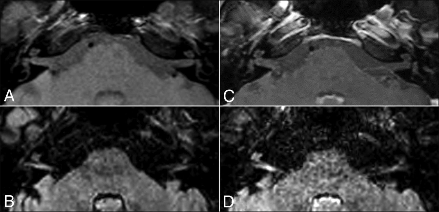
Inflammatory pattern in a 35-year-old man with right SSHL. The precontrast T1-weighted sequence (A) shows no signal abnormalities. The precontrast 3D-FLAIR sequence (B) shows a high signal in the right cranial nerves VII and VIII and in the middle and upper turns of the left cochlea. A postcontrast T1-weighted sequence (C) does not show enhancement, whereas a postcontrast 3D-FLAIR sequence (D) shows the cochlea and cranial nerves VII and VIII as markedly enhanced on the right side.

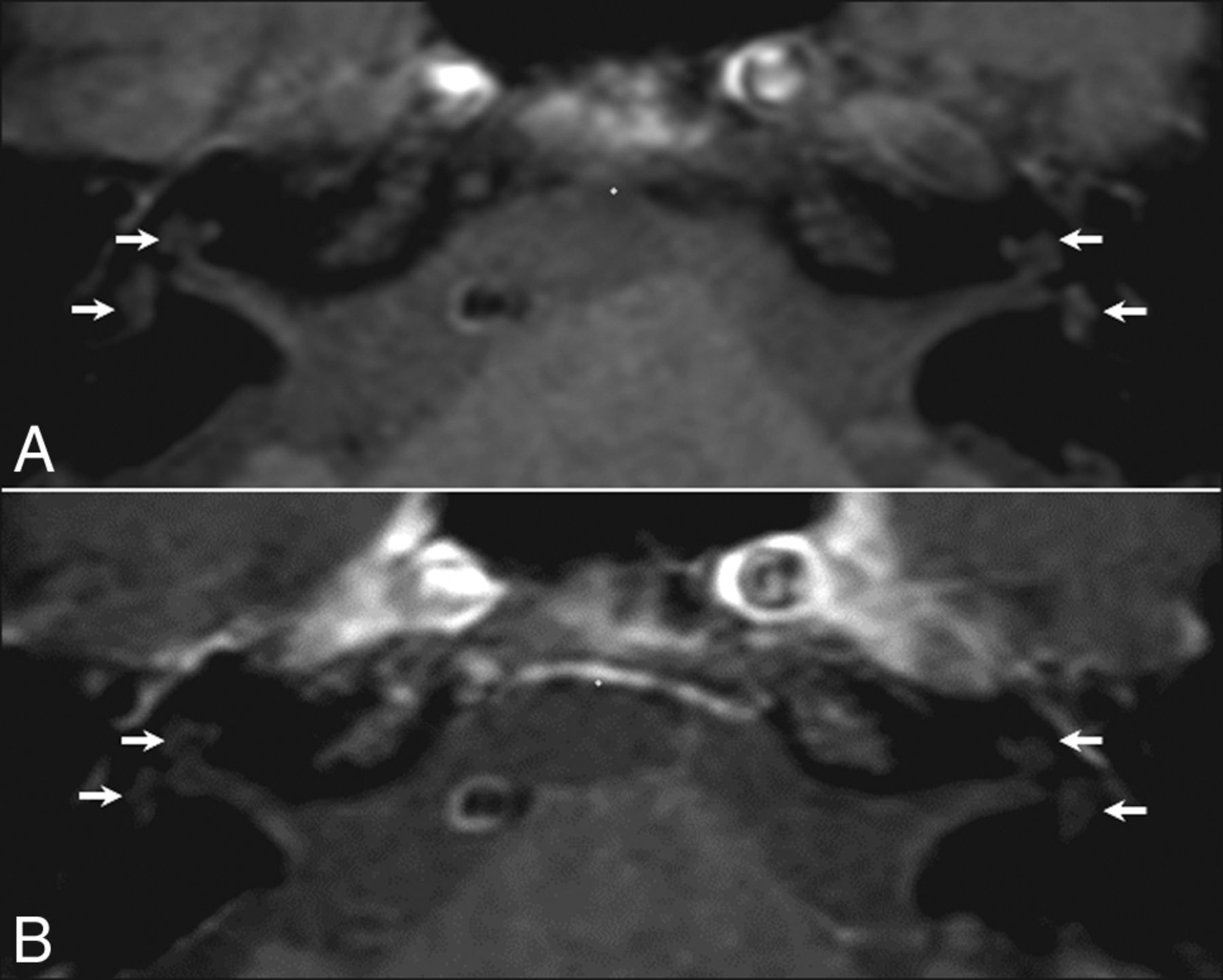
Regarding the vascular pattern, radiologists should be aware that pseudohyperintensity of the intralabyrinthine fluid can normally be detected on fat-suppressed T1-weighted images, which could hamper the diagnosis. This pseudohyperintensity has previously been described as an artifact25 and can be differentiated from methemoglobin because it is symmetrical and less evident on postcontrast fat-suppressed T1-weighted images (see Fig 3 for a detailed explanation).

Fig 3.
  • Download figure
  • Open in new tab
  • Download powerpoint
Fig 3.

Normal MR imaging findings in a 59-year-old man with right SSHL. The precontrast T1-weighted sequence (A) shows spontaneous hyperintense intralabyrinthine fluid in both inner ear structures (arrows), which is symmetric and less evident on the same sequence after contrast injection (B). This hyperintensity is an artifact caused by the altered dynamic range when the fat signal is subtracted, but also reflects an alteration in the visual appearance of signal intensity as the ambient contrast is changed (checker-shadow illusion).24

Recent studies have investigated the relationship between precontrast 3D-FLAIR signals and clinical findings regardless of the hyperintensity on precontrast T1-weighted sequences.9⇓–11,23 The rate of abnormalities on precontrast 3D-FLAIR images correlated with the level of hearing loss at onset, resulting in fewer abnormalities in patients with mild to moderate hearing loss compared with patients with profound hearing loss.10,14,15 However, Yoshida et al11 did not observe similar results. Inflammatory diseases can also affect the vestibule, the semicircular canals, or cranial nerve VIII, and precontrast 3D-FLAIR images can detect signal abnormalities in these structures (Fig 3).10⇓⇓⇓–14,23 Hyperintensity on precontrast 3D-FLAIR images of the vestibule or the semicircular canals has been associated with vertigo,11,14 which is reported in approximately 30% of SSHL cases.9

The 2 MR imaging patterns are not always associated with inner ear enhancement on postcontrast 3D-FLAIR, which is consistent with blood-labyrinth barrier breakdown.14 The advantage of postcontrast 3D-FLAIR images is that they identify patients with more severe blood-labyrinth barrier breakdown when signal abnormalities are subtle on precontrast 3D-FLAIR images.23 However, the significance of the inner ear enhancement is still unclear. Viral infection, immune-mediated inner ear disease, and perilymphatic fistulas have been suggested as possible causes of cochlear enhancement.26,27

Usually, the 3D T2-weighted steady-state free procession sequence does not show any pathologic findings23; nevertheless, it is essential to investigate the morphology of the inner ear structures, cranial nerve VIII, internal auditory canal, and cerebellopontine angle.

Prognostic Value of MR Imaging

To the best of our knowledge, no published studies have investigated the difference in prognosis and outcome between the 2 MR imaging patterns mentioned above, though a vascular pattern has been associated with a poor prognosis.28 On the contrary, the published studies have concentrated on the prognostic value of 3D-FLAIR abnormalities and reported conflicting results. Two studies showed that a high signal in the affected inner ear on precontrast 3D-FLAIR was associated with a poor prognosis.11,15 In 2 other studies, the hearing outcome was worse in patients with multiple-location hyperintensities on precontrast 3D-FLAIR (cochlea plus vestibule) than in patients with a single-subsite hyperintensity (cochlea only).12,29 A recent study by Liao et al23 showed that the more asymmetric FLAIR signal between the affected ear and the normal one and presence of a high signal beyond the cochlea indicated a poorer prognosis. Lee et al10 demonstrated that precontrast 3D-FLAIR abnormalities do not affect the prognosis when the initial hearing loss is mild to moderate, whereas such abnormalities represent a negative prognostic factor in patients with initial profound hearing loss. However, Berrettini et al14 failed to find a correlation between the severity of precontrast 3D-FLAIR abnormalities and hearing improvement. Lee et al9 reported that a high signal on precontrast 3D-FLAIR did not significantly affect the final hearing ability.

These inconsistent results are partially explained by methodologic differences, including the time span between SSHL onset and MR imaging, method of MR imaging assessment, pharmacologic protocols of drug administration during follow-up, length of follow-up, and assessment criteria used to determine hearing improvement.30 A meta-analysis by Gao and Chi30 that included studies of patients without primary treatment before temporal bone MR imaging concluded that precontrast 3D-FLAIR hyperintensity in the inner ear is associated with more severe initial hearing loss and a lower chance of recovery.

Current Management of SSHL and Perspectives in MR Imaging Research

The results of SSHL treatment are still largely unpredictable; a very large variability in responses has been reported, ranging from no response to total recovery.7,31 Moreover, the high rate of spontaneous recovery, which varies from 45%–65%,21 should be considered.

Although cochlear vascular microthrombosis has been hypothesized as the main pathogenic mechanism,32⇓–34 because there is no objective test that can detect the occlusion of microvessels, the pharmacologic treatment remains highly empirical, and its overall efficacy is controversial because of the absence of prospective double-blind studies.3 Pharmacologic treatment includes many drugs that, without a certain etiology of SSHL, are often prescribed in combination: oral and/or intratympanic corticosteroids, hyperbaric oxygen therapy, antivirals drugs, vasodilators, and vasoactive substances.

For decades, the “gold standard” treatment for SSHL has been the oral administration of corticosteroids.35,36 The exact mechanism by which steroids improve hearing is still unknown, though some of the major hypotheses are that they modulate cochlear function, decrease inflammation and edema, improve cochlear blood flow, and protect against cochlear ischemia. Although the evidence concerning the use of oral corticosteroids remains contradictory,37 it seems reasonable to offer them because of the potentially devastating disability caused by SSHL and the relatively low morbidity of the treatment.3 Intratympanic steroid treatment allows high steroid concentrations to be reached in the perilymph while avoiding the common side effects of systemic steroids. A recent meta-analysis has shown the benefits of intratympanic steroid treatment in combination with oral corticosteroids as the first-line therapy and salvage monotherapy in idiopathic SSHL.38 Hyperbaric oxygen treatment is also used as primary, adjuvant, or salvage therapy because it is thought to protect hair cells from ischemic damage by increasing oxygenation.39,40 However, cost/benefit issues, limited availability, and the absence of strong evidence because of the difficulties of organizing controlled randomized studies have halted its widespread application. Although there is no clear evidence supporting their use, antiviral drugs are often used in pharmacologic protocols for the treatment of SSHL because various viruses have been implicated in its etiology.41 Vasodilators and vasoactive substances are sometimes used because obstructed vascular flow to the cochlea is a theoretic cause of SSHL, but there is currently insufficient evidence to support their routine use.3

Future research should investigate the role of MR imaging with a 3D-FLAIR sequence in the therapeutic management of SSHL (On-line Figure). Before future trials can investigate different medical protocols guided by MR imaging findings, longitudinal studies should investigate the effects of early steroid treatment and the time interval between disease onset and scanning on the sensitivity of MR imaging. In our opinion, its optimal timing would be upon admission to the emergency department or on the same day as a clinical assessment by an audiologist or otolaryngologist. We hypothesize that precontrast and contrast-enhanced MR imaging abnormalities are more easily detected before steroid treatment is started, so MR imaging should be performed as soon as possible, and preferably before the masking effect of steroids becomes apparent.28 In line with this suggestion, Berrettini et al14 have reported that the time interval between SSHL onset and MR imaging tended to be shorter in patients with 3D-FLAIR abnormalities than in those without 3D-FLAIR abnormalities (P = .06). However, this has not been confirmed by other authors.

Conclusions

MR imaging with 3D-FLAIR sequences provides new insights into SSHL etiology and may change current clinical and therapeutic practices. Radiologists should therefore be trained to perform tailored temporal bone MR imaging in the case of SSHL and recognize the common findings and pitfalls of the technique to provide clinicians with information useful for patient management.

REFERENCES

  1. 1.↵
    1. Rauch SD
    . Clinical practice. Idiopathic sudden sensoneurinal hearing loss. N Eng J Med 2008;359:833–40 doi:10.1056/NEJMcp0802129 pmid:18716300
    CrossRefPubMed
  2. 2.↵
    1. Byl FM Jr.
    . Sudden hearing loss: eight years' experience and suggested prognostic table. Laryngoscope 1984;94:647–61 pmid:6325838
    PubMed
  3. 3.↵
    1. Lawrence R,
    2. Thevasagayam R
    . Controversies in the management of sudden sensorineural hearing loss: an evidence-based review. Clin Otolaryngol 2015;40:176–82 doi:10.1111/coa.12363 pmid:25521864
    CrossRefPubMed
  4. 4.↵
    1. Kuhn M,
    2. Heman-Ackah SE,
    3. Shaikh JA, et al
    . Sudden sensorineural hearing loss: a review of diagnosis, treatment, and prognosis. Trends Amplif 2011;15:91–105 doi:10.1177/1084713811408349 pmid:21606048
    CrossRefPubMed
  5. 5.↵
    1. Chau JK,
    2. Lin JR,
    3. Atashband S, et al
    . Systematic review of the evidence for the etiology of adult sudden sensorineural hearing loss. Laryngoscope 2010;120:1011–21 doi:10.1002/lary.20873 pmid:20422698
    CrossRefPubMed
  6. 6.↵
    1. Schreiber BE,
    2. Agrup C,
    3. Haskard DO, et al
    . Sudden sensorineural hearing loss. Lancet 2010;375:1203–11 doi:10.1016/S0140-6736(09)62071-7 pmid:20362815
    CrossRefPubMed
  7. 7.↵
    1. Conlin AE,
    2. Parnes LS
    . Treatment of sudden sensorineural hearing loss: I. A systematic review. Arch Otolaryngol Head Neck Surg 2007;133:573–81 doi:10.1001/archotol.133.6.573 pmid:17576908
    CrossRefPubMed
  8. 8.↵
    1. Stachler RJ,
    2. Chandrasekhar SS,
    3. Archer SM, et al
    . Clinical practice guideline: sudden hearing loss. Otolaryngol Head Neck Surg 2012;146:S1–35 doi:10.1177/0194599812436449 pmid:22383545
    CrossRefPubMed
  9. 9.↵
    1. Lee HY,
    2. Jung SY,
    3. Park MS, et al
    . Feasibility of three-dimensional fluid-attenuated inversion recovery magnetic resonance imaging as a prognostic factor in patients with sudden hearing loss. Eur Arch Otorhinolaryngol 2012;269:1885–91 doi:10.1007/s00405-011-1834-1 pmid:22083357
    CrossRefPubMed
  10. 10.↵
    1. Lee JI,
    2. Yoon RG,
    3. Lee JH, et al
    . Prognostic value of labyrinthine 3D-FLAIR abnormalities in idiopathic sudden sensorineural hearing loss. AJNR Am J Neuroradiol 2016;37:2317–22 doi:10.3174/ajnr.A4901 pmid:27516239
    Abstract/FREE Full Text
  11. 11.↵
    1. Yoshida T,
    2. Sugiura M,
    3. Naganawa S, et al
    . Three-dimensional fluid-attenuated inversion recovery magnetic resonance imaging findings and prognosis in sudden sensorineural hearing loss. Laryngoscope 2008;118:1433–37 doi:10.1097/MLG.0b013e318172ef85 pmid:18475208
    CrossRefPubMed
  12. 12.↵
    1. Ryu IS,
    2. Yoon TH,
    3. Ahn JH, et al
    . Three-dimensional fluid-attenuated inversion recovery magnetic resonance imaging in sudden sensorineural hearing loss: correlations with audiologic and vestibular testing. Otol Neurotol 2011;32:1205–09 doi:10.1097/MAO.0b013e31822e969f pmid:21921851
    CrossRefPubMed
  13. 13.↵
    1. Kim TY,
    2. Park DW,
    3. Lee YJ, et al
    . Comparison of inner ear contrast enhancement among patients with unilateral inner ear symptoms in MR images obtained 10 minutes and 4 hours after gadolinium injection. AJNR Am J Neuroradiol 2015;36:2367–72 doi:10.3174/ajnr.A4439 pmid:26316569
    Abstract/FREE Full Text
  14. 14.↵
    1. Berrettini S,
    2. Seccia V,
    3. Fortunato S, et al
    . Analysis of the 3-dimensional fluid-attenuated inversion-recovery (3D-FLAIR) sequence in idiopathic sudden sensorineural hearing loss. JAMA Otolaryngol Head Neck Surg 2013;139:456–64 doi:10.1001/jamaoto.2013.2659 pmid:23681028
    CrossRefPubMed
  15. 15.↵
    1. Zhu H,
    2. Ou Y,
    3. Fu J, et al
    . A comparison of inner ear imaging features at different time points of sudden sensorineural hearing loss with three-dimensional fluid-attenuated inversion recovery magnetic resonance imaging. Eur Arch Otorhinolaryngol 2015;272:2659–65 doi:10.1007/s00405-014-3187-z pmid:25097028
    CrossRefPubMed
  16. 16.↵
    1. Witsell DL,
    2. Khoury T,
    3. Schulz KA, et al
    . Evaluation of compliance for treatment of sudden hearing loss: a CHEER Network study. Otolaryngol Head Neck Surg 2016;155:48–55 doi:10.1177/0194599816650175 pmid:27371626
    CrossRefPubMed
  17. 17.↵
    1. Coelho DH,
    2. Thacker LR,
    3. Hsu DW
    . Variability in the management of idiopathic sudden sensorineural hearing loss. Otolaryngol Head Neck Surg 2011;145:813–17 doi:10.1177/0194599811412721 pmid:21690271
    CrossRefPubMed
  18. 18.↵
    1. Portmann M,
    2. Dauman R,
    3. Duriez F, et al
    . Modern diagnostic strategy for acoustic neuromas. Arch Otorhinolaryngol 1989;246:286–91 doi:10.1007/BF00463577 pmid:2590038
    CrossRefPubMed
  19. 19.↵
    1. Biavati MJ,
    2. Gross JD,
    3. Wilson WR, et al
    . Magnetic resonance imaging evidence of a focal pontine ischemia in sudden hearing loss and seventh nerve paralysis. Am J Otol 1994;15:250–53 doi:10.1016/0196-0709(94)90091-4 pmid:8172312
    CrossRefPubMed
  20. 20.↵
    1. Franklin DJ,
    2. Coker NJ,
    3. Jenkins HA
    . Sudden sensorineural hearing loss as a presentation of multiple sclerosis. Arch Otolaryngol Head Neck Surg 1989;115:41–45 doi:10.1001/archotol.1989.01860250043023 pmid:2909229
    CrossRefPubMed
  21. 21.↵
    1. Hagiwara M,
    2. Roland JT Jr.,
    3. Wu X, et al
    . Identification of endolymphatic hydrops in Ménière's disease utilizing delayed postcontrast 3D FLAIR and fused 3D FLAIR and CISS color maps. Otol Neurotol 2014;35:e337–42 doi:10.1097/MAO.0000000000000585 pmid:25251300
    CrossRefPubMed
  22. 22.↵
    1. Ryan M,
    2. Weissman JL,
    3. Kaylie D
    . Is Gadolinium contrast enhancement necessary in screening MRI for asymmetric sensorineural hearing loss? Laryngoscope 2015;125:783–84 doi:10.1002/lary.24871 pmid:25111873
    CrossRefPubMed
  23. 23.↵
    1. Liao WH,
    2. Wu HM,
    3. Wu HY, et al
    . Revisiting the relationship of three-dimensional fluid attenuation inversion recovery imaging and hearing outcomes in adults with idiopathic unilateral sudden sensorineural hearing loss. Eur J Radiol 2016;85:2188–94 doi:10.1016/j.ejrad.2016.10.005 pmid:27842665
    CrossRefPubMed
  24. 24.↵
    1. Yagi A,
    2. Sato N,
    3. Takahashi A, et al
    . Added value of contrast-enhanced CISS imaging in relation to conventional MR images for the evaluation of intracavernous cranial nerve lesions. Neuroradiology 2010;52:1101–09 doi:10.1007/s00234-010-0690-9 pmid:20383633
    CrossRefPubMed
  25. 25.↵
    1. Huynh TN,
    2. Johnson T,
    3. Poder L, et al
    . T1 pseudohyperintensity on fat-suppressed magnetic resonance imaging: a potential diagnostic pitfall. J Comput Assist Tomogr 2011;35:459–61 doi:10.1097/RCT.0b013e31822227c3 pmid:21765301
    CrossRefPubMed
  26. 26.↵
    1. Mark AS,
    2. Fitzgerald D
    . Segmental enhancement of the cochlea on contrast-enhanced MR: correlation with the frequency of hearing loss and possible sign of perilymphatic fistula and autoimmune labyrinthitis. AJNR Am J Neuroradiol 1993;14:991–96 pmid:8352175
    Abstract/FREE Full Text
  27. 27.↵
    1. Fitzgerald DC,
    2. Mark AS
    . Sudden hearing loss: frequency of abnormal findings on contrast-enhanced MR studies. AJNR Am J Neuroradiol 1998;19:1433–36 pmid:9763372
    Abstract
  28. 28.↵
    1. Lee JW,
    2. Park YA,
    3. Park SM, et al
    . Clinical features and prognosis of sudden sensorineural hearing loss secondary to intralabyrinthine hemorrhage. J Audiol Otol 2016;20:31–35 doi:10.7874/jao.2016.20.1.31 pmid:27144231
    CrossRefPubMed
  29. 29.↵
    1. Ramos HV,
    2. Barros FA,
    3. Yamashita H, et al
    . Magnetic resonance imaging in sudden deafness. Braz J Otorhinolaryngol 2005;71:422–26 doi:10.1016/S1808-8694(15)31193-9 pmid:16446954
    CrossRefPubMed
  30. 30.↵
    1. Gao Z,
    2. Chi FL
    . The clinical value of three-dimensional fluid-attenuated inversion recovery magnetic resonance imaging in patients with idiopathic sudden sensorineural hearing loss: a meta-analysis. Otol Neurotol 2014;35:1730–35 doi:10.1097/MAO.0000000000000611 pmid:25393973
    CrossRefPubMed
  31. 31.↵
    1. Conlin AE,
    2. Parnes LS
    . Treatment of sudden sensorineural hearing loss: II. A meta-analysis. Arch Otolaryngol Head Neck Surg 2007;133:582–86 doi:10.1001/archotol.133.6.582 pmid:17576909
    CrossRefPubMed
  32. 32.↵
    1. Passamonti SM,
    2. Di Berardino F,
    3. Bucciarelli P, et al
    . Risk factors for idiopathic sudden sensorineural hearing loss and their association with clinical outcome. Thromb Res 2015;135:508–12 doi:10.1016/j.thromres.2015.01.001 pmid:25619439
    CrossRefPubMed
  33. 33.↵
    1. Capaccio P,
    2. Ottaviani F,
    3. Cuccarini V, et al
    . Genetic and acquired prothrombotic risk factors and sudden hearing loss. Laryngoscope 2007;117:547–51 doi:10.1097/MLG.0b013e31802f3c6a pmid:17334320
    CrossRefPubMed
  34. 34.↵
    1. Quaranta N,
    2. De Ceglie V,
    3. D'Elia A
    . Endothelial dysfunction in idiopathic sudden sensorineural hearing loss: a review. Audiol Res 2016;6:151 doi:10.4081/audiores.2016.151 pmid:27588164
    CrossRefPubMed
  35. 35.↵
    1. Moskowitz D,
    2. Lee KJ,
    3. Smith HW
    . Steroid use in idiopathic sudden sensorineural hearing loss. Laryngoscope 1984;94:664–66 pmid:6717224
    PubMed
  36. 36.↵
    1. Wilson WR,
    2. Byl FM,
    3. Laird N
    . The efficacy of steroids in the treatment of idiopathic sudden hearing loss. A double-blind clinical study. Arch Otolaryngol 1980;106:772–76 doi:10.1001/archotol.1980.00790360050013 pmid:7002129
    CrossRefPubMed
  37. 37.↵
    1. Crane RA,
    2. Camilon M,
    3. Nguyen S, et al
    . Steroids for treatment of sudden sensorineural hearing loss: a meta-analysis of randomized controlled trials. Laryngoscope 2015;125:209–17 doi:10.1002/lary.24834 pmid:25045896
    CrossRefPubMed
  38. 38.↵
    1. Lavigne P,
    2. Lavigne F,
    3. Saliba I
    . Intratympanic corticosteroids injections: a systematic review of literature. Eur Arch Otorhinolaryngol 2016;273:2271–78 doi:10.1007/s00405-015-3689-3 pmid:26100030
    CrossRefPubMed
  39. 39.↵
    1. Fattori B,
    2. Berrettini S,
    3. Casani A, et al
    . Sudden hypoacusis treated with hyperbaric oxygen therapy: a controlled study. Ear Nose Throat J 2001;80:655–60 pmid:11579852
    PubMed
  40. 40.↵
    1. Narozny W,
    2. Sicko Z,
    3. Przewozny T, et al
    . Usefulness of high doses of glucocorticoids and hyperbaric oxygen therapy in sudden sensorineural hearing loss treatment. Otol Neurotol 2004;25:916–23. doi:10.1097/00129492-200411000-00010 pmid:15547420
    CrossRefPubMed
  41. 41.↵
    1. Awad Z,
    2. Huins C,
    3. Pothier DD
    . Antivirals for idiopathic sudden sensorineural hearing loss. Cochrane Database Syst Rev 2012;8:CD006987 doi:10.1002/14651858.CD006987.pub2 pmid:22895957
    CrossRefPubMed
  • Received January 5, 2017.
  • Accepted after revision March 20, 2017.
  • © 2017 by American Journal of Neuroradiology
View Abstract
PreviousNext
Back to top

In this issue

American Journal of Neuroradiology: 38 (8)
American Journal of Neuroradiology
Vol. 38, Issue 8
1 Aug 2017
  • Table of Contents
  • Index by author
  • Complete Issue (PDF)
Advertisement
Print
Download PDF
Email Article

Thank you for your interest in spreading the word on American Journal of Neuroradiology.

NOTE: We only request your email address so that the person you are recommending the page to knows that you wanted them to see it, and that it is not junk mail. We do not capture any email address.

Enter multiple addresses on separate lines or separate them with commas.
MR Imaging in Sudden Sensorineural Hearing Loss. Time to Talk
(Your Name) has sent you a message from American Journal of Neuroradiology
(Your Name) thought you would like to see the American Journal of Neuroradiology web site.
CAPTCHA
This question is for testing whether or not you are a human visitor and to prevent automated spam submissions.
Citation Tools
MR Imaging in Sudden Sensorineural Hearing Loss. Time to Talk
G. Conte, F. Di Berardino, C. Sina, D. Zanetti, E. Scola, C. Gavagna, L. Gaini, G. Palumbo, P. Capaccio, F. Triulzi
American Journal of Neuroradiology Aug 2017, 38 (8) 1475-1479; DOI: 10.3174/ajnr.A5230

Citation Manager Formats

  • BibTeX
  • Bookends
  • EasyBib
  • EndNote (tagged)
  • EndNote 8 (xml)
  • Medlars
  • Mendeley
  • Papers
  • RefWorks Tagged
  • Ref Manager
  • RIS
  • Zotero
Share
MR Imaging in Sudden Sensorineural Hearing Loss. Time to Talk
G. Conte, F. Di Berardino, C. Sina, D. Zanetti, E. Scola, C. Gavagna, L. Gaini, G. Palumbo, P. Capaccio, F. Triulzi
American Journal of Neuroradiology Aug 2017, 38 (8) 1475-1479; DOI: 10.3174/ajnr.A5230
del.icio.us logo Digg logo Reddit logo Twitter logo Facebook logo Google logo Mendeley logo
  • Tweet Widget
  • Facebook Like
  • Google Plus One
Purchase

Jump to section

  • Article
    • Abstract
    • ABBREVIATION:
    • Conclusions
    • REFERENCES
  • Figures & Data
  • Supplemental
  • Info & Metrics
  • References
  • PDF

Related Articles

  • PubMed
  • Google Scholar

Cited By...

  • Sudden Sensorineural Hearing Loss: A Diagnostic and Therapeutic Emergency
  • Crossref
  • Google Scholar

This article has not yet been cited by articles in journals that are participating in Crossref Cited-by Linking.

More in this TOC Section

  • Deep Learning for Synthetic CT from Bone MRI in the Head and Neck
  • Intraosseous Venous Malformations of the Head and Neck
  • Normalized Parameters of Dynamic Contrast-Enhanced Perfusion MRI and DWI-ADC for Differentiation between Posttreatment Changes and Recurrence in Head and Neck Cancer
Show more Head & Neck

Similar Articles

Advertisement

News and Updates

  • Lucien Levy Best Research Article Award
  • Thanks to our 2021 Distinguished Reviewers
  • Press Releases

Resources

  • Evidence-Based Medicine Level Guide
  • How to Participate in a Tweet Chat
  • AJNR Podcast Archive
  • Ideas for Publicizing Your Research
  • Librarian Resources
  • Terms and Conditions

Opportunities

  • Share Your Art in Perspectives
  • Get Peer Review Credit from Publons
  • Moderate a Tweet Chat

American Society of Neuroradiology

  • Neurographics
  • ASNR Annual Meeting
  • Fellowship Portal
  • Position Statements

© 2022 by the American Society of Neuroradiology | Print ISSN: 0195-6108 Online ISSN: 1936-959X

Powered by HighWire