
Article Figures & Data
Figures
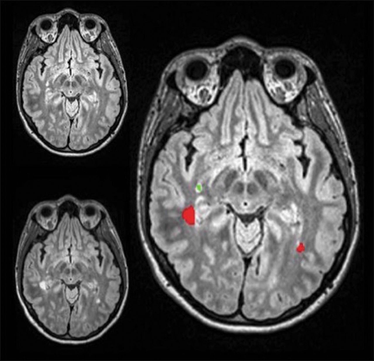
- Fig 1.
Prior FLAIR MR imaging in the upper left-hand corner, current FLAIR MR imaging in the lower left-hand corner, and the coregistered composite image on the right displaying new lesions in red, while lesions that regressed are green.
- Fig 2.
Left: Contrast-enhanced T1 MR imaging shows enhancement of a pre-existing lesion in the right centrum semiovale (arrow). Middle: The composite image from the coregistering software shows no growth of this corresponding lesion on FLAIR between the prior and current study (arrow). Additionally, 1 lesion had slightly regressed in the right posterior corona radiata (green on the composite image). Right: The composite image using the coregistering software at a more inferior level shows evidence of a new lesion in the splenium of the corpus callosum (arrow and red on composite image on the right).


Tables
Patients on IMT at the time of their most recent scan
Type of IMT No. of Patients Fingolimod 11 Glatiramer acetate 36 Interferon β-1a 23 Ocrelizumab 1 Dimethyl fumarate 1 Teriflunomide 9 Natalizumab 1 Note:—IMT indicates immune-modulating therapy.


Jump to section
Related Articles
Cited By...
- Accuracy of Noncontrast T2 SPACE in Active MS Cord Lesion Detection
- Nonlesional Sources of Contrast Enhancement on Postgadolinium "Black-Blood" 3D T1-SPACE Images in Patients with Multiple Sclerosis
- Comparison of Unenhanced and Gadolinium-Enhanced Imaging in Multiple Sclerosis: Is Contrast Needed for Routine Follow-Up MRI?