
Article Figures & Data
Figures
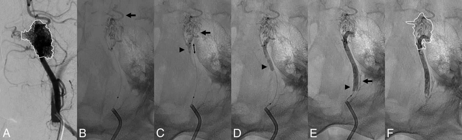
- Fig 1.
Representative embolization procedure with PHIL 25% LV. A, Before the embolization procedure, a diagnostic angiography is performed through a guiding catheter. The RM is delineated in the diagnostic angiography image with complete filling of the RM. B, X-ray after the first injection. After the first injection, most of the RM is already embolized. The injection is stopped because of embolization distal to the RM (arrow). C, X-ray after the second injection. The second injection leads to slight additional filling of the lateral parts of the RM (black arrow). The injection is stopped because of reflux (arrowhead). Retrospectively, this is the injection with which the maximal embolization extent was reached. Accordingly, the reflux distance is measured in this image (double arrow). X-rays after the fifth (D) and eighth (E) injections. No more filling of the RM is achieved with the following injections, which were all stopped because of reflux (arrowheads). After the eighth injection, the procedure is terminated because of reflux of the LEA into the APA (arrow). F, After termination of the procedure, the catheters are removed and the embolized portion of the RM is delineated. The area of the completely filled RM (A) and the area of the embolized RM (F) are related, resulting in the embolization extent.
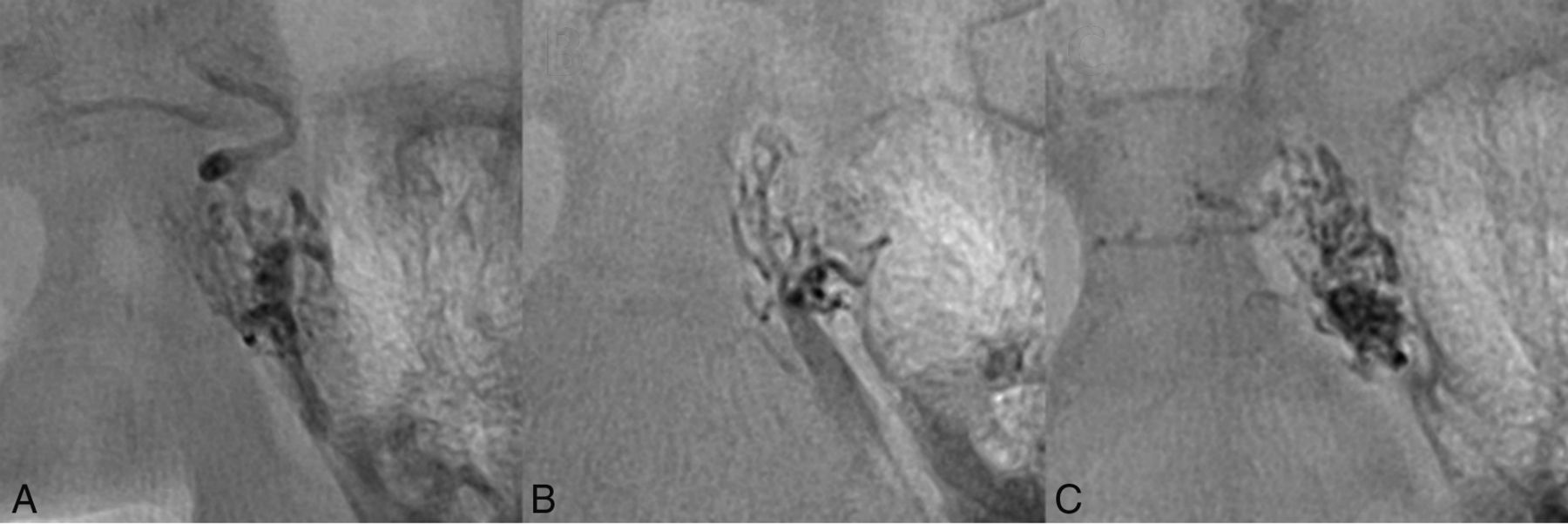
- Fig 2.
Visibility of the embolic agents. X-rays after the first injection shown for PHIL 25% LV (A), Squid 12 (B), and standard PHIL 25% (C). Note the adequate visibility of all 3 embolic agents.
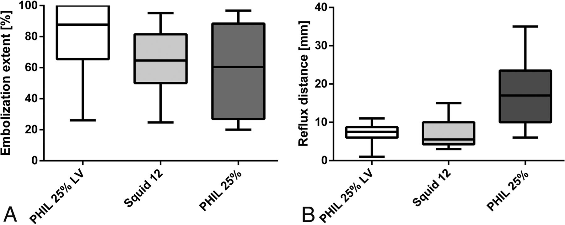
- Fig 3.
Illustration of embolization extent and reflux distance. A, The embolization extent tended to be higher for PHIL 25% LV, however, without reaching statistical significance. B, The reflux distance was significantly lower for the 2 extra-low-viscosity LEAs PHIL 25% LV and Squid 12 compared with standard PHIL 25%.
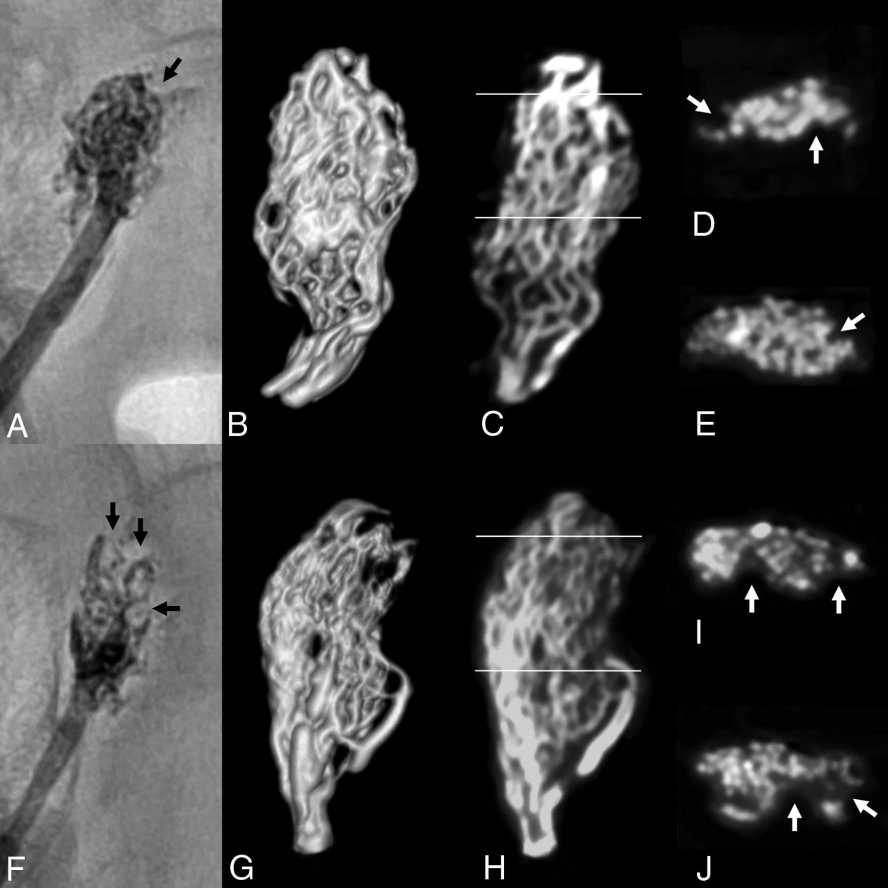
- Fig 4.
Distribution of the LEA in HR-DVT. The distribution of the LEA in HR-DVT is shown for 2 representative cases. A–E, PHIL 25% LV. F–J, Standard PHIL 25%. A and F, Postinterventional x-rays. B and G, Volume-rendering of the HR-DVT dataset. C and H, Coronal HR-DVT images. D, E, I, and J, Axial HR-DVT images of the middle (E and J) and the distal (D and I) parts of the RM (lines in C and H indicate the position of the axial planes). Note the perceptibility of single blood vessels of the RM in the high-resolution HR-DVT images. Well-circumscribed filling defects were identified in the axial HR-DVT images, which were particularly detected in the distal, only partially embolized parts of the RM (white arrows). The same filling defects were also seen in the 2D x-rays (black arrows).

Tables
Summary of the resultsa
PHIL 25% LV Squid 12 Standard PHIL 25% P Value Total procedure time (s) 395 (310–528) 436 (430–501) 328 (239–479) .386b Required volume of embolic agent (mL) 0.7 (0.7–0.9) 0.8 (0.7–0.9) 0.7 (0.6–0.7) .121b Visibility 3 (3–3) 3 (3–3) 3 (3–3) NA Forward flow control 5 (5–5) 5 (5–5) 5 (5–5) .335b Embolization extent (%) 87.7 (68.0–100) 64.6 (52.2–73.0) 60.4 (27.0–75.9) .146b Reflux distance (mm) 8 (6–8) 6 (5–10) 17 (14–21) .011b >.999c .049d .017e Events of embolization distal to the RM per procedure (No.) 0 (0–1) 0 (0–0) 0 (0–0) .527b Note:—NA indicates that the P value was not available because all values are identical.
↵a Data are presented as median (lower quartile–upper quartile).
↵b Kruskal-Wallis test.
↵c Post hoc Dunn test, PHIL 25% LV vs Squid 12.
↵d Post hoc Dunn test, PHIL 25% LV vs standard PHIL 25%.
↵e Post hoc Dunn test, Squid 12 vs. standard PHIL 25%.


Jump to section
Related Articles
Cited By...
- LIQUID - Treatment of high-grade dural arteriovenous fistulas with Squid liquid embolic agent: a prospective, observational multicenter study
- Experimental investigation of transvenous embolization of arteriovenous malformations using different in vivo models
- Visibility of liquid embolic agents in fluoroscopy: a systematic in vitro study
- Visibility of liquid embolic agents in fluoroscopy: a systematic in vitro study
- Experimental investigation of transvenous embolization of arteriovenous malformations using different in vivo models
- First clinical multicenter experience with the new Scepter Mini microballoon catheter
- Imaging Artifacts of Liquid Embolic Agents on Conventional CT in an Experimental in Vitro Model
- Intermixed Dimethyl-Sulfoxide-Based Nonadhesive Liquid Embolic Agents Delivered Serially via the Same Microcatheter for Cerebral AVM Treatment