Advertisement

Main menu

  • Home
  • Content
    • Current Issue
    • Publication Preview--Ahead of Print
    • Past Issue Archive
    • Case of the Week Archive
    • Classic Case Archive
    • Case of the Month Archive
  • For Authors
  • About Us
    • About AJNR
    • Editors
    • American Society of Neuroradiology
  • Submit a Manuscript
  • Podcasts
    • Subscribe on iTunes
    • Subscribe on Stitcher
  • More
    • Subscribers
    • Permissions
    • Advertisers
    • Alerts
    • Feedback
  • Other Publications
    • ajnr

User menu

  • Subscribe
  • Alerts
  • Log in

Search

  • Advanced search
American Journal of Neuroradiology

American Journal of Neuroradiology

  • Subscribe
  • Alerts
  • Log in

Advanced Search

  • Home
  • Content
    • Current Issue
    • Publication Preview--Ahead of Print
    • Past Issue Archive
    • Case of the Week Archive
    • Classic Case Archive
    • Case of the Month Archive
  • For Authors
  • About Us
    • About AJNR
    • Editors
    • American Society of Neuroradiology
  • Submit a Manuscript
  • Podcasts
    • Subscribe on iTunes
    • Subscribe on Stitcher
  • More
    • Subscribers
    • Permissions
    • Advertisers
    • Alerts
    • Feedback
  • Follow AJNR on Twitter
  • Visit AJNR on Facebook
  • Follow AJNR on Instagram
  • Join AJNR on LinkedIn
  • RSS Feeds
Research ArticlePediatrics

Persisting Embryonal Infundibular Recess in Morning Glory Syndrome: Clinical Report of a Novel Association

A. D'Amico, L. Ugga, R. Cuocolo, M. Cirillo, A. Grandone and R. Conforti
American Journal of Neuroradiology May 2019, 40 (5) 899-902; DOI: https://doi.org/10.3174/ajnr.A6005
A. D'Amico
aFrom the Department of Advanced Biomedical Sciences (A.D., L.U., R. Cuocolo), University of Naples “Federico II,” Naples, Italy
  • Find this author on Google Scholar
  • Find this author on PubMed
  • Search for this author on this site
  • ORCID record for A. D'Amico
L. Ugga
aFrom the Department of Advanced Biomedical Sciences (A.D., L.U., R. Cuocolo), University of Naples “Federico II,” Naples, Italy
  • Find this author on Google Scholar
  • Find this author on PubMed
  • Search for this author on this site
  • ORCID record for L. Ugga
R. Cuocolo
aFrom the Department of Advanced Biomedical Sciences (A.D., L.U., R. Cuocolo), University of Naples “Federico II,” Naples, Italy
  • Find this author on Google Scholar
  • Find this author on PubMed
  • Search for this author on this site
  • ORCID record for R. Cuocolo
M. Cirillo
bDipartimento di Scienze Mediche, Chirurgiche, Neurologiche (M.C.)
  • Find this author on Google Scholar
  • Find this author on PubMed
  • Search for this author on this site
  • ORCID record for M. Cirillo
A. Grandone
cMetaboliche e dell'Invecchiamento, Dipartimento della Donna (A.G.), del Bambino e di Chirurgia Generale e Specialistica
  • Find this author on Google Scholar
  • Find this author on PubMed
  • Search for this author on this site
  • ORCID record for A. Grandone
R. Conforti
dDipartimento di Medicina di Precisione (R. Conforti), University of Campania “Luigi Vanvitelli,” Naples, Italy.
  • Find this author on Google Scholar
  • Find this author on PubMed
  • Search for this author on this site
  • ORCID record for R. Conforti
  • Article
  • Figures & Data
  • Info & Metrics
  • References
  • PDF
Loading

Article Figures & Data

Figures

  • Fig 1.
    • Download figure
    • Open in new tab
    • Download powerpoint
    Fig 1.

    Sagittal contrast-enhanced T1-weighted (B) and high-resolution T2-weighted (D) images with corresponding stylized anatomic drawings (A and C) of patient 1 (upper row) and patient 2 (lower row) depicting the main findings reported in the text, including PEIR.

  • Fig 2.
    • Download figure
    • Open in new tab
    • Download powerpoint
    Fig 2.

    Coronal T2-weighted images of patient 1 (A) and patient 2 (B) showing thickening of the optic chiasm (arrows).
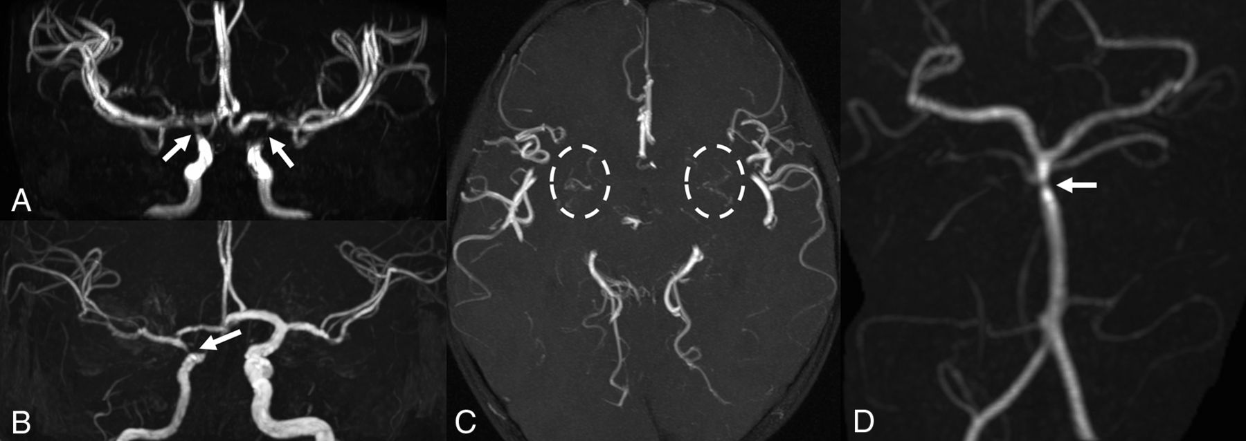
  • Fig 3.
    • Download figure
    • Open in new tab
    • Download powerpoint
    Fig 3.

    MR angiography. Patient 1 (A and C) has bilateral focal stenoses of the internal carotid arteries (arrows) with thin collateral vessels in the lenticulostriate regions (dashed circles). Patient 2 shows unilateral stenosis of the right internal carotid (B) and basilar (D) arteries (arrows).

PreviousNext
Back to top

In this issue

American Journal of Neuroradiology: 40 (5)
American Journal of Neuroradiology
Vol. 40, Issue 5
1 May 2019
  • Table of Contents
  • Index by author
  • Complete Issue (PDF)
Advertisement
Print
Download PDF
Email Article

Thank you for your interest in spreading the word on American Journal of Neuroradiology.

NOTE: We only request your email address so that the person you are recommending the page to knows that you wanted them to see it, and that it is not junk mail. We do not capture any email address.

Enter multiple addresses on separate lines or separate them with commas.
Persisting Embryonal Infundibular Recess in Morning Glory Syndrome: Clinical Report of a Novel Association
(Your Name) has sent you a message from American Journal of Neuroradiology
(Your Name) thought you would like to see the American Journal of Neuroradiology web site.
CAPTCHA
This question is for testing whether or not you are a human visitor and to prevent automated spam submissions.
Citation Tools
Persisting Embryonal Infundibular Recess in Morning Glory Syndrome: Clinical Report of a Novel Association
A. D'Amico, L. Ugga, R. Cuocolo, M. Cirillo, A. Grandone, R. Conforti
American Journal of Neuroradiology May 2019, 40 (5) 899-902; DOI: 10.3174/ajnr.A6005

Citation Manager Formats

  • BibTeX
  • Bookends
  • EasyBib
  • EndNote (tagged)
  • EndNote 8 (xml)
  • Medlars
  • Mendeley
  • Papers
  • RefWorks Tagged
  • Ref Manager
  • RIS
  • Zotero
Share
Persisting Embryonal Infundibular Recess in Morning Glory Syndrome: Clinical Report of a Novel Association
A. D'Amico, L. Ugga, R. Cuocolo, M. Cirillo, A. Grandone, R. Conforti
American Journal of Neuroradiology May 2019, 40 (5) 899-902; DOI: 10.3174/ajnr.A6005
del.icio.us logo Digg logo Reddit logo Twitter logo Facebook logo Google logo Mendeley logo
  • Tweet Widget
  • Facebook Like
  • Google Plus One
Purchase

Jump to section

  • Article
    • Abstract
    • ABBREVIATIONS:
    • Discussion
    • Conclusions
    • Acknowledgments
    • REFERENCES
  • Figures & Data
  • Info & Metrics
  • References
  • PDF

Related Articles

  • No related articles found.
  • PubMed
  • Google Scholar

Cited By...

  • No citing articles found.
  • Crossref
  • Google Scholar

This article has not yet been cited by articles in journals that are participating in Crossref Cited-by Linking.

More in this TOC Section

Pediatrics

  • Arterioectatic Spinal Angiopathy of Childhood: Clinical, Imaging, Laboratory, Histologic, and Genetic Description of a Novel CNS Vascular Pathology
  • Brain Abnormalities in Patients with Germline Variants in H3F3: Novel Imaging Findings and Neurologic Symptoms Beyond Somatic Variants and Brain Tumors
  • Asymmetry Matters: Diffusion Tensor Tractography of the Uncinate Fasciculus in Children with Verbal Memory Deficits
Show more Pediatrics

Head & Neck

  • Glymphatic System in Ocular Diseases: Evaluation of MRI Findings
  • Do We Need Gadolinium-Based Contrast Agents for Routine MRI Surveillance of Unoperated Pituitary Macroadenoma?
  • Adding MR Diffusion Imaging and T2 Signal Intensity to Neck Imaging Reporting and Data System Categories 2 and 3 in Primary Sites of Postsurgical Oral Cavity Carcinoma Provides Incremental Diagnostic Value
Show more Head & Neck

Similar Articles

Advertisement

News and Updates

  • Lucien Levy Best Research Article Award
  • Thanks to our 2021 Distinguished Reviewers
  • Press Releases

Resources

  • Evidence-Based Medicine Level Guide
  • How to Participate in a Tweet Chat
  • AJNR Podcast Archive
  • Ideas for Publicizing Your Research
  • Librarian Resources
  • Terms and Conditions

Opportunities

  • Share Your Art in Perspectives
  • Get Peer Review Credit from Publons
  • Moderate a Tweet Chat

American Society of Neuroradiology

  • Neurographics
  • ASNR Annual Meeting
  • Fellowship Portal
  • Position Statements

© 2022 by the American Society of Neuroradiology | Print ISSN: 0195-6108 Online ISSN: 1936-959X

Powered by HighWire