Advertisement

Main menu

  • Home
  • Content
    • Current Issue
    • Publication Preview--Ahead of Print
    • Past Issue Archive
    • Case of the Week Archive
    • Classic Case Archive
    • Case of the Month Archive
  • For Authors
  • About Us
    • About AJNR
    • Editors
    • American Society of Neuroradiology
  • Submit a Manuscript
  • Podcasts
    • Subscribe on iTunes
    • Subscribe on Stitcher
  • More
    • Subscribers
    • Permissions
    • Advertisers
    • Alerts
    • Feedback
  • Other Publications
    • ajnr

User menu

  • Subscribe
  • Alerts
  • Log in

Search

  • Advanced search
American Journal of Neuroradiology

American Journal of Neuroradiology

  • Subscribe
  • Alerts
  • Log in

Advanced Search

  • Home
  • Content
    • Current Issue
    • Publication Preview--Ahead of Print
    • Past Issue Archive
    • Case of the Week Archive
    • Classic Case Archive
    • Case of the Month Archive
  • For Authors
  • About Us
    • About AJNR
    • Editors
    • American Society of Neuroradiology
  • Submit a Manuscript
  • Podcasts
    • Subscribe on iTunes
    • Subscribe on Stitcher
  • More
    • Subscribers
    • Permissions
    • Advertisers
    • Alerts
    • Feedback
  • Follow AJNR on Twitter
  • Visit AJNR on Facebook
  • Follow AJNR on Instagram
  • Join AJNR on LinkedIn
  • RSS Feeds
Research ArticlePediatrics

Comparison of CSF and MRI Findings among Neonates and Infants with E coli or Group B Streptococcal Meningitis

S.F. Kralik, M.K. Kukreja, M.J. Paldino, N.K. Desai and J.G. Vallejo
American Journal of Neuroradiology August 2019, 40 (8) 1413-1417; DOI: https://doi.org/10.3174/ajnr.A6134
S.F. Kralik
aFrom the Department of Radiology (S.F.K., M.K.K., M.J.P., N.K.D.), Texas Children's Hospital, Houston, Texas
bIndiana University Riley Children's Hospital (S.F.K.), Indianapolis, Indiana
  • Find this author on Google Scholar
  • Find this author on PubMed
  • Search for this author on this site
  • ORCID record for S.F. Kralik
M.K. Kukreja
aFrom the Department of Radiology (S.F.K., M.K.K., M.J.P., N.K.D.), Texas Children's Hospital, Houston, Texas
  • Find this author on Google Scholar
  • Find this author on PubMed
  • Search for this author on this site
  • ORCID record for M.K. Kukreja
M.J. Paldino
aFrom the Department of Radiology (S.F.K., M.K.K., M.J.P., N.K.D.), Texas Children's Hospital, Houston, Texas
  • Find this author on Google Scholar
  • Find this author on PubMed
  • Search for this author on this site
  • ORCID record for M.J. Paldino
N.K. Desai
aFrom the Department of Radiology (S.F.K., M.K.K., M.J.P., N.K.D.), Texas Children's Hospital, Houston, Texas
  • Find this author on Google Scholar
  • Find this author on PubMed
  • Search for this author on this site
  • ORCID record for N.K. Desai
J.G. Vallejo
cDepartment of Pediatrics (J.G.V.), Baylor College of Medicine, Houston, Texas.
  • Find this author on Google Scholar
  • Find this author on PubMed
  • Search for this author on this site
  • ORCID record for J.G. Vallejo
  • Article
  • Figures & Data
  • Info & Metrics
  • References
  • PDF
Loading

Article Figures & Data

Figures

  • Tables
  • Fig 1.
    • Download figure
    • Open in new tab
    • Download powerpoint
    Fig 1.

    A 12-day-old neonate with GBS meningitis. Axial DWI demonstrates multifocal areas of diffusion restriction (ADC not shown) in the parenchyma, consistent with infarcts and cerebritis.

  • Fig 2.
    • Download figure
    • Open in new tab
    • Download powerpoint
    Fig 2.

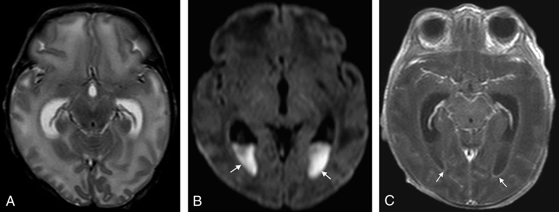
    A 19-day-old neonate with E coli meningitis. A, Axial T2-weighted image demonstrates disproportionate enlargement of the lateral ventricles relative to the sulci, consistent with hydrocephalus. B, Axial DWI demonstrates areas of diffusion restriction (ADC not shown) in the ventricles (arrows), consistent with intraventricular purulent material. C, Axial T1-weighted postcontrast image demonstrates abnormal ependymal enhancement along the lateral ventricles (arrows).

Tables

  • Figures
  • Comparison of CSF and MRI findings in patients with GBS and E coli meningitisa

    FindingGBS (n = 57)E coli (n = 50)P Valueb
    CSF corrected WBC count (cells/μL)3116 ± 50584770 ± 126841.00
    CSF glucose (mg/dL)35.2 ± 21.834.0 ± 24.61.00
    CSF total protein (mg/dL)517.8 ± 867.0910.7 ± 1414.21.00
    Leptomeningeal enhancement49%46%1.00
    Cerebritis21%7%.77
    Ependymal enhancement7%28%.063
    Abscess/granuloma2%2%1.00
    Subdural effusion49%26%.24
    Hemorrhage33%46%1.00
    Extra-axial purulent material37%38%1.00
    Intraventricular purulent material9%22%.84
    Sinus thrombosis11%4%1.00
    Hydrocephalus0%22%.0014
    Infarct40%14%.038
    • ↵a CSF values are listed as mean ± SD.

    • ↵b P value is adjusted for multiple comparisons (n = 14).

PreviousNext
Back to top

In this issue

American Journal of Neuroradiology: 40 (8)
American Journal of Neuroradiology
Vol. 40, Issue 8
1 Aug 2019
  • Table of Contents
  • Index by author
  • Complete Issue (PDF)
Advertisement
Print
Download PDF
Email Article

Thank you for your interest in spreading the word on American Journal of Neuroradiology.

NOTE: We only request your email address so that the person you are recommending the page to knows that you wanted them to see it, and that it is not junk mail. We do not capture any email address.

Enter multiple addresses on separate lines or separate them with commas.
Comparison of CSF and MRI Findings among Neonates and Infants with E coli or Group B Streptococcal Meningitis
(Your Name) has sent you a message from American Journal of Neuroradiology
(Your Name) thought you would like to see the American Journal of Neuroradiology web site.
CAPTCHA
This question is for testing whether or not you are a human visitor and to prevent automated spam submissions.
Citation Tools
Comparison of CSF and MRI Findings among Neonates and Infants with E coli or Group B Streptococcal Meningitis
S.F. Kralik, M.K. Kukreja, M.J. Paldino, N.K. Desai, J.G. Vallejo
American Journal of Neuroradiology Aug 2019, 40 (8) 1413-1417; DOI: 10.3174/ajnr.A6134

Citation Manager Formats

  • BibTeX
  • Bookends
  • EasyBib
  • EndNote (tagged)
  • EndNote 8 (xml)
  • Medlars
  • Mendeley
  • Papers
  • RefWorks Tagged
  • Ref Manager
  • RIS
  • Zotero
Share
Comparison of CSF and MRI Findings among Neonates and Infants with E coli or Group B Streptococcal Meningitis
S.F. Kralik, M.K. Kukreja, M.J. Paldino, N.K. Desai, J.G. Vallejo
American Journal of Neuroradiology Aug 2019, 40 (8) 1413-1417; DOI: 10.3174/ajnr.A6134
del.icio.us logo Digg logo Reddit logo Twitter logo CiteULike logo Facebook logo Google logo Mendeley logo
  • Tweet Widget
  • Facebook Like
  • Google Plus One
Purchase

Jump to section

  • Article
    • Abstract
    • ABBREVIATIONS:
    • Materials and Methods
    • Results
    • Discussion
    • Conclusions
    • Footnotes
    • REFERENCES
  • Figures & Data
  • Info & Metrics
  • References
  • PDF

Related Articles

  • No related articles found.
  • PubMed
  • Google Scholar

Cited By...

  • No citing articles found.
  • Crossref
  • Google Scholar

This article has not yet been cited by articles in journals that are participating in Crossref Cited-by Linking.

More in this TOC Section

  • Neuroradiologic Phenotyping of Galactosemia: From the Neonatal Form to the Chronic Stage
  • Impact of Prematurity on the Tissue Properties of the Neonatal Brain Stem: A Quantitative MR Approach
  • Neuroimaging Offers Low Yield in Children Positive for SARS-CoV-2
Show more PEDIATRICS

Similar Articles

Advertisement

News and Updates

  • Lucien Levy Best Research Article Award
  • Thanks to our 2020 Distinguished Reviewers
  • Press Releases

Resources

  • Evidence-Based Medicine Level Guide
  • How to Participate in a Tweet Chat
  • AJNR Podcast Archive
  • Ideas for Publicizing Your Research
  • Librarian Resources
  • Terms and Conditions

Opportunities

  • Share Your Art in Perspectives
  • Get Peer Review Credit from Publons
  • Moderate a Tweet Chat

American Society of Neuroradiology

  • Neurographics
  • ASNR Annual Meeting
  • Fellowship Portal
  • Position Statements

© 2021 by the American Society of Neuroradiology | Print ISSN: 0195-6108 Online ISSN: 1936-959X

Powered by HighWire