Advertisement

Main menu

  • Home
  • Content
    • Current Issue
    • Publication Preview--Ahead of Print
    • Past Issue Archive
    • Case of the Week Archive
    • Classic Case Archive
    • Case of the Month Archive
  • For Authors
  • About Us
    • About AJNR
    • Editors
    • American Society of Neuroradiology
  • Submit a Manuscript
  • Podcasts
    • Subscribe on iTunes
    • Subscribe on Stitcher
  • More
    • Subscribers
    • Permissions
    • Advertisers
    • Alerts
    • Feedback
  • Other Publications
    • ajnr

User menu

  • Subscribe
  • Alerts
  • Log in

Search

  • Advanced search
American Journal of Neuroradiology

American Journal of Neuroradiology

  • Subscribe
  • Alerts
  • Log in

Advanced Search

  • Home
  • Content
    • Current Issue
    • Publication Preview--Ahead of Print
    • Past Issue Archive
    • Case of the Week Archive
    • Classic Case Archive
    • Case of the Month Archive
  • For Authors
  • About Us
    • About AJNR
    • Editors
    • American Society of Neuroradiology
  • Submit a Manuscript
  • Podcasts
    • Subscribe on iTunes
    • Subscribe on Stitcher
  • More
    • Subscribers
    • Permissions
    • Advertisers
    • Alerts
    • Feedback
  • Follow AJNR on Twitter
  • Visit AJNR on Facebook
  • Follow AJNR on Instagram
  • Join AJNR on LinkedIn
  • RSS Feeds
LetterLETTER

Intraoperative MR and Synthetic Imaging

M.I. Vargas, B.M.A. Delattre, P. Vayssiere, M. Corniola and T. Meling
American Journal of Neuroradiology February 2020, 41 (2) E4-E6; DOI: https://doi.org/10.3174/ajnr.A6373
M.I. Vargas
aDivision of Neuroradiology
  • Find this author on Google Scholar
  • Find this author on PubMed
  • Search for this author on this site
  • ORCID record for M.I. Vargas
B.M.A. Delattre
bDivision of Radiology
  • Find this author on Google Scholar
  • Find this author on PubMed
  • Search for this author on this site
  • ORCID record for B.M.A. Delattre
P. Vayssiere
cDivision of Neurosurgery Geneva University Hospital and Faculty of Medicine of Geneva Geneva, Switzerland
  • Find this author on Google Scholar
  • Find this author on PubMed
  • Search for this author on this site
  • ORCID record for P. Vayssiere
M. Corniola
cDivision of Neurosurgery Geneva University Hospital and Faculty of Medicine of Geneva Geneva, Switzerland
  • Find this author on Google Scholar
  • Find this author on PubMed
  • Search for this author on this site
  • ORCID record for M. Corniola
T. Meling
cDivision of Neurosurgery Geneva University Hospital and Faculty of Medicine of Geneva Geneva, Switzerland
  • Find this author on Google Scholar
  • Find this author on PubMed
  • Search for this author on this site
  • ORCID record for T. Meling
  • Article
  • Figures & Data
  • Info & Metrics
  • References
  • PDF
Loading

In the past 2 decades, intraoperative MRI (ioMRI) has been implemented in surgery for pituitary tumors, low-grade gliomas,1 epilepsy, and deep brain stimulation. In neuro-oncological surgery, the adjunct of ioMRI improves the extent of resection, which eventually results in improved overall and progression-free survival.2 However, despite encouraging results, there are no specific sequences dedicated to ioMRI to our knowledge.

In the neuroradiologist’s point of view, one of the main difficulties is to find the most useful and time-effective sequence to guide the surgeon in determining the existence of residual tumor tissue. Often, multiple series before and after the administration of a contrast agent are used. In our opinion, this time-consuming step is not compatible with a surgical context in which time for imaging is limited. This restricts the neuroradiologist, especially in complex cases. One potential solution might reside in the use of so-called synthetic imaging (SyntheticMR; SyntheticMR, Linköping, Sweden), which allows us to obtain 1) different types of contrast, and 2) quantitative information, such as T1 and T2 mapping of the tissues.3,4

The use of SyntheticMR in low-grade tumors could represent a real shift of paradigm. Due to edema, air, blood, and immediate postoperative changes, these lesions do not allow a clear delimitation of the tumor border solely by visual analysis, and remaining tumor may thus be missed. Having multiple contrasts in a single sequence by using SyntheticMR may aid the interpretation, in a reduced acquisition time (7 min). The quantitative values may also be of great interest; indeed, synthetic imaging provides several types of cartography, such as cerebral cortex, CSF, white matter, and other tissues, as shown in Fig 1. Quantitative values of T1, T2, and proton density of the tissues are also available. It has already been shown that relaxation rates can be used to grade gliomas,5 but, at that time, there were no sequences with short acquisition times. Recently, this finding was confirmed with an MR fingerprinting technique.6 The latter sequence is under development and therefore, not fully available, contrary to SyntheticMR. In Fig 2, we show that T2 measured with SyntheticMR in the case of a World Health Organization grade I left occipital meningioma is different between the lesion and the cerebral cortex. In our opinion, this principle could be used to help delineate remnant tumor tissue also in low-grade tumors.

FIG 1.
  • Download figure
  • Open in new tab
  • Download powerpoint
FIG 1.

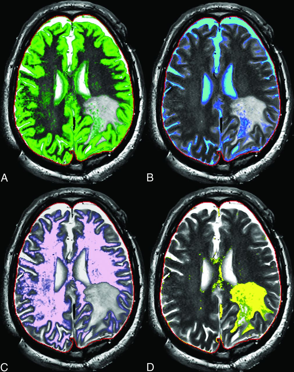
Intraoperative Synthetic MR cartographies illustrating gray matter (green in A), CSF (blue in B), white matter (pink in C), and edema (yellow in D).

FIG 2.
  • Download figure
  • Open in new tab
  • Download powerpoint
FIG 2.

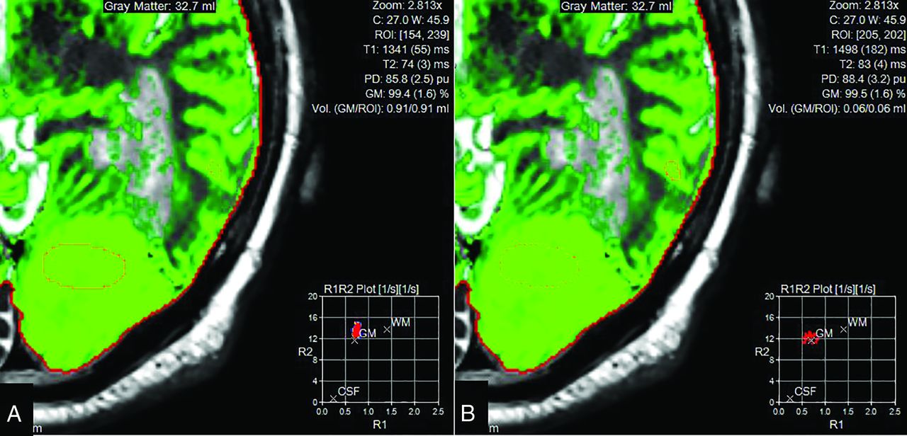
Region of interest in the tumor (A), showing decreased T2 values in rapport to gray matter (B) and, on the contrary, T1 values similar to the gray matter.

We, therefore, think that SyntheticMR could be beneficial for ioMRI by providing multiple contrasts and relaxation-rate quantification to give a reliable interpretation of the presence of residual tumor, thus improving patient care.

Acknowledgement

The authors thank IMRIS. The SyntheticMR sequence (MDME) was provided by Karolinska University Hospital, developed in collaboration with Synthetic MR AB and Frederik Testud, Siemens Healthcare AB, Sweden.

Footnotes

  • Disclosures: None.

References

  1. 1.↵
    1. Coburger J,
    2. Merkel A,
    3. Scherer M, et al
    . Low-grade glioma surgery in intraoperative magnetic resonance imaging: results of a multicenter retrospective assessment of the German study group for intraoperative magnetic resonance imaging. Neurosurgery 2016;78:775–86 doi:10.1227/NEU.0000000000001081 pmid:26516822
    CrossRefPubMed
  2. 2.↵
    1. Senft C,
    2. Bink A,
    3. Franz K, et al
    . Intraoperative MRI guidance and extent of resection in glioma surgery: a randomised, controlled trial. Lancet Oncol 2011;12:997–1003 doi:10.1016/S1470-2045(11)70196-6 pmid:21868284
    CrossRefPubMed
  3. 3.↵
    1. Warntjes JBM,
    2. Leinhard OD,
    3. West J, et al
    . Rapid magnetic resonance quantification on the brain: Optimization for clinical usage. Magn. Reson. Med. 2008;60:320–9 doi:10.1002/mrm.21635 pmid:18666127
    CrossRefPubMed
  4. 4.↵
    1. Granberg T,
    2. Uppman M,
    3. Hashim F, et al
    . Clinical Feasibility of Synthetic MRI in Multiple Sclerosis: A Diagnostic and Volumetric Validation Study. Am. J. Neuroradiol. 2016;37:1023–9 doi:10.3174/ajnr.A4665 pmid:26797137
    Abstract/FREE Full Text
  5. 5.↵
    1. Newman S,
    2. Haughton VM,
    3. Yetkin Z, et al
    . TI, T2 and proton density measurements in the grading of cerebral gliomas. Eur Radiol 1993;49–52
  6. 6.↵
    1. Badve C,
    2. Yu A,
    3. Dastmalchian S, et al
    . MR fingerprinting of adult brain tumors: initial experience. AJNR Am J Neuroradiol 2017;38:492–99 doi:10.3174/ajnr.A5035 pmid:28034994
    Abstract/FREE Full Text
  • © 2020 by American Journal of Neuroradiology
PreviousNext
Back to top

In this issue

American Journal of Neuroradiology: 41 (2)
American Journal of Neuroradiology
Vol. 41, Issue 2
1 Feb 2020
  • Table of Contents
  • Index by author
  • Complete Issue (PDF)
Advertisement
Print
Download PDF
Email Article

Thank you for your interest in spreading the word on American Journal of Neuroradiology.

NOTE: We only request your email address so that the person you are recommending the page to knows that you wanted them to see it, and that it is not junk mail. We do not capture any email address.

Enter multiple addresses on separate lines or separate them with commas.
Intraoperative MR and Synthetic Imaging
(Your Name) has sent you a message from American Journal of Neuroradiology
(Your Name) thought you would like to see the American Journal of Neuroradiology web site.
CAPTCHA
This question is for testing whether or not you are a human visitor and to prevent automated spam submissions.
Citation Tools
Intraoperative MR and Synthetic Imaging
M.I. Vargas, B.M.A. Delattre, P. Vayssiere, M. Corniola, T. Meling
American Journal of Neuroradiology Feb 2020, 41 (2) E4-E6; DOI: 10.3174/ajnr.A6373

Citation Manager Formats

  • BibTeX
  • Bookends
  • EasyBib
  • EndNote (tagged)
  • EndNote 8 (xml)
  • Medlars
  • Mendeley
  • Papers
  • RefWorks Tagged
  • Ref Manager
  • RIS
  • Zotero
Share
Intraoperative MR and Synthetic Imaging
M.I. Vargas, B.M.A. Delattre, P. Vayssiere, M. Corniola, T. Meling
American Journal of Neuroradiology Feb 2020, 41 (2) E4-E6; DOI: 10.3174/ajnr.A6373
del.icio.us logo Digg logo Reddit logo Twitter logo CiteULike logo Facebook logo Google logo Mendeley logo
  • Tweet Widget
  • Facebook Like
  • Google Plus One
Purchase

Jump to section

  • Article
    • Acknowledgement
    • Footnotes
    • References
  • Figures & Data
  • Info & Metrics
  • References
  • PDF

Related Articles

  • No related articles found.
  • PubMed
  • Google Scholar

Cited By...

  • No citing articles found.
  • Crossref
  • Google Scholar

This article has not yet been cited by articles in journals that are participating in Crossref Cited-by Linking.

More in this TOC Section

  • Acute Stroke Imaging in Transfer Patients Who Received Recent Intravenous Iodinated Contrast at an Outside Facility: An Unrecognized Problem That Deserves More Attention
  • Primary Angiitis of the CNS with Unremarkable Vessel Wall MR Imaging: How the “T1 Shinethrough” Effect on SWI Adds to the Detection of Gadolinium Enhancement of Small Intraparenchymal Brain Vessels
  • Seeing What We Expect to See in COVID-19
Show more LETTERS

Similar Articles

Advertisement

News and Updates

  • Lucien Levy Best Research Article Award
  • Thanks to our 2020 Distinguished Reviewers
  • Press Releases

Resources

  • Evidence-Based Medicine Level Guide
  • How to Participate in a Tweet Chat
  • AJNR Podcast Archive
  • Ideas for Publicizing Your Research
  • Librarian Resources
  • Terms and Conditions

Opportunities

  • Share Your Art in Perspectives
  • Get Peer Review Credit from Publons
  • Moderate a Tweet Chat

American Society of Neuroradiology

  • Neurographics
  • ASNR Annual Meeting
  • Fellowship Portal
  • Position Statements

© 2021 by the American Society of Neuroradiology | Print ISSN: 0195-6108 Online ISSN: 1936-959X

Powered by HighWire