
Article Figures & Data
Figures
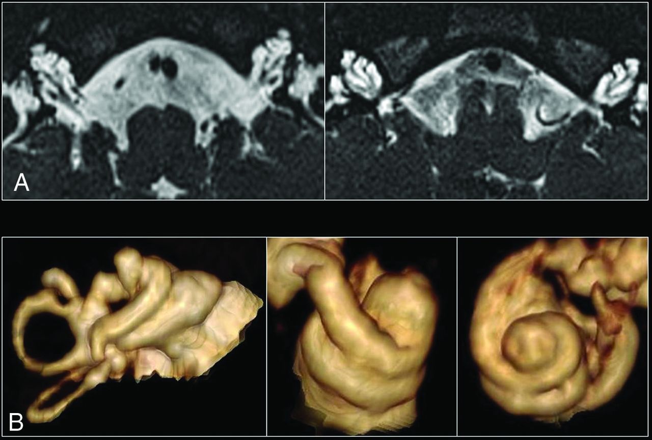
- Fig 1.
A, High-resolution axial T2 images of both inner ears in a patient with incomplete partition type III. The 3 main features include a dilated internal auditory canal, incomplete separation of the basal turn of the cochlea from the internal auditory canal, with an absent lamina cribrosa and modiolus. B, 3D reconstructions of several cochleae with incomplete partition type III, which demonstrate the presence of interscalar septa and cochlear turns.
- Fig 2.
A, Axial T2 image of a normal hypothalamus at the level of the optic radiations. B–G, Axial T2 images of the hypothalamus in a patient with DFNX2, which demonstrate progressive folding of the ventromedial hypothalamus. Note the presence of bilateral clefts in most cases, with external clefts being more easily recognizable (arrows). Internal clefts are also noted on B and C (arrowheads).
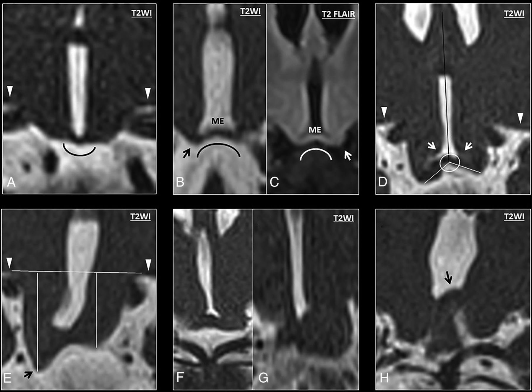
- Fig 3.
A, Coronal T2 anatomy of a normal hypothalamus showing the classic convex morphology of the medial eminence. B–H, High-resolution coronal T2 images show progressive bending of the ventromedial hypothalamus (from mild to severe). B, High-resolution coronal T2 image shows abnormal concavity at the junction between the ventromedial hypothalamus and the medial eminence, with a low-lying ventromedial hypothalamus (black arrow) in relation to the medial eminence (ME). These findings show an abnormal concave morphology of the hypothalamus in relation to the pituitary gland. C, Coronal T2 FLAIR image of another patient with DFNX2, which demonstrates isointense signal compared with the adjacent globus pallidus. Note again the characteristic bending of the ventromedial hypothalamus and its caudal location in relation to the medial eminence (white arrow). D, Measurement of the angle between the tip of the ventromedial hypothalamus and the septum pellucidum (white arrows). E, Measurement of the craniocaudal length of the ventromedial hypothalamus at the lowest point (black arrow) in relation to the basal forebrain (horizontal line). The basal forebrain (white arrowheads) is also indicated on images A and D. F and G, In some cases, the low-lying hypothalamus and the folding are so severe that some hypothalamic segments appear masslike, though the overall appearance is in keeping with diffuse folding. H, Severe hypothalamic bending in a patient with DFNX2, which shows cranial folding of the hypothalamus (black arrow) apart from the typical low-lying or hanging infundibular nucleus (not shown in this image).
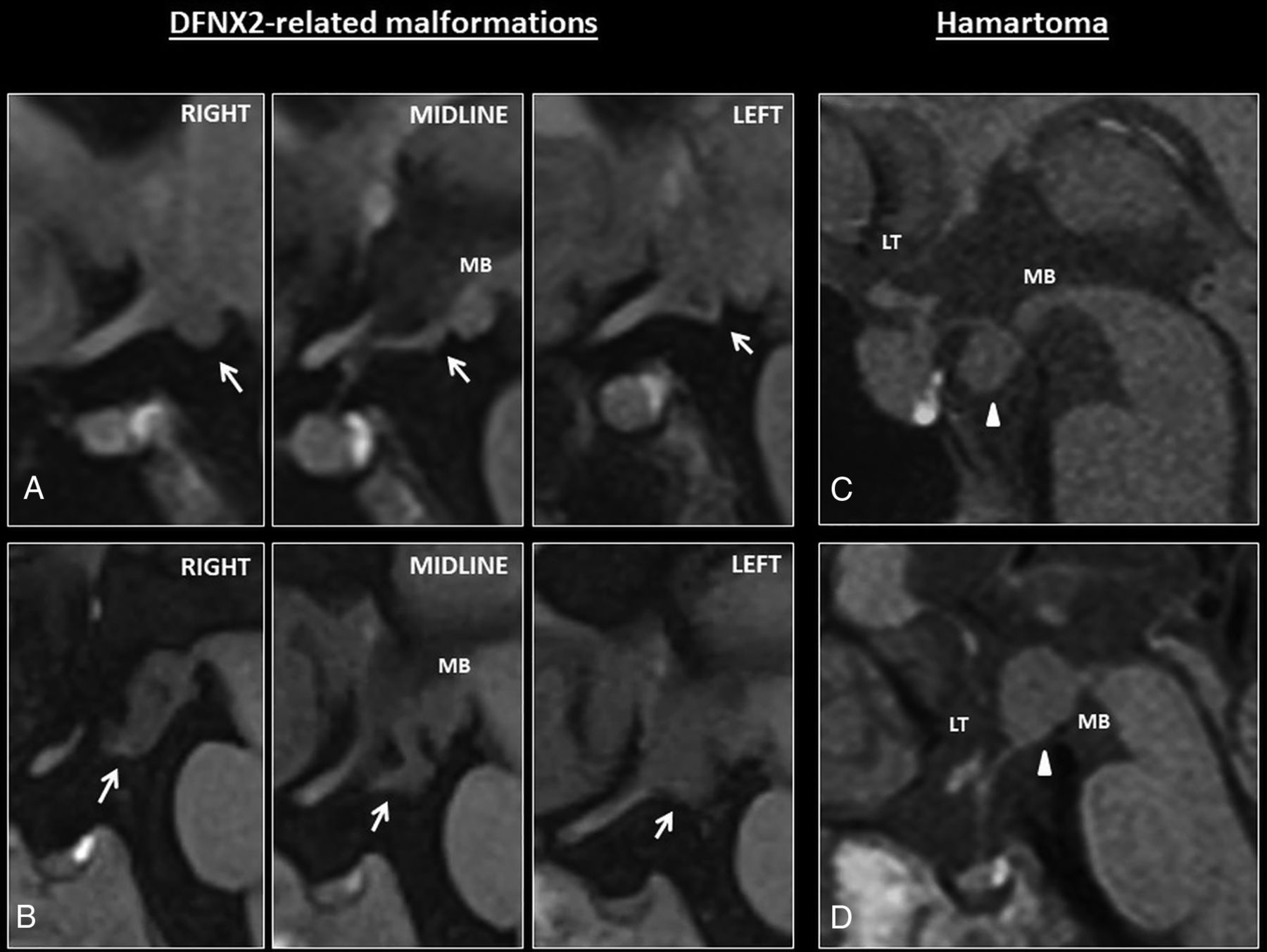
- Fig 4.
A, Sagittal T1 images show mild hypothalamic folding in a patient with DFNX2 (white arrows). This is usually apparent on one of the sagittal slices, such as the left/right one on these series; however, coronal images are better to depict subtle hypothalamic folding. Note the T1-isointense signal of the hypothalamic folding. B, The folding is apparent on all the sagittal images through the hypothalamus. Note again the T1-isointense signal compared with the adjacent brain parenchyma. C–D, Hypothalamic hamartomas tend to be masslike rather than cause hypothalamic folding. They usually arise from the tuber cinereum protruding caudally toward the suprasellar cistern or grow within the third ventricle (white arrowheads) and tend to involve adjacent structures such as the mamillary bodies. MB indicates mamillary bodies; LT, lamina terminalis.


Tables
Proposed MR imaging features of hypothalamic malformations in patients with DFNX2 compared with age-matched controlsa
Patients with DFNX2 (n = 10) Age-Matched Controls (n = 69) Axial T2 Folded appearance 70% (P < .001; κ = 0.95) 0% Bilateral abnormal internal or external cleft 60% (P < .001; κ = 0.83) 0% Coronal T2 Concave medial eminence 75% (P < .001; κ = 0.78) 0% Right hypothalamic-septum angle 115.5° ± 17.34° (P < .001) 79° ± 8.05° ICC = 0.88 (95% CI, 0.75–0.95) Left hypothalamic-septum angle 113.87° ± 16.87° (P < .001) 81.87° ± 9.32° ICC = 0.77 (95% CI, 0.58–0.89) Right forebrain-hypothalamus length (mm) 7.15 ± 3.02 (P < .001) 4.26 ± 0.7 ICC = 0.92 (95% CI, 0.70–0.97) Left forebrain-hypothalamus length (mm) 6.9 ± 2.1 (P < .001) 4.21 ± 0.73 ICC = 0.87 (95% CI, 0.70–0.95) Note:—κ indicates the Cohen κ coefficient.
↵a Data are mean values ± standard deviation. P values correspond to Wilcoxon/Mann-Withney test for differences in means of DFNX2 versus age-matched controls.


Jump to section
Related Articles
Cited By...
- No citing articles found.