Advertisement

Main menu

  • Home
  • Content
    • Current Issue
    • Publication Preview--Ahead of Print
    • Past Issue Archive
    • Case of the Week Archive
    • Classic Case Archive
    • Case of the Month Archive
  • For Authors
  • About Us
    • About AJNR
    • Editors
    • American Society of Neuroradiology
  • Submit a Manuscript
  • Podcasts
    • Subscribe on iTunes
    • Subscribe on Stitcher
  • More
    • Subscribers
    • Permissions
    • Advertisers
    • Alerts
    • Feedback
  • Other Publications
    • ajnr

User menu

  • Subscribe
  • Alerts
  • Log in

Search

  • Advanced search
American Journal of Neuroradiology

American Journal of Neuroradiology

  • Subscribe
  • Alerts
  • Log in

Advanced Search

  • Home
  • Content
    • Current Issue
    • Publication Preview--Ahead of Print
    • Past Issue Archive
    • Case of the Week Archive
    • Classic Case Archive
    • Case of the Month Archive
  • For Authors
  • About Us
    • About AJNR
    • Editors
    • American Society of Neuroradiology
  • Submit a Manuscript
  • Podcasts
    • Subscribe on iTunes
    • Subscribe on Stitcher
  • More
    • Subscribers
    • Permissions
    • Advertisers
    • Alerts
    • Feedback
  • Follow AJNR on Twitter
  • Visit AJNR on Facebook
  • Follow AJNR on Instagram
  • Join AJNR on LinkedIn
  • RSS Feeds
Research ArticleHead & Neck

The Pharyngolaryngeal Venous Plexus: A Potential Pitfall in Surveillance Imaging of the Neck

P.M. Bunch, R.T. Hughes, E.P. White, J.R. Sachs, B.A. Frizzell and C.M. Lack
American Journal of Neuroradiology May 2021, 42 (5) 938-944; DOI: https://doi.org/10.3174/ajnr.A7033
P.M. Bunch
aFrom the Department of Radiology (P.M.B., E.P.W., J.R.S., C.M.L.), Wake Forest School of Medicine, Winston Salem, North Carolina
  • Find this author on Google Scholar
  • Find this author on PubMed
  • Search for this author on this site
  • ORCID record for P.M. Bunch
R.T. Hughes
bDepartment of Radiation Oncology (R.T.H., B.A.F.), Wake Forest School of Medicine, Winston Salem, North Carolina.
  • Find this author on Google Scholar
  • Find this author on PubMed
  • Search for this author on this site
  • ORCID record for R.T. Hughes
E.P. White
aFrom the Department of Radiology (P.M.B., E.P.W., J.R.S., C.M.L.), Wake Forest School of Medicine, Winston Salem, North Carolina
  • Find this author on Google Scholar
  • Find this author on PubMed
  • Search for this author on this site
  • ORCID record for E.P. White
J.R. Sachs
aFrom the Department of Radiology (P.M.B., E.P.W., J.R.S., C.M.L.), Wake Forest School of Medicine, Winston Salem, North Carolina
  • Find this author on Google Scholar
  • Find this author on PubMed
  • Search for this author on this site
  • ORCID record for J.R. Sachs
B.A. Frizzell
bDepartment of Radiation Oncology (R.T.H., B.A.F.), Wake Forest School of Medicine, Winston Salem, North Carolina.
  • Find this author on Google Scholar
  • Find this author on PubMed
  • Search for this author on this site
  • ORCID record for B.A. Frizzell
C.M. Lack
aFrom the Department of Radiology (P.M.B., E.P.W., J.R.S., C.M.L.), Wake Forest School of Medicine, Winston Salem, North Carolina
  • Find this author on Google Scholar
  • Find this author on PubMed
  • Search for this author on this site
  • ORCID record for C.M. Lack
  • Article
  • Figures & Data
  • Info & Metrics
  • References
  • PDF
Loading

Article Figures & Data

Figures

  • Tables
  • FIG 1.
    • Download figure
    • Open in new tab
    • Download powerpoint
    FIG 1.

    Transverse histopathologic section of the hypopharynx of a full-term fetus at the level of the cricoid cartilage shows the locations of the PCVP and the PPVP. Reproduced from Butler5 with permission from BMJ Publishing Group Ltd.

  • FIG 2.
    • Download figure
    • Open in new tab
    • Download powerpoint
    FIG 2.

    Labeled (A) and unlabeled (B) axial neck CT images with contrast at the level of the cricoarytenoid joints in a patient without visible PLVP demonstrate the expected locations of the PCVP (asterisk, A) between the larynx anteriorly and the hypopharyngeal mucosa (dashed line, A) posteriorly, and the PPVP (pound sign, A) between the hypopharyngeal mucosa anteriorly and the inferior constrictor musculature (solid line, A) posteriorly. Axial neck CT images with contrast in 2 additional patients (C and D) demonstrate visible PCVP (arrows, C) and visible PPVP (arrows, D).

  • FIG 3.
    • Download figure
    • Open in new tab
    • Download powerpoint
    FIG 3.

    Axial contrast-enhanced neck CT images obtained before (A and C) and after (B and D) definitive radiation therapy for laryngeal squamous cell carcinoma (arrow, A). Posttreatment images demonstrate a substantial decrease in size of the treated tumor (arrow, B) as well as prominent PCVP (circle, D) that was not definitively identifiable on the baseline pretreatment neck CT (circle, C). The prominent PCVP (circle, D) was described as suspicious for progressive neoplasm but confirmed to be vascular after 22 months of imaging follow-up.

  • FIG 4.
    • Download figure
    • Open in new tab
    • Download powerpoint
    FIG 4.

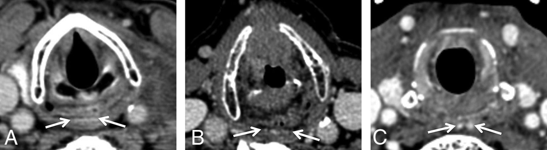
    Axial contrast-enhanced neck CT images obtained in 3 different patients demonstrate representative images of the PCVP (arrows, A–C) at the cricoid cartilage level (A), the arytenoid cartilage level (B), and the supra-arytenoid level (C).

  • FIG 5.
    • Download figure
    • Open in new tab
    • Download powerpoint
    FIG 5.

    Axial contrast-enhanced neck CT images in 3 different patients demonstrate representative examples of bilobed (arrows, A), dot-dash (arrows, B), and linear (arrows, C) PCVP morphology.

  • FIG 6.
    • Download figure
    • Open in new tab
    • Download powerpoint
    FIG 6.

    Axial contrast-enhanced neck CT images in 3 different patients demonstrate representative examples of linear (arrows, A), dot-dash (arrows, B), and bilobed (arrows, C) PPVP morphology.

Tables

  • Figures
    • View popup
    Table 1:

    Characteristics of the study groups

    Laryngeal CancerLymphomaP-Value
    Sex
        Male33 (67%)33 (67%)1.00
        Female16 (33%)16 (33%)
    Age (yr)
        Mean [SD]58.9 [11.0]58.9 [11.1]1.00
        Minimum2525
        Maximum8384
    Smoking history
        Yes47 (96%)26 (53%)<.001
        No2 (4%)23 (47%)
    Radiation dose (Gy)
        Median70.0NA
        Minimum64.0NA
        Maximum70.2NA
    Neck CTs
        Total222226
        Per patient (mean) (SD)4.5 (2.8)4.6 (2.7).88
        Minimum22
        Maximum1614
    Months of CT follow-upa
        Total10051835
        Mean (SD)20.5 (13.5)37.4 (31.4)<.001
        Minimum41
        Maximum59162
    • Note:—NA indicates not applicable.

    • ↵a For patients with laryngeal cancer, follow-up ended when either no more neck CTs were available or the patient underwent salvage laryngectomy.

    • View popup
    Table 2:

    Visibility of PLVP within the study groups with respect to patient characteristics

    Laryngeal CancerLymphoma
    PLVP Visible?
    YesNoPYesNoP
    Sex
        Male2310.502310.29
        Female133142
    Age (yr)
        Mean [SD]58.4 [11.5]60.3 [10.0].6059.3 [11.6]57.6 [9.6].64
    Smoking history
        Yes34131.00179.10
        No20203
    Radiation dose (Gy)
        Median (range)70.0 (64.0–70)70.0 (65.3–70.2).26NANANA
    • Note:—NA indicates not applicable.

    • View popup
    Table 3:

    Visibility of PCVP within the study groups with respect to patient characteristics

    Laryngeal CancerLymphoma
    PCVP Visible?
    YesNoPYesNoP
    Sex
        Male2013.362310.29
        Female124142
    Age (yr)
        Mean [SD]57.3 [10.9]61.9 [10.8].1759.3 [9.6]57.6 [11.6].64
    Smoking history
        Yes3017.54179.10
        No20203
    Radiation dose (Gy)
        Median (range)70.0 (64.0–70)70.0 (65.3–70.2).57NANANA
    • Note:—NA indicates not applicable.

    • View popup
    Table 4:

    Visibility of PPVP within the study groups with respect to patient characteristics

    Laryngeal CancerLymphoma
    PPVP Visible?
    YesNoPYesNoP
    Sex
        Male12211.001023.01
        Female511115
    Age (yr)
        Mean [SD]59.0 [13.9]58.8 [9.3].9662.6 [10.9]56.1 [10.6].04
    Smoking history
        Yes16311.00818.09
        No111310
    Smoking history
        Median (range)70.0 (64.0–70)70.0 (65.3–70.2).06NANANA
    • Note:—NA indicates not applicable.

PreviousNext
Back to top

In this issue

American Journal of Neuroradiology: 42 (5)
American Journal of Neuroradiology
Vol. 42, Issue 5
1 May 2021
  • Table of Contents
  • Index by author
  • Complete Issue (PDF)
Advertisement
Print
Download PDF
Email Article

Thank you for your interest in spreading the word on American Journal of Neuroradiology.

NOTE: We only request your email address so that the person you are recommending the page to knows that you wanted them to see it, and that it is not junk mail. We do not capture any email address.

Enter multiple addresses on separate lines or separate them with commas.
The Pharyngolaryngeal Venous Plexus: A Potential Pitfall in Surveillance Imaging of the Neck
(Your Name) has sent you a message from American Journal of Neuroradiology
(Your Name) thought you would like to see the American Journal of Neuroradiology web site.
CAPTCHA
This question is for testing whether or not you are a human visitor and to prevent automated spam submissions.
Citation Tools
The Pharyngolaryngeal Venous Plexus: A Potential Pitfall in Surveillance Imaging of the Neck
P.M. Bunch, R.T. Hughes, E.P. White, J.R. Sachs, B.A. Frizzell, C.M. Lack
American Journal of Neuroradiology May 2021, 42 (5) 938-944; DOI: 10.3174/ajnr.A7033

Citation Manager Formats

  • BibTeX
  • Bookends
  • EasyBib
  • EndNote (tagged)
  • EndNote 8 (xml)
  • Medlars
  • Mendeley
  • Papers
  • RefWorks Tagged
  • Ref Manager
  • RIS
  • Zotero
Share
The Pharyngolaryngeal Venous Plexus: A Potential Pitfall in Surveillance Imaging of the Neck
P.M. Bunch, R.T. Hughes, E.P. White, J.R. Sachs, B.A. Frizzell, C.M. Lack
American Journal of Neuroradiology May 2021, 42 (5) 938-944; DOI: 10.3174/ajnr.A7033
del.icio.us logo Digg logo Reddit logo Twitter logo Facebook logo Google logo Mendeley logo
  • Tweet Widget
  • Facebook Like
  • Google Plus One
Purchase

Jump to section

  • Article
    • Abstract
    • ABBREVIATIONS:
    • MATERIALS and METHODS
    • RESULTS
    • DISCUSSION
    • CONCLUSIONS
    • References
  • Figures & Data
  • Info & Metrics
  • References
  • PDF

Related Articles

  • No related articles found.
  • PubMed
  • Google Scholar

Cited By...

  • No citing articles found.
  • Crossref
  • Google Scholar

This article has not yet been cited by articles in journals that are participating in Crossref Cited-by Linking.

More in this TOC Section

  • Phosphaturic Mesenchymal Tumor
  • Microstructural Visual Pathway White Matter Alterations in Primary Open-Angle Glaucoma: A Neurite Orientation Dispersion and Density Imaging Study
  • Iodine Maps from Dual-Energy CT to Predict Extrathyroidal Extension and Recurrence in Papillary Thyroid Cancer Based on a Radiomics Approach
Show more HEAD & NECK

Similar Articles

Advertisement

News and Updates

  • Lucien Levy Best Research Article Award
  • Thanks to our 2021 Distinguished Reviewers
  • Press Releases

Resources

  • Evidence-Based Medicine Level Guide
  • How to Participate in a Tweet Chat
  • AJNR Podcast Archive
  • Ideas for Publicizing Your Research
  • Librarian Resources
  • Terms and Conditions

Opportunities

  • Share Your Art in Perspectives
  • Get Peer Review Credit from Publons
  • Moderate a Tweet Chat

American Society of Neuroradiology

  • Neurographics
  • ASNR Annual Meeting
  • Fellowship Portal
  • Position Statements

© 2022 by the American Society of Neuroradiology | Print ISSN: 0195-6108 Online ISSN: 1936-959X

Powered by HighWire