Article Text

Abstract
OBJECTIVE Focal lesions limited to the splenium of the corpus callosum (SCC) are rare and little is known about their aetiology. Three patients were examined for presurgical evaluation in epilepsy with a transient lesion in the SCC and a pathophysiological hypothesis is presented.
METHODS Three patients were identified with a circumscribed lesion in the centre of the corpus callosum. Follow up MRI was performed, the medical records examined retrospectively, and the literature reviewed.
RESULTS The patients showed identical lesions in the SCC with reduced T1 and increased T2 signal intensity and an unaffected marginal hemline of a few mm. Patients were asymptomatic and control MRIs showed complete normalisation within 2 months. Patients had been treated with antiepileptic drugs (AEDs) without signs of toxicity. In all patients AEDs were rapidly reduced for diagnostic purposes, but only one had psychomotor seizures, 5 days before imaging.
CONCLUSIONS A transient lesion in the SCC has so far only been described in 13 patients with epilepsy and has been interpreted either as reversible demyelination due to AED toxicity or transient oedema after secondary generalised seizures. The data confirm neither of these hypotheses. A transient lesion in the SCC seems to be a non-specific end point of different disease processes leading to a vasogenic oedema. This suggests, in these patients, a multifactorial pathology triggered by transient effects of AEDs on arginine vasopressine and its function in fluid balance systems in a condition of vitamin deficiency. The complete and rapid reversibility in all cases without specific intervention is emphasised and any invasive diagnostic or therapeutic approach is discouraged.
- corpus callosum
- anticonvulsant drugs
- brain oedema
- argipressine
Statistics from Altmetric.com
Focal imaging abnormalities of the corpus callosum are rare but have been described in various clinical conditions. We have detected a circumscribed lesion confined to the splenium of the corpus callosum in three patients during presurgical evaluation of therapy for refractory epilepsy. An identical lesion has been reported, but the pathophysiological mechanisms have not been identified.1-2 We have reviewed the literature on splenial lesions and present a new hypothesis about the underlying mechanisms.
Case reports
PATIENT 1
A 34 year old woman had an unclassified epilepsy syndrome with partial and generalised features. Her history was otherwise unremarkable with the exception of eight pregnancies in a period of 10 years.
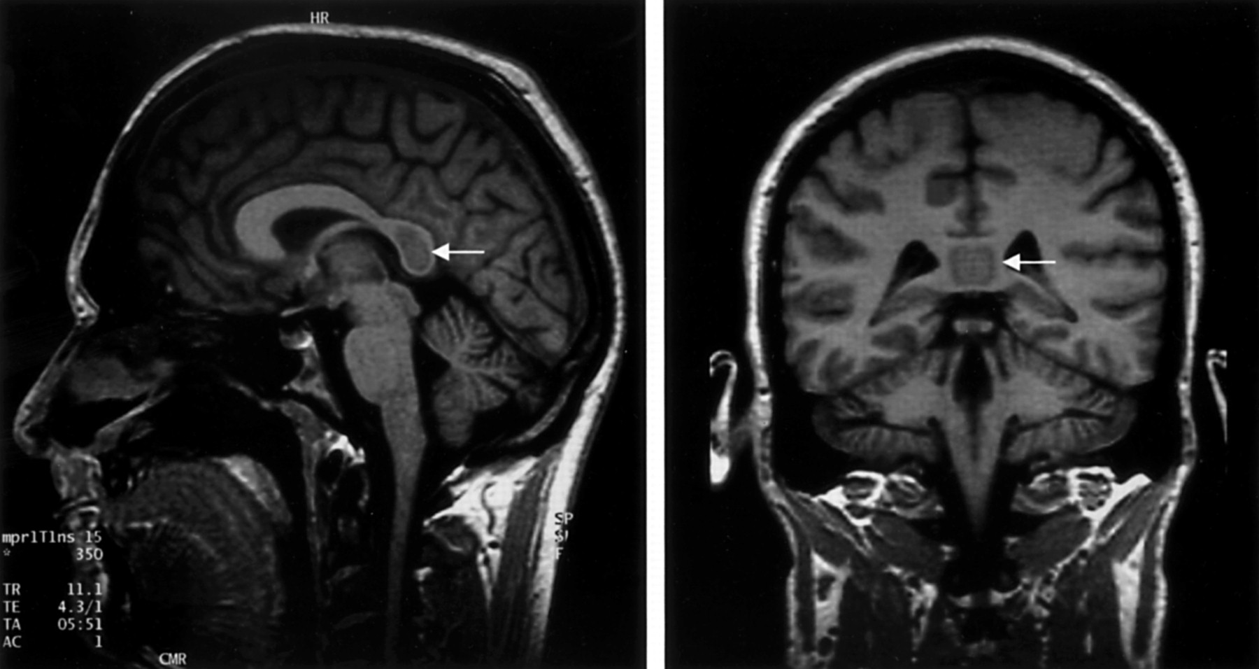
During non-invasive monitoring for 10 days she did not show any ictal event and her EEG was normal even though we had stopped phenytoin medication (serum concentration 21.8 mg/l). Brain MRI showed a hypointense signal in the splenium of the corpus callosum on the T1 weighted and inversion recovery scans that was hyperintense in the T2 weighted as well as in FLAIR images (fig 1).

Circumscribed lesion of the corpus callosum in a 34 year old woman with epilepsy (patient 1), detected after 10 seizure free days of video-EEG monitoring and withdrawal of phenytoin therapy. The lesion is confined to the central areas of the splenium and shows a hypointense signal on T1 weighted images (left) and a hyperintense signal on T2 weighted images (right).
These characteristics suggested an abnormally high water content in these areas. The lesion was not associated with further white matter abnormalities and left about 2 mm of the marginal area around it unaffected. The lesion was asymptomatic and transient disappearing within a few weeks without any specific intervention. Brain MRI taken 1 year before had been completely normal. A diagnostic investigation did not disclose any neurological symptom and routine laboratory tests, including CSF analysis and a search for typical CNS infections, were all normal. Brain CT obtained 17 days after the detection of the lesion was normal and another MRI 5 days later demonstrated complete disappearance of the lesion.
PATIENT 2
This 34 year old man had four to six epileptic seizures a year. They started with prolonged auras and progressed to generalised tonic-clonic seizures. During 10 days of monitoring the EEG showed left and right temporal slowing and rare sharp waves over the right temporal region. He had no seizure despite withdrawal of phenytoin (serum concentration 9.3 mg/l) and lamotrigine (serum concentration 0.47 mg/l). Brain MRI 2 days after the start of a carbamazepine therapy showed an identical splenial lesion as patient 1 (fig 2) as well as a right temporal tumour and an arachnoid cyst in the left anterior temporal region.

Circumscribed lesion of the corpus callosum in a 34 year old man with symptomatic focal epilepsy (patient 2), detected after 10 seizure free days of video-EEG monitoring and changes of anticonvulsive medication. Note the unaffected marginal hemline.
Routine laboratory tests showed increased serum liver enzymes (ALT 52 U/l, GGT 558 U/l) and megalocytic erythrocytes (mean corpuscular volume 93.8 fl). The patient reported prior alcohol misuse but had allegedly stopped the misuse years before. Brain MRI 6 months later demonstrated that the splenial lesion had completely resolved without intervention.
Summary of clinical findings in three patients with transient splenial lesion
PATIENT 3
This 33 year old patient had a temporal lobe epilepsy due to right hippocampal sclerosis. He had complex partial seizures with auras. Routine laboratory tests showed megalocytic eythrocytes (mean corpuscular volume 98.9 fl). During the presurgical evaluation we stopped carbamazepine medication (serum concentration 8.7 mg/l) and reduced primidone (serum concentrations: primidone 2.8 mg/l; phenobarbital 8.9 mg/l). After several days the patient had six short psychomotor seizures within 24 hours. We increased the dose of primidone and found raised serum concentrations 4 days later (primidone 12.2 mg/l; phenobarbital 11.2 mg/l). That day we also performed MRI that demonstrated a lesion in the splenium similar to that of patients 1 and 2. Eight weeks later MRI disclosed complete resolution with unchanged medication. A summary of results for the three patients is shown in the table.
Discussion
A circumscribed lesion in the splenium of the corpus callosum is a rare finding and little is known about its aetiology. We describe three patients with different epilepsy syndromes and different seizure types, each showing an identical lesion in the splenium. Patients had a normal clinical neurological status and we could demonstrate spontaneous resolution of the lesions within 8 weeks in two of them. We cannot exclude a mild disconnection syndrome, but the rapid and spontaneous normalisation of the MR findings makes severe neuronal functional deficits unlikely.
A similar lesion has already been described in 13 epileptic patients. Chason et al described seven patients with temporal lobe epilepsy examined during presurgical evaluation.1 They interpreted the lesion as transient focal oedema due to transcallosal seizure spread in secondary generalised seizures. The history of our patients does not confirm this hypothesis, as none of the patients had a secondarily generalised seizure in the relevant period before imaging. In addition, the role of the corpus callosum in secondarily generalised seizures with an epileptogenic focus in the temporal lobe is controversial.3
Kim et al reported the same lesion in six patients with focal epilepsy but without a detailed description of the clinical conditions under which the MRI was obtained.2They classified the finding as demyelination and hypothesised that anticonvulsive drug toxicity could be the cause. Particularly, they attributed higher phenytoin doses or vigabatrin medication to the phenomenon and ascribed the reversibility, found in the two patients who were reexamined with MRI, to the withdrawal of their medication. The clinical records of our patients do not support this hypothesis either and we agree with Tennison that various points in this theory lack evidence and plausibility.4 So far, a lesion confined to the splenium has been described in 16 epileptic patients by three independent groups that accordingly reported no clinical signs and complete reversibility of this alteration.
Transient focal lesions in patients with epilepsy have been described after focal status epilepticus and exceptionally after single seizures and were interpreted as focal brain oedema.5-9 In all cases it was predominantly located within the grey matter and never in the corpus callosum. Oedema in the corpus callosum as part of more widespread white matter lesions has been shown in hypertensive encephalopathy,10 in eclampsia, and in cerebral malaria.11-12 Focal oedema in the corpus callosum is also a typical finding in post-traumatic MR studies, most commonly within the posterior body and splenium.13-15
Similar lesions, however, which show the same preponderance in the splenium, have only been described twice. Ogura et al reported an identical lesion in a 7 year old girl with haemolytic-uraemic syndrome and mild encephalopathy that also showed rapid spontaneous normalisation.16 This syndrome is known to be a microangiopathic disorder and there is little doubt that the findings represent a focal brain oedema.
The splenium was also shown to be a predilection site for white matter oedema in a series of nine men with high altitude cerebral oedema.17 In these severely symptomatic patients the splenial abnormalities were part of more widespread lesions, but the authors noted a very strong preponderance of splenial manifestations. There is convincing evidence that in acute mountain sickness these lesions represent a vasogenic oedema.17-20
In summary, a transient lesion in the splenium of the corpus callosum seems to be a non-specific end point of different disease processes leading to a vasogenic oedema.
It is noteworthy that our patients as well as the seven patients reported by Chason et al had undergone presurgical seizure monitoring before imaging.1 Among all patients with epilepsy examined by MRI, only a minority had undergone seizure monitoring before, but this situation is reported in a clear majority of the patients who show the splenial lesion. For that reason, the specific conditions of seizure monitoring might be associated with this finding. Seizure frequency and the rate of secondarily generalised seizures is commonly higher during monitoring, hypothesised as being the underlying cause by Chason et al.1 However, the data of our seizure free patients disprove this concept. Additionally, withdrawal and redosing of drugs is performed more abruptly during monitoring. This could contribute to transient oedema mediated by the influence of anticonvulsive drugs (AEDs) on fluid-balance systems, namely, arginine-vasopressin (AVP). It has been demonstrated that AVP regulates regional cerebral blood flow,21 influences brain water content, and can contribute to brain oedema formation.22-23 Different AEDs have been shown to interact with AVP.24 Both clinical experience and experimental studies showed that carbamazepine can enhance the antidiuretic effect of AVP.25-27 The reduction of serum AVP concentrations after several weeks of carbamazepine therapy25-26 can thus be interpreted as an adaptive reaction of the fluid balance system to the influence of carbamazepine. It was also shown that phenytoin injections can antagonise the antidiuretic effect of AVP, an effect that was not seen with continuous oral phenytoin therapy and was attributed to rapid achievement of high serum concentrations.28 In conclusion, there is evidence that in continuous AED therapy the fluid balance system adapts to the influences of anticonvulsive agents. We hypothesise, however, that in a situation of abrupt serum concentration changes, there might be a short period of dysequilibrium causing a syndrome of inappropriate antidiuresis that could contribute to brain oedema in a predelection site. In our patients, falling serum concentrations of phenytoin (patients 1 and 2) and rising concentrations of carbamazepine (patient 2) both could have caused enhanced AVP effects. For patient 3, it is remarkable that primidone was shown to have a similar influence on AVP serum concentrations as carbamazepine.24 This concept could be tested by performing a water loading test in patients while making changes in their anticonvulsive medication.
Concepts about pathophysiological mechanisms for a transient brain oedema cannot explain the localisation of our findings if we do not assume that the splenium of the corpus callosum is especially sensitive to vasogenic oedema. Studies about post-traumatic lesions in the corpus callosum generally explain the prevalence of callosal injury by its vulnerability to shearing forces.13-14 As this mechanism seems unlikely in our patients, it is of particular interest that altered vasopressin concentrations and a syndrome of inappropriate antidiuresis are well documented complications after brain trauma.29-31 Anatomical studies in the corpus callosum did not find a different fibre density or principally different fibre composition in the splenium compared with other regions of the corpus callosum.32-34 Kakou et aldescribed the splenium as the only region where the vertebrobasilar system intervenes in the vascularisation of the corpus callosum that is primarily supplied by the carotid system.35 In our opinion, none of these data can explain a special vulnerability of the splenium towards focal oedema.
In fact, we suppose that if not based on individual susceptibility, the splenial lesions in our patients can only be interpreted as a consequence of a multifactorial pathology. Further investigation could be guided by the pathological mechanisms in Marchiafava-Bignami disease, which shows an identical pattern of central demyelination in the splenium.36-38 This disease is thought to be a multifactorial process, probably mediated by vitamin deficiency.37 It is noteworthy that all our patients presented with risk factors or signs of vitamin deficiency apart from the general risk associated with their anticonvulsive medication. Patient 1 reported eight pregnancies within a period of 10 years before we detected the lesion, a situation that often causes vitamin deficiency, especially low folate concentrations. Patient 2 showed markedly increased liver function tests and megalocytic erythrocytes, a typical sign of vitamin B12 or folate deficiency. In patient 3 laboratory tests found megalocytic erythrocytes. So in all our cases, a vitamin deficient status is likely, but not proved.
A similar multifactorial aetiology based on vitamin deficiency with additional trigger mechanisms has also been hypothesised for central pontine myelinolysis, another demyelinating disease often associated with alcohol misuse.39-41 This concept is supported by a report of a 15 month old boy with severe folate deficiency who had central pontine myelinolysis after mild hyponatremia.42
In conclusion, vitamin deficiency is probably a condition that facilitates white matter oedema and demyelination. The predilection site and form of manifestation—for example, pontine, extrapontine, or splenial lesion—would, however, be determined by various specific trigger mechanisms. More clinical data about other patients are required to test these hypotheses.
Acknowledgments
This work was supported by a grant from the Deutsche Forschungsgemeinschaft (DFG-Eb 111/2–2).
