Article Text

Abstract
Objectives To assess the possibility that diffusion tensor imaging (DTI) can detect white matter damage in mild traumatic brain injury (mTBI) patients via systematic review and meta-analysis.
Methods DTI studies that compared mTBI patients and controls were searched using MEDLINE, Web of Science, and EMBASE, (1980 through April 2012).
Results A comprehensive literature search identified 28 DTI studies, of which 13 independent DTI studies of mTBI patients were eligible for the meta-analysis. Random effect model demonstrated significant fractional anisotropy (FA) reduction in the corpus callosum (CC) (p=0.023, 95% CIs −0.466 to −0.035, 280 mTBIs and 244 controls) with no publication bias and minimum heterogeneity, and a significant increase in mean diffusivity (MD) (p=0.015, 95% CIs 0.062 to 0.581, 154 mTBIs and 100 controls). Meta-analyses of the subregions of the CC demonstrated in the splenium FA was significantly reduced (p=0.025, 95% CIs −0.689 to −0.046) and MD was significantly increased (p=0.013, 95% CIs 0.113 to 0.950). FA was marginally reduced in the midbody (p=0.099, 95% CIs −0.404 to 0.034), and no significant change in FA (p=0.421, 95% CIs −0.537 to 0.224) and MD (p=0.264, 95% CIs −0.120 to 0.438) in the genu of the CC.
Conclusions Our meta-analysis revealed the posterior part of the CC was more vulnerable to mTBI compared with the anterior part, and suggested the potential utility of DTI to detect white matter damage in the CC of mTBI patients.
- Corpus callosum
- fractional anisotropy
- human
- systematic review
- meta-analysis
This is an open-access article distributed under the terms of the Creative Commons Attribution Non-commercial License, which permits use, distribution, and reproduction in any medium, provided the original work is properly cited, the use is non commercial and is otherwise in compliance with the license. See: http://creativecommons.org/licenses/by-nc/2.0/ and http://creativecommons.org/licenses/by-nc/2.0/legalcode.
Statistics from Altmetric.com
Introduction
Mild traumatic brain injury (mTBI) is one of the most controversial neurologic injuries, as there is no obvious biological marker. Diffusion tensor imaging (DTI) has been considered as a potential biomarker in mTBI patients with otherwise normal neuroimaging,1 as it can detect white matter microstructure changes. The most commonly used scalar invariant derived from DTI is fractional anisotropy (FA), which quantifies the orientation and integrity of white matter tracts. A decrease in FA may indicate axonal degradation and discontinuities with excess water between tracts or in perivascular spaces, which may also occur in mTBI.2
In the field of DTI studies of mTBI patients, the corpus callosum (CC) has been repeatedly investigated. Because a number of neuroimaging studies of moderate and severe traumatic brain injury demonstrated that the CC was one brain region that consistently experiences some of the largest amounts of deformation of any brain region,3–5 and DTI studies demonstrated decreased FA in the CC.6 ,7 Further, both linear and angular acceleration may damage callosal fibers, which may lead to microstructural changes that can be identified in neuroimaging studies, particularly DTI.8 In addition, postmortem studies revealed histological lesions in the CC (reviewed in refs9 ,10).
Although a number of DTI studies with mTBI patients have investigated brain damage, they yielded inconsistent results. While some studies reported an increase or no change in FA following mTBI,11 other studies reported a significant reduction in FA.12
Thus, we hypothesised that the microstructure brain damage in mTBI patients can be detected by DTI; moreover, we hypothesised that also mTBI patients show similar location and direction of microstructure brain damage to that with moderate to severe traumatic brain injury, that is, FA is reduced in the CC and IC in mTBI patients. To investigate the hypotheses, we carried out a systematic review of the literature of DTI studies of mTBI patients and conducted a meta-analysis of DTI studies of mTBI patients, and have located a priori-defined region of interest (ROI). Although we have a hypotheses, we do not confine ROI in the CC and IC, but performed a meta-analysis wherever it was possible, for the entire brain, to examine whether DTI can be a biomarker of mTBI.
Methods
Systematic review
Data sources
DTI studies that examined the FA of mTBI patients compared with control subjects were obtained through the computerised databases of MEDLINE, Web of Science and EMBASE. The search terms used in the systematic screening were ‘brain injury’, ‘axonal injury’ and ‘trauma’, which were also combined with the terms ‘tensor’, ‘TBSS’, ‘tract-based spatial statistics’ (TBSS) and ‘tractography’. Two reviewers (YA and RI) performed independent screenings of the titles and abstracts of the studies to identify relevant studies to be included. Reference lists of included articles were also examined to search additional studies to be included.
Whole brain voxel-based analysis and ROI analysis
There are two major different approaches to examine microstructure damage from DTI data. The first one is the voxelwise whole brain analysis (WBA), such as voxel-based analysis (VBA)13 or TBSS14 approaches. Typically, they only provide details of areas that are significantly different between groups. The other one is ROI analysis, including that which utilised tractography for the definition of ROI.15 They do not investigate globally, but provide values of ROIs even when there was no significant difference.
In the current review, we performed systematic screening of DTI studies of mTBI, and we divide studies into two sections, based on the difference in the approach. Voxel-based global approaches, such as VBA studies and TBSS studies, were assigned to the section on WBA studies. A priori-defined local approaches, such as ROI studies and tractography studies, were sorted as ROI studies.
Selection criteria for database
We sought articles for database (1) in peer-reviewed journals between 1980 and April 2012, and (2) which studied brain DTI in mTBI patients compared with a control group. The literature search was performed without language restrictions.
Meta-analysis
Heterogeneity of WBA studies
There are two kinds of WBA studies, TBSS and VBA. As they differ from each other in the process of registration and smoothing,14 ,16 results from these different studies are not unitable. Further, though reliable methods of meta-analysis of VBA have already been established,17 ,18 a reliable method of meta-analysis of TBSS has rarely been introduced. Thus, we can conduct a meta-analysis of only VBA studies.
As described bellow, the current systematic review yielded only five VBA studies whose thresholds completely differ with each other, which is not suitable and sufficient to conduct a meta-analysis. Thus, we will conduct a meta-analysis of ROI studies in the current work.
Meta-analysis of ROI studies
Selection of studies for meta-analysis
In addition to selection criteria for the database, we further imposed the following criteria for inclusion into the meta-analysis: (1) studies that utilised tractography or ROI methods; (2) studies included that reported sufficient data to allow for effect-size calculations; (3) had recruited more than three participants for each group and (4) reported no detectable change in the ROI by conventional imaging such as MRI and CT. To ensure the meta-analysis was sufficiently powered, brain region values were included if there were two or more studies reporting more than three datasets with sufficient data in total. If studies did not report sufficient data, we emailed the corresponding author to obtain further information. In cases where the author did not respond, we excluded the study from our analysis.
Data extraction
To perform the meta-analyses, we defined a standardised mean difference (SMD) as the effect-size statistic, which is defined as the difference between the mean of the experiment group and that of the comparison group divided by the pooled SD.19 In the current meta-analyses, the mean of DTI values in mTBI patients was subtracted from those in the control groups in each ROI, respectively, and divided by the pooled SD of both. Meta-analysis of Observational Studies in Epidemiology guidelines for conducting and reporting meta-analysis of observational studies were followed.20
Statistical analysis
All meta-analyses were performed using the Comprehensive Meta-Analysis version 2 software (©2006, Biostat, Inc., Englewood, New Jersey, USA). Standardised mean difference was calculated and used for the effect sizes. For meta-analysis, a single-effect size was computed from one comparison. In studies that used multiple ROIs from a single region, for example, the CC, the weighted average effect size was calculated. In studies that reported DTI values in the left and right hemispheres,21–23 we calculated the mean of two effect sizes from the left and right hemispheres and integrated it in the analysis.
In studies that compared three groups, such as one control and two mTBI groups, the number of participants in the control group was divided into two groups to avoid duplicate counting.24 ,25
Furthermore, to perform sensitivity analysis of the CC, the datasets were assigned into three groups according to the CC location: genu, midbody and splenium. We employed random effect models for all the meta-analyses to minimise the potential heterogeneity between studies, such as variation in location of ROI, number of excitation (NEX) and b factor. We focused on five major DTI-derived invariants, including FA, apparent diffusion coefficient (ADC), mean diffusivity (MD), axial diffusivity (AD), and radial diffusivity. We performed a meta-analysis of the invariants of which more than three studies have been reported.
Sensitivity analysis
We performed a one-way sensitivity analysis to test the robustness of the results from the meta-analysis by excluding one dataset at a time. By doing this, we can assess whether any single dataset is responsible for the result.
Further, the effect of potential confounders were tested by sensitivity analysis in specified subgroups excluding studies with potential confounds. These potential confounds included comorbid psychiatric disorders, such as post-traumatic stress disorder, medication or substance misuse, including alcohol and field strength of MR scanner. In studies that did not mention the existence of major psychiatric disorder, we recognised them as the studies without major psychiatric disorder.
Meta-regression analysis
The meta-analysis revealed significant differences between mTBI patients and control individuals with a sufficient number of datasets.26 To investigate the effect of possible modifiers or abnormalities, we performed meta-regression analyses to examine the relationship between mean duration of time between the point of injury and imaging, male ratio, and the mean age and SMD for the FA values in the whole CC and splenium. The regression was examined using the Comprehensive Meta-Analysis 2 software.
Assessment of between-study heterogeneity
Between-study heterogeneity was assessed using the I2 statistics. Thresholds for the interpretation of I2 were based on previous studies suggesting that 0% to 50% represents mild heterogeneity, 50% to 75% moderate heterogeneity, and 75% to 100% considerable heterogeneity.
Publication bias
Publication bias was assessed qualitatively by visual inspection of funnel plots, and quantitatively by linear regression analysis. Based on previous literature, this calculation was tested with datasets of at least 10.26 A significance difference (p<0.10) suggested the studies were heterogeneous.25
Data synthesis
Twenty demographic, clinical and methodological variants, including the number of participants, number of male participants, mean age, diagnostic criteria of mTBI, psychiatric comorbidity, medication and substance misuse, strength of magnetic field (Tesla), b factor, number of excitation, echo time (TE), repetition time, type of values reported by the study and main findings were extracted for both, WBA and ROI studies and, additionally, four and one characteristics of imaging were obtained for WBA and ROI studies, respectively, as shown in online supplementary tables 1-1 and 1-2. The datasets, participants, mean differences, 95% CI, Z-value, p value, and I2 score are shown in table 1.
Meta-analysis by tract and value
Results
Study selection for database
The literature search produced 592 articles, of which 68 DTI studies were identified as potential candidates for the database. Twenty studies were excluded because they included data from moderate and severe traumatic brain injury, which could not be separated from the mTBI data. Seven studies were discarded because they were review articles or did not report original data. Six studies were excluded because they did not compare mTBI patients with control subjects. Two studies were not DTI studies. Two studies were not included in the database due to overlap of data.27 ,28 Thus, 28 studies were included in the database. Among these 28 studies, 10 studies utilised whole brain voxelwise analysis and were included in the WBA section, whereas 19 studies were assigned to the ROI section, and are shown in online supplementary tables 1–1, and 1-2. The process of study selection is shown in figure 1.

Process of study selection.
Study selection for meta-analyses
From the 19 studies in the section on ROI studies, three studies were excluded because they did not report sufficient data to calculate effect size.29–31 Two studies were excluded because they located the ROI only in the fornix and cingulum.32 ,33 As only two studies with two datasets reported DTI values from the cingulum with sufficient data to calculate effect size, and one reported DTI values from fornix, cingulum and fornix were excluded from the meta-analysis.
Additionally, one study was discarded because it utilised an original statistical analysis with percentile approach, as it was unable to calculate an effect size.34 Therefore, 13 studies were included in the meta-analysis.12 ,21–25 35–41
Characteristics of included studies
Section of WBA studies
Nine studies involving 189 mTBI patients and 154 controls were identified in this section.2 ,11 ,34 ,41–47 Five utilised the TBSS approach,41 ,43 ,45–47 whereas five2 ,11 ,34 ,42 ,44 adopted the voxel-based WBA. Thresholds adopted in these studies differ considerably; one utilised quantile analysis,34 five studies41 ,43 ,45–47 adopted threshold at p<0.05 and one2 at p<0.01 multiple comparison corrected, the other did not correct multiple comparison. Eight2 ,11 ,34 ,41–43 ,46 ,47 utilised a 3-T scanner and two44 ,45 used a 1.5-T scanner. Although the included studies demonstrated inconsistent results, three TBSS studies41 ,43–45 with the same threshold did not reveal any significant difference in FA values between mTBI patients and controls, whereas one study demonstrated diffuse FA reduction.47 (online supplementary table 1-1)
Section of ROI studies
Thirteen studies with 15 independent comparisons, 280 mTBI patients and 244 controls were included in the meta-analysis.12 ,21–25 35–41 Twelve patients were recruited, whose GCS score ranged from 13 to 15.12 ,21–25 35–40 Seven12 ,21 ,22 ,25 ,35 ,37 ,40 utilised a 1.5-T MRI scanner and six23 ,24 ,36 ,38 ,39 ,41 used a 3-T MRI scanner. All investigated FA value, whereas three examined ADC22 ,40 ,41 and four examined MD.24 ,25 ,37 ,38 Two reported AD and radial diffusivity.35 ,37 The duration between the event and scan ranged from three 3 days12 to eight 8 years.37 Nine out of 13 studies recruited mTBI patients without past history of major psychiatric problems,21–23 ,25 ,35 ,36 ,39–41 the remaining four studies did not mention any history of major psychiatric problems among mTBI patients.
Meta-analysis of DTI measures in the CC and IC of mTBI patients
Corpus callosum
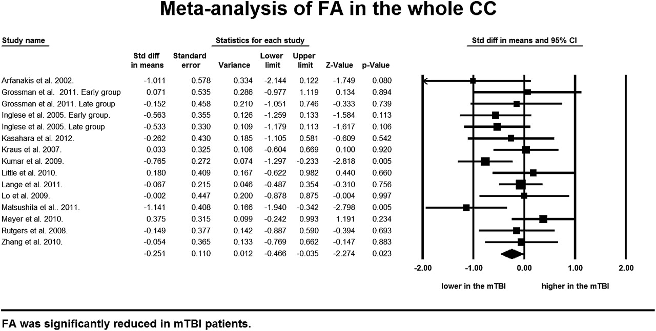
Thirteen studies with 15 datasets that recruited 280 mTBI patients and 244 controls were integrated in the meta-analytical differences in FA value in the CC, and showed a significant FA decrease in mTBI patients (p=0.023) without heterogeneity and no publication bias (Table 1, figure 2).12 ,21–25 35–41 Three studies with 39 mTBI patients and 35 controls demonstrated no significant difference in ADC values (p=0.198) (table 1).22 ,40 ,41 Finally, four studies with six independent datasets involving 154 mTBI patients and 100 controls revealed a significant increase in MD in mTBI patients (p=0.015) without heterogeneity (table 1).24 ,25 ,37 ,38

Forest plot of FA in the CC Standardised mean differences (SMD) for FA measures in the CC between mTBI patients and control subjects. The forest plot displays SMD and 95% confidential intervals.
Genu of the corpus callosum
Eleven studies with 12 datasets involved 248 mTBI patients, and 215 controls were integrated using a random effect model, and demonstrated no significant difference between mTBI and controls in FA value in the genu of the CC (p=0.450) (Table 1, online supplementary figure 1).12 ,21–23 ,25 ,36–41 Three studies with 39 mTBI patients and 35 controls demonstrated no significant differences in ADC values (p=0.115) (table 1),22 ,40 ,41 while three studies with four comparison groups showed no significant changes in MD (p=0.264) (table 1).27 ,37 ,38
Midbody of the corpus callosum
In nine studies which recruited 197 mTBI patients and 156 controls, a marginal reduction in FA values was observed in the midbody of the CC (p=0.099) (Table 1, online supplementary figure 2).12 ,23 ,35–41
Splenium of the corpus callosum
Thirteen studies with 15 datasets with 280 mTBI patients and 244 controls revealed a significant reduction in FA values in mTBI patients (p=0.025) without publication bias and moderate heterogeneity (Table 1, online supplementary figure 3).12 ,21–25 35–41 Three studies with 39 mTBI patients and 35 controls revealed no significant differences in ADC values (p=0.358) (table 1).22 ,40 ,41 A significant increase in MD (p=0.013) was demonstrated in mTBI patients using the random effect model in four studies with six independent datasets involving 154 mTBI patients and 100 controls with moderate heterogeneity (table 1).24 ,25 ,37 ,38
Sensitivity analyses
To test the robustness of the findings, that the FA value is significantly reduced and MD is significantly increased in the whole CC and the splenium of the CC in mTBI patients, a one-way sensitivity analysis of each meta-analysis was performed. In the CC, all the one-way sensitivity analyses preserved a marginal reduction of FA with 12 out of 15 replicating a significant reduction of FA (online supplementary table 2), while five out of six analyses replicated a significant MD increment (online supplementary table 2). In the splenium of the CC, all the one-way sensitivity analyses preserved a marginal reduction of FA with 12 out of 15 replicating a significant reduction of FA, while four out of six analyses replicated a significant MD increment (online supplementary table 2).
Sensitivity analysis performed in the specified-subgroups without major psychiatric problems demonstrated significant FA reduction in the whole CC (p=0.019) and splenium (p=0.016), and MD increase in the whole CC (p=0.042) and splenium (p=0.026) (online supplementary table 3). Sensitivity analysis of studies with a 1.5-T scanner also preserved significanct FA reduction in the whole CC and splenium (p<0.001, p=0.003) and MD increment in the whole CC and splenium (p=0.011, p=0.022) (online supplementary table 3). On the other hand, sensitivity analyses of studies without medication or substance misuse did not show significant difference in FA or MD in the whole CC and splenium (online supplementary table 3).
Meta-regression
To investigate the effect of FA reduction in the whole CC and splenium of mTBI patients, we conducted meta-regressions with three potential modifiers; duration of time between the point of injury, male ratio, and imaging and mean age of participants. Meta-regression analysis revealed no significant effect of duration of time (p=0.83), male ratio (p=0.70) and mean age (p=0.38) on reduction of FA in the whole CC in mTBI patients. However, the mean age of mTBI patients had a marginally significant effect on reduction of FA in the splenium (p=0.06) (figure 3), but no significant effect of male ratio (p=0.31), and duration of time (p=0.46) (online supplementary table 4).

The relationship between effect size for FA and age of study participants The effect size from each comparison of VOIs is plotted by the mean age of mTBI patients. The line of best fit shows a gradual but substantial decrease of FA.
Discussion
To the best of our knowledge, this is the first meta-analysis of DTI studies of mTBI patients to demonstrate significantly reduced FA and significantly increased MD in the CC compared with controls, without publication bias and with no heterogeneity. In addition, a subanalysis based on the subregions of the CC demonstrated a significant decrease of FA and significant increase of MD in the splenium, a marginal reduction of FA in the midbody and no changes in FA and MD in the genu of the CC. The viability of the FA reduction and increase in MD in the whole CC and splenium were examined by a one-way sensitivity analysis of each meta-analysis. Sensitivity analyses further demonstrated that these findings were preserved in specified subgroups with studies without psychiatric disorders.
Of the types of DTI studies, the hypothesis-free, whole brain analyses, such as VBA or TBSS approaches are useful to investigate the overall changes in white matter, rather than specific white matter tracts or areas.13 ,14 In contrast, the other type of DTI study, the ROI analysis, which investigates a priori-defined region or tractography, is useful to investigate specific tracts and defined ROI, and report invariances derived from tensor imaging. Thus, selecting DTI studies with a priori-defined design analysis is rational when we have an a priori hypothesis that DTI in the CC may be the potential tool to detect microstructure damage in the white matter following mTBI.
The significant FA reduction demonstrated by our meta-analysis suggests the potential diagnostic ability of DTI in mTBI patients. Our findings have provoked the assumption that previous studies were unable to demonstrate significant differences because of the small number of participants and varying duration after trauma.
In keeping with the findings of a previous study that reported unmyelinated axons in the CC after animal TBI,48 we found that a reduced FA value indicates differences in cellular membrane integrity, fiber myelination, fiber diameter and directionality in the CC after mTBI. The CC is the main fiber tract that connects the hemispheres and is topographically organised. The genu of the CC represent fibers from the prefrontal cortex; the midbody is composed of fibers from the premotor, motor, parietal and superior temporal cortex; and the splenium represents fibers from the inferior temporal and occipital cortex. There are some possible reasons why the CC is vulnerable to mTBI. First, the CC is a very organised area of the brain, with axons predominantly oriented in one direction, is inherently anisotropic and has higher FA values compared with less organised areas of white matter. Second, as the CC connects both hemispheres, external accelerational forces at a lateral or oblique-lateral angle of rotation can cause injury to the CC.49 In addition, the CC has been recognised as a region frequently injured by shear strain,50 as shown in victims of shaken baby syndrome.51 ,52
The meta-analysis clearly demonstrated selective FA reduction in the posterior part of the CC, such as it reached significance in the splenium and a marginal difference in the midbody, as well as selective MD increases in the splenium. MD is another invariant that is often reported in DTI studies, which is a measure of the average molecular diffusion independent of any tissue directionality and is affected by cellular size and integrity.1 Results, such as lower FA and higher MD, imply that the posterior part of the CC is more vulnerable to injury than the anterior part like the genu. The vulnerability of the posterior part of the CC compared with the anterior part has been repeatedly reported by imaging studies of brain injury.50 ,53 ,54 One MRI study reported that 80% of CC injury was distributed in the posterior part in patients with a moderate to severe TBI.55 ,56 Though the mean age of the included studies was limited, ranging from 27 to 42, it had a marginally significant effect on the FA reduction in the splenium (p=0.06). Although we are unable to offer a compelling explanation for this relationship, it may potentially be due to age-related changes in white matter maturity. Previous studies have reported that FA is increased during childhood and adolescence, and reaches its peak value at around age 30, then gradually decreases.56 An external trauma may accelerate the loss of FA. To test this relationship, a large-scale original study is required.
There are a number of clinical and methodological confounds that may effect the result. For example, sexual dimorphism of the CC in DTI has been reported.57 Although meta-regression showed no significant relation between male ratio and effect size, male ratio was not available from all the studies. Thus, the finding should be interpreted with caution. Medication and substance misuse also influence results.58 Though no studies reported participants of substance misuse, some studies did not clarify that they excluded individuals with substance abuse, and sensitivity analysis excluding them did not show significant difference. Thus, the potential effect of medication or substance abuse is not completely assessed. With regard to strength of magnetic field, although sensitivity analysis with studies which utilised a 1.5-T scanner preserved the findings, sensitivity analysis with a 3-T scanner did not show significance. As microstructure damage is more detectable in stronger magnetic field, this result is against the hypothesis.
Limitation
Our study, however, does have several methodological considerations and limitations that must be acknowledged. Due to the nature of a meta-analysis, we can make statistical analysis only at the level of studies, with no way of confirming that the mTBI participants of the included studies actually exhibited the findings presented. We found considerable heterogeneity between studies that may attribute to methodological issues. Some of the included studies utilised a 1.5-T MR scanner, while others used a 3-T. In addition, the NEX, b factor and number of directions are considerably different. Although we employed a random effect model to tolerate heterogeneity, the different methodologies integrated from these studies may be criticised. Although we conducted a comprehensive literature search and meta-analyses of the CC, IC and corona radiate, there is the possibility that other brain areas could also present white matter changes that were not investigated in this meta-analysis.59
In addition, we cannot deny the probable existence of the data we could not find. Further, though we have demonstrated significant FA reductions in the CC of mTBI patients with a large effect size, the large total number of participants could help the small differences reach significance. Additionally, it has been repeatedly reported that mTBI patients were at risk of developing mental health problems, such as depression and substance misuse, versus those mental health problems that are common among individuals who have mTBI.60 And it is widely known that those mental health problems alter DTI values.61 Although sensitivity analysis with studies without major psychiatric disorders preserved the significance of the findings, due to lack of sufficient number of studies that investigated mental health problems among mTBI patients, it was impossible to examine whether the DTI change demonstrated any result from pure injury or was related to psychological problems that may be the cause and sequel of injury.
Finally, it is a pivotal point that diagnostic tools of mTBI are changing. Almost all the included studies recruited patients with a GCS scored between 13 and 15, however, there has been a recent argument on the definition of mTBI, and whether it should include patients with a GCS score of 13.62
Conclusion
In conclusion, our findings from this meta-analysis of DTI studies of mTBI patients clearly demonstrate a significant FA reduction in the CC without publication bias and heterogeneity. The results provide strong evidence that DTI can detect microstructural damage in the white matter of mTBI patients, highlighting its potential utility in clinical settings.
Acknowledgments
We thank all the researchers who participated in this study, especially Dr Keiji Hashimoto, Dr Kazumi Kasahara and Dr Andy Mayer who kindly shared undisclosed data.
References
Supplementary materials
Supplementary Data
This web only file has been produced by the BMJ Publishing Group from an electronic file supplied by the author(s) and has not been edited for content.
Files in this Data Supplement:
- Data supplement 1 - Online Table 1-1
- Data supplement 2 - Online Table 1-2
- Data supplement 3 - Online Table 2
- Data supplement 4 - Online Table 3
- Data supplement 5 - Online Table 4
Footnotes
Competing interests None.
Provenance and peer review Not commissioned; externally peer reviewed.
Data sharing statement Dr KH and Dr KK had let us share SD of FA values in their study. Dr AM had let us share mean and SD of FA values in their study.