Article Text

Abstract
As endovascular approaches to intracranial aneurysm (IA) treatment continue to evolve, the use of intracranial stents has advanced from an infrequent adjunct to potential curative monotherapy. Early results of endovascular therapy for IAs have clearly underscored large aneurysm size, low coil packing density and wide aneurysm neck as factors limiting successful long term obliteration. Intracranial stents were originally introduced as adjuncts to mitigate these limitations by facilitating tighter coil packing and preventing coil herniation. As evidence of their utility as flow diversion devices and as catalysts for aneurysm neck remodeling surfaced, their potential as standalone therapy was realised and is currently under close scrutiny. Here we review the evolution of stents in the treatment of IAs, from balloon expandable stents, to self-expanding stents, to the exciting advances in monotherapeutic flow diverting stents, amalgamating occlusion rates and reviewing complication rates.
- Cerebrovascular
- Subarachnoid Haemorrhage
Statistics from Altmetric.com
Introduction
Since the International Subarachnoid Aneurysm Trial,1 traditional endovascular therapy via coil occlusion of cerebral aneurysm domes has been endorsed with considerable enthusiasm. Nevertheless, coil occlusion used alone is limited by recanalisation and in large or wide necked aneurysms.2 To address this limitation, adjunctive balloon assistance3 and permanent stent placement have served as scaffolds to prevent coil prolapse out of wide necked aneurysms and improve packing density.4 ,5 The former technique can be limited by coil prolapse after balloon deflation, parent vessel injury leading to dissection or even rupture, as well as thromboembolic events.3 Stent placement serves as a long term option that may also divert flow from the aneurysm, a concept that has recently been expounded upon as a means of curative stent monotherapy for aneurysms. As we explore the past 14 years of intracranial stent placement in the treatment of intracranial aneurysms (IAs), we will highlight the evolution of the concept while underscoring occlusion rates balanced against the added risks of stent placement—namely, embolic ischaemic events, haemorrhagic complications and in-stent stenosis.
Balloon expandable coronary stents
Higashida et al 6 were the first to report the use of stent supported coil embolisation for the treatment of an IA in 1997. Without dedicated intracranial stent designs, subsequent early case series employed balloon expandable coronary stents in the treatment of wide necked, large or fusiform aneurysms (see supplementary tables A and B, available online only).7–12 Lanzino et al 9 published the first case series of patients treated with stent assisted coiling of their aneurysms in 1999. They described 10 patients with vertebrobasilar or proximal internal carotid artery (ICA) aneurysms, achieving a modest 30% initial complete occlusion rate, and a 33% (2/6 patients) occlusion rate for patients followed for at least 3 months. It is important not to compare these rates to the ostensibly higher ones reported for sole coiling of IAs1 as aneurysms in these series represent a unique subset of aneurysms that would be ‘uncoilable’ (ie, 0% occlusion rate) if managed by coiling alone.
Results
In the largest series detailing the application of coronary stents in the treatment of IAs and atherosclerosis in 124 patients, Lylyk et al 11 reported an impressive initial 75% and delayed 93% obliteration rate for IAs. They reported an overall 10.9% morbidity and 6.3% death rate, with a 10% stent deployment failure rate. Deployment failure occurred only in aneurysms distal to the carotid siphon, as was also seen in the series of Han et al. 7 Across six coronary stent series reviewed (see supplementary tables A and B, available online only), the overall initial obliteration rate was 72%, increasing to 83% at follow-up.7–12 Recanalisation was seen in 11% of aneurysms overall. Deployment failures occurred in 8% of cases overall. Guidewire perforation occurred in 4% and vessel rupture in 3% of cases. Stent migration occurred in 5% of cases and delayed stenosis in 4% of cases. Long term neurological morbidity was seen in 8% of patients and overall mortality was 5% across these six series.
Stent monotherapy and early flow diversion
The concept of stent monotherapy as a means of flow diversion and resultant curative thrombosis of aneurysms was born early with coronary stents as they have approximately 15% metal surface coverage. This endovascular correlate to the surgical bypass relies on a theoretical shift in the intra-aneurysmal flow pattern as a result of turbulence, reducing the intra-aneurysmal vortex, resulting in stasis and thrombosis.13 It can thus be limited by a branch–vessel arising from the aneurysm, not only from the perspective of symptomatic occlusion of the vessel but also from continued flow through the vessel, preventing requisite flow stagnation and thrombosis of the aneurysm.
Zenteno et al 13 treated 20 fusiform or wide necked saccular posterior circulation aneurysms with stent monotherapy, 18 with balloon expandable coronary stents. One patient died as a result of rehaemorrhage; the remainder were reported as modified Rankin Scale score of 0 at follow-up. At the 1 year angiographic follow-up, 14/18 (78%) patients had complete angiographic occlusion of their aneurysm and only 1/18 (6%) patients demonstrated aneurysm recanalisation.
Stent monotherapy with coronary stents further developed with the concept of the stent within a stent technique to increase the overall metal surface coverage and resultant flow diversion.14 Saatci et al 15 reported a series of 25 aneurysms treated with covered stents (Jostent; Jomed International, Helsingborg, Sweden). Twenty-three of 25 aneurysms (92%) demonstrated immediate exclusion. Although all patients developed early post-intervention worsening headaches, all symptoms as a result of mass effect (nine cranial nerve palsies) improved thereafter.
Early limitations of balloon mounted coronary stents included their relative rigidity that generally contraindicated access in cases of proximal vessel tortuosity and in most cases distal to the carotid siphon. Necessary balloon inflation to deploy the stent poses a risk of thromboemboli, vessel dissection and even vessel rupture. Intimal hyperplasia as a result of endothelial injury from inflation may also lead to delayed in-stent stenosis.
Neuroform stent
Taking advantage of improved memory shape capability, self-expanding nitinol stents are dedicated intracranial stents created to mitigate the risks of delivery and requisite balloon inflation. They were designed with the goal of easier safer deliverability and manoeuvrability. Notably, they had generally less metal surface coverage than their coronary stent precursors, potentially relatively limiting their flow diverting capabilities.
Evolution of the Neuroform stent
The first of these stents, the Neuroform stent (Boston Scientific Neurovascular, Fremont, California, USA), was approved by the Food and Drug Administration (FDA) in 2002 for the treatment of IAs. Dedicated to intracranial usage, this flexible, self-expanding, open cell stent is available in diameters of 2.5–4.5 mm and lengths of 10–30 mm. It is made up of 6–8 linked radiolucent cells comprised of a nickel–titanium alloy (nitinol) with a high degree of elasticity, deformity and shape memory. It has four radiopaque markers at each end and 6.5–9.5% metal surface coverage, allowing for easy adjunctive coiling through its interstices. However, this flexible open cell design does make it more prone to migration or herniation into the aneurysm. In addition, its open cell design provided a weaker scaffold for coil support with some cases of coil herniation. It cannot be easily retrieved. The stent has gone through several iterations from 2002, primarily evolving in the form of ease of delivery and deployment.16 The first Neuroform stent was delivered via a non-braided catheter through which a 0.014 inch microguidewire and stabiliser was passed. One year later, the Neuroform 2 design employed a hydrophilic braided microcatheter, and in the following years, the Neuroform 2 Treo and Neuroform 3 employed a third connector to the central portion of the stent itself. The Neuroform 2 Treo also has three connectors instead of two between segments, decreasing the area of open cells and increasing the scaffold strength of the stent. The Neuroform 3 also employs a braided stabiliser catheter which reduces friction on deployment. The most current Neuroform EZ stent can be delivered through a variety of microcatheters, including Renegade Hi-Flo (Stryker) and the Marksman (Covidien).
Results
Supplementary table C (available online only) summarises the prominent series detailing at least 20 patients treated with the Neuroform stent5 ,16–35 (figure 1, illustrative case). Across these 19 series, 1408 patients with 1618 aneurysms were treated. Deployment failures occurred in 7% of cases overall, largely associated with using the original Neuroform 1.27 Complete obliteration rates, highly dependent on the selection of aneurysms treated, were overall 53% initially and 66% at follow-up. Recanalisation was seen in 16% of aneurysms. Stent migration was infrequent (approximately 2% overall). A phenomenon under considerable scrutiny, delayed in-stent stenosis, was rare—occurring in 32/802 cases (4%) with angiographic follow-up. Only five of these 32 cases (16%, 0.6% of all cases) were symptomatic.

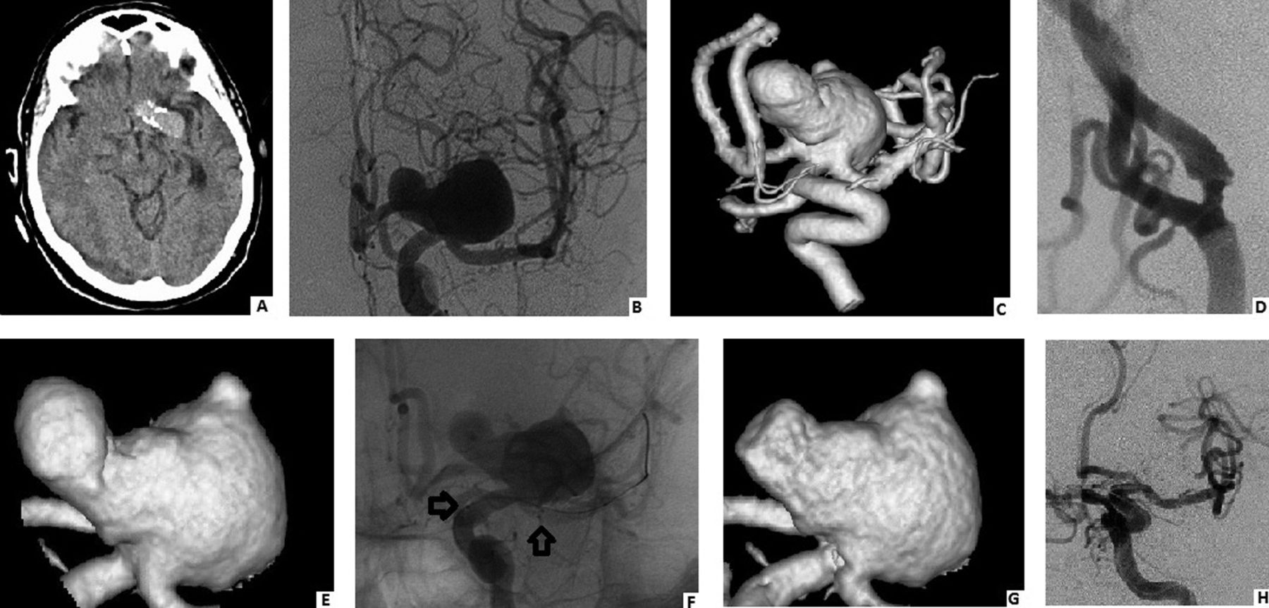
Illustrative case of Neuroform stent use. This 65-year-old woman presented with a seizure. CT suggested a large calcified aneurysm (A). This was confirmed on angiography which demonstrated a large bilobed internal carotid artery (ICA) terminus aneurysm (B, anteroposterior (AP) view) with a wide neck (C). The patient also had a proximal ICA stenosis (D, AP view of ICA bifurcation). Reconstructed images of the aneurysm demonstrated at least two daughter domes (E). A Neuroform stent was placed (F, arrows), and after placement, the aneurysm demonstrated a decrease in size (G, compare with E). The aneurysm was then coiled with excellent angiographic results (H, AP view after coiling).
Symptomatic thromboembolic events occurred in 8% of cases overall. In a study devoted to the analysis of thromboembolic complications after Neuroform stenting, Lessne et al 36 noted an overall rate of 5% (12/224 cases) that increased to 20% among cases with subarachnoid haemorrhage (3.1% if unruptured). This was the strongest factor associated with post-stent thromboembolic events; these patients did not receive a preprocedural clopidogrel dosage which was given in unruptured cases. Other factors included smaller stent calibre and patients with a history of hypertension. Series devoted to the study of stenting in the setting of subarachnoid haemorrhage have had mixed results—in one series with 33 patients undergoing stent–coil treatment of ruptured aneurysms, there was only one intraprocedural thrombotic event and no other reported peri- or postprocedural complications.24 In another with 61 cases, the overall complication rate was 21% with a reported 30 day mortality of 20%—this included one case of an aneurysm that rebled after treatment.31
Intracranial haemorrhage was observed in 3% of cases overall (includes guidewire perforation, vessel rupture and aneurysm rupture). As mentioned above, owing to the delivery design of the Neuroform, potential distal migration and resultant guidewire perforation can occur. Notably, Raslan et al 29 noted that four procedure related haemorrhages occurred with the earlier, more difficult to deliver, Neuroform 1.
Permanent neurological morbidity was seen in 4% of cases and mortality in 2% of cases overall. Although not providing more detailed subgroup analysis, a recent large study of 135 cases employing the Neuroform stent reported similar overall neurological morbidity and death rates of 5% and 3%, respectively.37
Neuroform stent monotherapy
Despite its low metal surface coverage, the flow diverting potential of the Neuroform has been used in the management of pseudoaneurysms. Fiorella et al 38 noted a 50% complete resolution rate after a mean of 9.8 months of follow-up of 10 ruptured intradural pseudoaneurysms treated with Neuroform stent monotherapy. One intraprocedural thrombosis was noted with resultant infarcts, and at a follow-up, two asymptomatic in-stent stenoses were observed. A recent report by Suh et al 39 demonstrated the feasibility of the stent within a stent technique, enhancing flow diversion by stent overlap in the treatment of 11 ruptured vertebrobasilar aneurysms. Early obliteration was seen in three lesions, but at follow-up, nine lesions were completely obliterated.
Technical advances with the Neuroform stent
Several advances in the stent–coil technique were made with the Neuroform stent. Cultivation of the Y stenting technique to provide a scaffold for wide necked basilar, middle cerebral artery (MCA) and ICA bifurcation aneurysms began early.40–42 Two stents are placed, each emanating from the same proximal parent artery (eg, basilar, M1 or ICA) but ending in a different distal efferent artery (eg, P1, M2 or M1 and A1), bridging the bifurcation40–42 (figure 2). In one early series of seven basilar apex aneurysms treated with this technique as an adjunct to coiling, all aneurysms were reported as completely or near completely embolised with only two transient neurological complications.41 Similarly suited for bifurcation aneurysms, the ‘waffle cone’ technique was also developed. In this approach, a single stent is placed into the aneurysm directly, using the portion of the stent protruding into the aneurysm as a concomitant reconstructed aneurysm neck and scaffold for subsequent coil embolisation.43

Illustrative case of Y stent technique with two Neuroform stents. This 60-year-old woman was found to have an 11×9.7 mm, anteriorly projecting, broad based aneurysm of a distal dysplastic basilar segment incorporating the origin of both posterior cerebral and superior cerebellar arteries (A, three-dimensional reconstruction). Aneurysm treatment was staged, beginning with initial stent placement (B, arrows denote stent markers) followed by coiling with 19 Guglielmi detachable coils leading to successful occlusion (C, D; arrows denote stent markers).
Enterprise stent
Succeeding the Neuroform stent with FDA approval in 2007, the Enterprise stent (Cordis) is a closed cell nitinol design, affording a stronger scaffold, minimising coil prolapse and also allowing for stent recapture and repositioning after up to 70% deployment. This allows for the ‘semi-deployment’ technique where the stent is partially deployed during coiling of the aneurysm to allow for subsequent retraction and repositioning for optimal final stent positioning after coiling.
The stent has a positioning marker on the delivery wire and four tantalum radiopaque markers at each end which flare to enhance apposition to the parent vessel wall. Although it has a similar percentage of metal surface coverage as the Neuroform, its closed cell design provides a stronger radial force for aneurysm neck scaffolding. It is available in only one diameter, 4.5 mm, and four lengths—14 mm, 22 mm, 28 mm and 37 mm.
The stent can be delivered directly via standard microcatheters. This decreases the potential risk of small vessel perforation as a result of distal migration of the guidewire. Furthermore, one can employ relatively small microcatheters to access communicating arteries less than 1 mm, in contrast with the Neuroform stent where transport across vessels less than 2 mm is generally contraindicated. This has allowed for the propagation of the horizontal stent technique, allowing for coiling of wide necked basilar apex and ICA terminus aneurysms.44 The stent is navigated through the circle of Willis—through the posterior communicating artery for basilar apex aneurysms and through the contralateral ICA and anterior communicating artery for ICA terminus aneurysms. Siddiqui et al 44 reported a series of eight aneurysms treated with this method, traversing communicating arteries ranging in size from 0.8 to 1.5 mm with the stent. This contrasts with early attempts to perform the horizontal stent technique with Neuroform stents that were limited to communicating arteries at least 2 mm in diameter.45
Results
This experience, along with other reports of the Enterprise stent, are summarised in supplementary table D (available online only).5 ,44 ,46–55 Across the 11 series detailing 443 patients with 457 aneurysms, deployment failures were seen in only 1% of cases overall. Although not receiving particular attention in these larger series, an increasing number of brief reports detailing early and delayed proximal migration of Enterprise stents have surfaced, particularly in the context of stenting from the basilar artery into P1.48 This may be in part due to the closed cell design encouraging migration proximally when there is a difference in parent vessel size between the stent's proximal and distal ends.
Initial complete obliteration and follow-up obliteration rates were 68% and 54%, respectively (see supplementary table D, available online only). The greater early obliteration rate may be a result of the fact that early and late obliteration rates were often not both reported in each series; a comparison may thus be deceiving. The largest series recently reported by Mocco et al based on a 10 centre USA registry with 213 consecutive patients,49 ,50 reported a 40% obliteration rate for 110 aneurysms. Eighty-eight per cent of aneurysms were at least 90% obliterated however. Focusing on delayed stent stenosis, this large series noted a relatively low (3%) rate.50 All three cases were asymptomatic and the worst case (80% stenosis) resolved spontaneously. Across all studies, delayed in-stent stenosis was seen in only 3% of cases and was asymptomatic. Recanalisation was seen in 11% of cases overall. Remarkably, overall symptomatic thromboembolic and haemorrhagic complications were seen in 2% of cases each. Permanent neurological morbidity and mortality was seen in 2% and 1% of cases overall, respectively. In the large series by Mocco et al, there was an early 6% temporary morbidity and 2.8% permanent morbidity rate.49 ,50 Of nine thromboembolic complications, seven occurred in a delayed fashion, always in the context of cessation of double antiplatelet therapy.50 Procedural mortality was seen in three cases due to haemorrhagic complications (2% of cases overall)—stratified however, this rate was 0.8% for unruptured aneurysms and 12% in ruptured cases.
Insight into post-stenting thromboembolic events
Although symptomatic thromboembolic complications are ostensibly rare, asymptomatic and potential delayed events may occur, potentially a result of relatively poorer apposition of the stiffer Enterprise stent to the parent vessel wall around curves. Employing early postprocedure 3T-MR angiography, Heller et al 56 noted flow signal outside the susceptibility artifact of the stent struts in 18/33 patients with the Enterprise stent and in 0/25 patients with the Neuroform stent. This phenomenon, dubbed the ‘crescent’ sign, had a remarkable correlation with postprocedural diffusion weighted imaging (DWI) positivity, potentially a result of emboli from the inadequately apposed stent or from the Enterprise's Parylene coating.56 Sixteen of the 58 patients had DWI positivity after stent placement, although none had permanent neurological sequelae as a result. Twelve had a positive ‘crescent’ sign (and were thus treated with Enterprise stents). These 12 patients had 60 foci of DWI positivity. Three of the four patients with DWI positivity that did not have a crescent sign were treated with the Enterprise stent, and the one patient with DWI positivity treated with a Neuroform stent had undergone a balloon test occlusion during the same procedure prior to stent placement. Each of these patients had only one focus of DWI positivity.56 This contrast is a provocative finding that may have considerable implications for Enterprise stent usage around sharper bends in future studies. To mitigate rates of incomplete stent apposition, this same group has recently illustrated the potential efficacy of a ‘dynamic push–pull’ delivery technique, entailing the combination of a microcatheter pull back and delivery microwire push to deliver the stent.57 This method may have further promise in its application to other closed cell stents.
Leo stent
The Leo stent (Balt, Montmorency, France), not available in North America, was the first self-expandable intracranial stent that allowed for resheathing and repositioning at up to 90% of deployment as a result of its closed cell design. It additionally was easily visualised via two radiopaque wires extending along its length, allowing for clear visualisation of its diameter and length as well as the parent vessel. In contrast with other intracranial stents that are laser cut from nitinol hypotubes, it is braided from a single nitinol wire with closed cells that can change in size.58 It also has greater metal surface coverage than other stents designed primarily for stent assisted coiling (Neuroform, Enterprise, Solo/Solitaire AB), with the most distinct flow diverting haemodynamic effect.58 It has been successfully used as monotherapy with reports detailing progressive thrombosis of fusiform MCA and basilar aneurysms.59 However, its homogeneous wall structure, stiffness and requisite usage of a larger stiffer microcatheter make navigation, particularly in the context of acute curves, quite difficult. Furthermore, if deployed around a curve, stretching of its homogeneous closed cell design may prevent full contact with the aneurysm neck.
Results
Three series dedicated to its adjunctive usage with coiling are reported in the literature and are provided in supplementary table E (available online only).59–61 For 64 cases, deployment failure was seen in three cases (5%) and stent migration in one case (2%). Initial complete occlusion was 67% overall, and long term complete occlusion was 85% across two series. Recanalisation was seen in 10% of cases. Symptomatic thromboembolic events were seen in 14% of cases overall, although no permanent neurological morbidity was reported. Another series reporting only morbidity and mortality for Leo stent assisted coiling cases noted a higher 15% neurological morbidity and 11% death rate for 27 cases.37 Combining these 27 cases with the 64 cases in supplementary table E, the overall morbidity and mortality rates would be 4% and 3%, respectively.
Solo/Solitaire AB stent
The Solo stent, now referred to as the Solitaire AB (ev3, Irvine, California, USA), also not available in North America for stent assisted coiling, was introduced soon after the Leo as the first fully retrievable intracranial stent. Some have used this as a particular advantage in ‘stent remodelling’ approaches wherein the stent is used with the same intent as a balloon without the interruption of blood flow.62 ,63 The stent is laser cut, and comprised of nitinol in a honeycomb pattern that provides added flexibility and easier delivery via a standard 0.018 or 0.021 inch microcatheter. It is available in 4 mm or 6 mm diameters with 15–20 mm and 30 mm lengths, respectively. It is detached electrolytically from a nitinol push wire in a fashion akin to coils. Although it has only 5–7% metal surface coverage, its radial strength is greater than other stents designed for stent assisted coiling.58 The gap length between struts is somewhat wider than the Enterprise stent (4 mm×3 mm), allowing for greater facility of stent assisted coiling and Y stenting.62 ,63 Another noted advantage is decreased thrombogenicity, with some deploying the stent without adjunctive clopidogrel.62 Visibility is limited however, as it is marked via a proximal coil and three distal markers.
Results
Across five series detailing 102 aneurysms in 96 patients, initial complete occlusion was seen in 55% of cases with occlusion at follow-up, increasing to 90% (see supplementary table F, available online only).62–66 Deployment failure was reported in only two cases (2%), and recanalisation was seen in 10% of cases overall; there were no reported cases of delayed in-stent stenosis. Thromboembolic events were reported in 3% of cases overall and haemorrhagic complications in 1%. Permanent neurological morbidity and mortality were reported in only one case each (1%).
SILK flow diverter
The SILK flow diverter (SFD; Balt Extrusion, Montmorency, France) is a closed cell mesh cylinder comprised of 48 braided nitinol strands and 35 μm platinum microfilaments with a high resultant metal surface coverage (approximately 35%). As a closed cell stent, it can be retrieved and/or repositioned at up to 90% deployment. The stent is unsheathed from its own delivery microcatheter (Vasco 21; Balt) via pushing the delivery wire and retrieving the microcatheter, allowing for expansion and compensation of foreshortening. It is available in 2–5 mm diameters and in 15–40 mm lengths. The stent is quite flexible but has a relatively lower radial force than other closed cell stents such as the Enterprise, allowing for potential stent migration and even vessel occlusion in stenotic vessels. Adjunctive stenting with other stents with greater radial force is thus sometimes performed.67 ,68
As a result of its low porosity, the SFD reduces wall shear stress and induces stasis of blood in the aneurysm dome, encouraging thrombosis.68–76 It additionally encourages neointimal growth across the neck of the aneurysm, leading to an overall effect of vessel wall reconstruction and correction of the haemodynamic disturbance.68–76 Notably however, the SFD never received regulatory approval as a monotherapeutic flow diverter.
Results
Supplementary table G (available online only) details the results from 10 series employing the SFD.68–77 Deployment failure was seen in 3% of cases and deployment difficulty in 11% of cases overall. Adjunctive coiling was employed in 22% of cases. Complete obliteration was seen in 72% of cases, with the series with the longest follow-up presenting an 86% obliteration rate after a mean follow-up of 13.2 months.62 No recanalisation was reported. Postprocedural parent artery occlusion was seen in 5% of cases and embolic events in 7% of cases. In a series devoted to basilar artery aneurysms to evaluate the effect of the SFD on basilar perforators, ischaemic events were ultimately seen in 25% of cases.73
Across the nine series, in-stent stenosis was reported for 10% of cases. Intracranial haemorrhage was seen in 3% of cases, and overall neurological morbidity and mortality were 6% and 4%, respectively. Notably, in the series by Wagner et al,76 all six patients with late complications had ophthalmic or supraclinoid aneurysms, perhaps underscoring the ICA tortuosity in this region as a potential culprit as a result of poor stent apposition to the vessel wall in a mechanism analogous to that described by Heller et al 56 for the Enterprise stent.
Delayed aneurysm rupture
Sobering reports of delayed aneurysm rupture following SFD stent therapy have surfaced.67 ,77 ,78 A recent retrospective analysis described 13 cases from 12 centres of delayed haemorrhage following SILK stent monotherapy.67 Ten patients bled within 3 months of treatment and three at 3–5 months. Remarkably, two of the aneurysms were reported to be completely thrombosed and 10 had partially thrombosed. Theorised mechanisms include insufficient flow diversion resulting in a detrimental more dangerous intra-aneurysmal flow pattern and organising luminal thrombus leading to aneurysm wall degradation via secreted proteases. The latter is supported by the finding that most ruptured aneurysms in the series were thin walled with extensive fast developing thrombus. Turowski et al 78 distinguish between red and white thrombi in their case report of a delayed aneurysm rupture. The former is unorganised and unstable with a greater associated lytic enzyme activity, potentially serving as a culprit of aneurysm wall breakdown and rupture. Kulcsar et al 67 identified risk factors for delayed rupture—symptomatic aneurysms suggesting wall instability and large aneurysm size or high aspect ratio suggesting an ability to contain large rapidly accumulating thrombi. They concluded that these potentially high risk aneurysms may be better treated with adjunctive coiling to potentially stabilise the thrombus. Indeed, in March 2010, an urgent field safety notice was released by the manufacturer (Balt Extrusion, Montmorency, France), advising to use the SFD only with adjunctive coil embolisation. As illustrated in our reviewed reports, this adjunct may ultimately reduce the chance of aneurysm dome shrinkage/disappearance.69
Pipeline embolisation device
The pipeline embolisation device (PED; ev3, Irvine, California, USA) is another flow diverting stent, approved by the FDA in 2011. It is a braided mesh cylinder of 48 woven 75% cobalt/25% platinum alloy microfilaments, affording 30% metal surface coverage. It is available in lengths up to 20 mm with diameters of 2.5–5 mm in 0.25 mm increments. It has a closed cell design with a greater radial force than the SFD, and it can be retrieved up to full deployment. It is attached to a pusher wire, compatible with a 3 F microcatheter. It is unsheathed while holding the delivery wire, and once the unsheathed segment expands, the delivery wire is rotated clockwise to release the distal end. The proximal segment is deployed via forward pressure on the delivery wire.
Results
Across 11 series detailing 414 patients with 482 aneurysms treated with the PED, deployment failure was seen in only 2% of cases and complete obliteration in 85% (see supplementary table H, available online only).77 ,79–87 No recanalisation was reported. Cases of early aneurysm obliteration were rare; however series with longer follow-up generally reported increasing rates of complete obliteration. In the large series by Lylyk et al,81 17/18 cases (94%) with at least 1 year of follow-up demonstrated complete obliteration. This was a gradual increase from 56% of cases at 3 months of follow-up and 93% of cases at 6 months of follow-up. The only case that did not demonstrate complete obliteration at 1 year had been previously stented with another stent type, potentially limiting the therapeutic effect of the PED by impairing its apposition to the parent vessel, allowing for endoleaks around the construct and disrupting the overgrowth of neointima.81 The authors thus recommend avoiding the employment of other stenting devices if flow diversion therapy is anticipated in the future. Importantly however, this series was comprised of primarily proximal aneurysms. The authors also closely evaluated delayed in-stent stenosis, reporting it for 7/38 cases (18%). It was fortunately asymptomatic in all cases, requiring no treatment. In 3/7 cases, it spontaneously improved. Overall, across our reviewed series, in-stent stenosis was seen in 7% of cases and parent artery thrombosis in 2%.
The early series of Szikora et al 83 reported a similarly high rate of complete obliteration (94%) at only 6 months of follow-up. This report underscored the phenomenal effect of involution of large and giant treated aneurysm, resulting in resolution of mass effect. This study also carefully examined the effect of PED placement on side branch flow. Patency of side branches is theorised to be maintained as a result of flow being driven by a pressure gradient between the parent artery and the downstream venous system.83 ,84 In this series, 28 visible side branches were covered, having an estimated diameter of 0.5–1 mm, including the ophthalmic, posterior communicating, anterior choroidal, anterior inferior cerebellar artery and posterior inferior cerebellar artery. One ophthalmic artery (of 17) was immediately occluded and resulted in a retinal branch occlusion. Two ophthalmic arteries, each covered by three or four devices, were asymptomatically occluded at the 6 month follow-up.83 Another study with a mean follow-up of 8.7 months of 19 ophthalmic arteries covered with the PED demonstrated occlusion in four cases, all asymptomatic.88. Not surprisingly, the authors did note that ophthalmic artery occlusion correlated with the number of PEDs covering the artery. Nevertheless, placement of even one PED in a baseline atheromatous or dolichoectatic vessel may not only limit its effect by poor vessel wall apposition, it may also significantly increase the risk of perforator compromise and resultant stroke.
Across our reviewed series, embolic events were reported in 3% of cases and haemorrhagic complications in 4% of cases. Of 10 reported haemorrhages, four were remote lobar parenchymal haemorrhages and three were aneurysm ruptures. Of the three aneurysm ruptures, two were from ruptured aneurysms treated with the PED.87 Permanent neurological morbidity and mortality were 2% and 3%, respectively.
Application to ruptured aneurysms
Of particular note is one recent series of 11 ruptured aneurysms treated with the PED; two aneurysms rebled, although 8/9 with a 6 month follow-up were occluded.87 This approach remains in its infancy, as scrutiny has also turned to the de novo rupture of previously unruptured aneurysms treated with the PED.89 A comparison of ruptured and unruptured giant aneurysms treated with the PED reported three cases of paraclinoidal giant aneurysms that ruptured within 1 week of treatment.89 Notably, two cases had proximal stenoses that were opened with angioplasty and/or stenting prior to PED delivery. In each case associated with rupture, there was an increase in pressure within the aneurysm sac following stent placement. In two cases, this may have been a result of treating the proximal parent vessel stenosis; in another case, the authors rationalise that the placement of the stent resulted in increased resistance across the segment as a result of flow diversion with a resultant increase in intra-aneurysmal pressure.89
Remote lobar haemorrhage
Another unusual finding is that of delayed remote lobar intraparenchymal haemorrhage after the use of the PED.90 Although the mechanism of this phenomenon is as yet unknown, some have postulated the possibilities of haemorrhagic infarction or alteration of the course of the parent artery leading to increased haemodynamic stress.90 Thus although overall complications with the PED compared with the SFD are lower, longer follow-up is needed to best quantify and potentially provide a mechanism for these serious complications.
Discussion
Clinical applications and decision making
The purpose of the treatment of IAs is to prevent aneurysm rupture and/or alleviate aneurysm mass effect. With limited clinical follow-up and the resultant rarity of reported aneurysm rupture/re-rupture in endovascular series, aneurysm occlusion and recanalisation rates are often employed as surrogates for aneurysm rupture rate/risk. Although these rates can be improved with adjunctive stenting in appropriate cases,4 ,5 ,37 the clear impact on risk of aneurysm rupture remains to be elucidated. From the perspective of alleviating aneurysm mass effect, adjunctive stenting in itself is not particularly viewed as an avenue to improve this treatment goal. On the other hand, flow diverting stent monotherapy has been shown to allow for the involution of the aneurysm dome and resultant alleviation of mass effect.69 ,83 In one series of 56 aneurysms treated with the SFD, 17 aneurysm sacs disappeared while 29 decreased in size.69 Notably, only one of these 46 aneurysms were treated with adjunctive coiling. In another series, 6/6 patients with mass effect symptoms demonstrated significant improvement or resolution of their symptoms after treatment with the PED.83
It is important to underscore that adjunctive or therapeutic stenting should only be employed when indicated—that is, for wide necked, fusiform or large aneurysms that require treatment and cannot be managed at lower risk via surgical clipping or sole coiling. A recent series of 216 aneurysms treated with stent assisted coiling demonstrated a significant increase in immediate and delayed occlusion rates and a decrease in recurrence rates for aneurysms treated with stent assistance.37 Nevertheless, morbidity and mortality increased for this cohort, from 3.8% to 7.4% and from 1.2% to 4.6%, respectively. These rates are further inflated when stent assisted coiling is employed for ruptured aneurysms.22 ,36 ,91
It is important to reinforce that true long term studies of stent assisted coiling do not exist. This is particularly emboldened when considering flow diverting stent monotherapy with its limited follow-up and accumulating reports of peculiar delayed aneurysm rupture and intraparenchymal haemorrhage. Even when considering complex cavernous aneurysms, the 4% mortality rate reported for 76 cavernous aneurysms treated with flow diversion in a large Italian multicentre study encourages pause when evaluating these aneurysms for flow diverting treatment.77 An ethical cognisance of these reports encourages the use of stent assisted coiling and flow diverting stent monotherapy in challenging aneurysms of the posterior circulation. In contrast, placement of a stent in a patient with an MCA bifurcation or posterior communicating artery aneurysm that may be amenable to surgical treatment, especially if ruptured, is not advisable. This contrasts with industrial pressure to ‘push the limit’ and attempt to treat generically low surgical risk aneurysms with adjunctive or flow diverting stent monotherapy. At least at this time, committing a patient to antiplatelet therapy and a permanent endoluminal construct with unknown long term results should not be considered. The undeniable paradox of dual antiplatelet usage while desiring aneurysm thrombosis, particularly in the context of flow diverting stent monotherapy, should also be kept in mind. This is particularly emboldened in the context of stent usage for ruptured aneurysms. Although extreme exceptions such as complex posterior circulation aneurysms may warrant this measure, their application to anterior circulation aneurysms amenable to clipping or balloon assisted coiling, particularly in the context of clear evidence of significant potential haemorrhagic complications,91 should be avoided. Illustrated in the series of Tumialan et al 91 with 11 patients with ruptured aneurysms treated with stent assisted coiling, 6/7 patients that required subsequent ventriculostomies or ventriculoperitoneal shunting suffered haemorrhagic complications with a 50% death rate, again reinforcing elevated complications rates after stenting of ruptured aneurysms.
Although both adjunctive stent assisted coiling and flow diverting stent monotherapy have achieved FDA approval, crucial post-approval studies to evaluate long term obliteration rates and more importantly complication rates as a result of implanting a permanent endoluminal construct are crucial. On the other hand, patients with high risk aneurysms that require treatment but require off label usage of these devices should only be considered if no true alternative exists. One study has demonstrated the relative safety and feasibility of employing flow diverting stents beyond the circle of Willis71; however, long term follow-up will be necessary in evaluating the safety of this off label usage of these stents.
Outcome science and aneurysm stenting
Although we employ the term ‘stent’ to refer to both standard intracranial coil assisting stents and flow diverters, it is important to distinguish the two terms. A flow diverter is an endoluminal construct designed to affect the flow field without the primary role of supporting a coil mass or maintaining artery patency. Although both properties are desirable characteristics, they are the primary features of a standard coil assisting intracranial stent. A review of intracranial stents and flow diverters is provided in table 1 and our tabulated results from supplementary tables A-H (available online only) are summarised in table 2. Results provided in supplementary tables A–H include pooled values obtained from series that we obtained from a PubMed search for English language articles up until May 2012 using the terms ‘aneurysm,’ ‘stent,’ ‘Neuroform,’ ‘Enterprise,’ “Leo,’ ‘Solo,’ ‘Solitaire,’ ‘Silk’ and Pipeline.’ We incorporated results from only case series to minimise publication bias of unusual/exceptional case reports. From a terse perusal, results are quite promising with exceedingly low complication rates. However, we could not account for varying antiplatelet regimens and endovascular techniques across series, an important consideration when pooling outcomes and complication rates. In addition, it is important to emphasise that these results are certainly influenced by both reporting and publication bias. The vast majority of our reviewed series had a mean follow-up of less than 1 year, and in numerous series late follow-up was lost in an ample percentage of patients.72 This may very well deflate reported recanalisation rates as well as delayed complication rates. Furthermore, given that outcomes are largely self-assessed, minimally symptomatic events may be discounted and occlusion rates inflated. This largely feeds into the publication bias of these studies that ultimately inflates our pooled rates to reflect those of the most experienced operators. In addition to the greater weight given to larger series, results were primarily from single centre studies in a highly competitive advancing field. These limitations strongly encourage the development of prospective multi-institutional registries with, most importantly, third party adjudication of outcomes, to ultimately perform clinical trials assessing obliteration rates and complications for stent assisted treatment of aneurysms. In the interim, these pooled results are better used for trends and for comparison between stents, rather than for the afforded absolute pooled rates.
Intracranial stents designed for aneurysm treatment
Summary of results from stent assisted coil embolisation/flow diverting stent monotherapy across the series reviewed in supplementary tables A–H (available online)
From a deployment perspective, it would seem that there is relative progress in comparing the Enterprise with the Neuroform, the Solo/Solitaire AB with the Leo, and the PED with the SFD, largely a function of the requisite delivery method, but also potentially a function of increasing operator experience. Occlusion rates are difficult to compare in light of extremely heterogeneous treatment populations, although they nevertheless appear similar for most stents, excepting the higher follow-up occlusion rates for the Solitaire AB and ostensible improvement in the PED compared with the SFD. Recanalisation rates and delayed stenosis rates were somewhat lower for the Enterprise stent compared with the Neuroform, potentially a function of its higher radial force. Although recanalisation rates were exceedingly low for the flow diverting stents, in-stent stenosis rates seem to be collectively higher. Thromboembolic events and long term morbidity and mortality diminish in progression from the Neuroform to the Enterprise and from the SFD to the PED, although in the former comparison, this may be in part due to increased operator experience and less relevant for the Neuroform 3 design. In addition, the recent study by Heller et al 56 underscores that silent thromboembolic events may in fact occur more frequently with the Enterprise stent owing to poorer vessel wall apposition.
In considering various stent options, it is important not to adapt a ‘one stent fits all’ approach. Certain devices are better suited for particular scenarios, parent vessel tortuosities and aneurysm morphologies. From the perspective of parent vessel size, the Leo or 6 mm Solitaire AB may be best for larger parent vessels. The 4 mm Solitaire AB, Neuroform or Enterprise stents may be better employed in medium vessels, while smaller vessels (2 mm) may better accommodate the Neuroform stent. In cases where the stent must be navigated through small access vessels (eg, horizontal stenting), the Enterprise may be an ideal choice. In light of the recent study by Heller et al,56 and considering the intuitive greater flexibility of an open cell stent, wide necked or large paraclinoidal aneurysms with sharp carotid siphons may best be managed with the Neuroform stent to facilitate good parent vessel wall apposition.
From the perspective of aneurysm morphology, the long length of the Enterprise stent makes it an ideal choice for fusiform aneurysms in cases where flow diverting monotherapy is not entertained. The technique of employing multiple overlapping Enterprise stents and coils has been successfully illustrated by Jeon et al. 46 If an added flow diverting ‘kick’ is desired to augment coiling, the very well visualised Leo stent with greater metal surface coverage may be a consideration. In cases of bifurcation aneurysms being managed with Y stenting, the Solitaire AB, with its larger cell size, may be most desirable. Its larger cell size and greater radial force are also advantageous in cases where coiling is to be performed in the same sitting as stenting or in cases of delayed coil herniation. Although reports of delayed aneurysm rupture have slowed the fervent enthusiasm for flow diverting stents, they nevertheless likely remain the best choice for fusiform or symptomatic dolichoectatic lesions. Interestingly, a dichotomy in delayed haemorrhage following flow diversion has subtly arisen with the SILK stent, more so associated with delayed aneurysm rupture and the PED more so associated with remote lobar haemorrhage. This may underscore a difference in their haemodynamic impact along with the possibility that the accumulation of clot and proteases occurs more swiftly after SILK flow diversion. As we gain greater insight into their haemodynamic effects and with continued progress in their development, the excitement for flow diverting stents will hopefully resume.
References
Supplementary materials
Supplementary Data
This web only file has been produced by the BMJ Publishing Group from an electronic file supplied by the author(s) and has not been edited for content.
Files in this Data Supplement:
- Data supplement 1 - Online appendix
Footnotes
-
Contributors Both authors have directly participated in acquiring and reviewing the data, along with drafting, revising and reviewing this final submitted manuscript. KUF, as corresponding author, carried out overall study supervision.
-
Competing interests None.
-
Provenance and peer review Not commissioned; externally peer reviewed.