Article Text

Abstract
Diagnosing ingested dental prostheses can be difficult and delays in treatment may result in serious complications. Patients often present with a vague history and very few reliable clinical signs. In addition, the fact that dental plates are often radiolucent may lead to the diagnosis being overlooked with disastrous consequences. A case of successful diagnosis and treatment is presented, and the importance of a high index of clinical suspicion to avoid the morbidity and mortality associated with missed impacted dentures is discussed.
- entures
- ingestion
- foreign body
- oesophagus
Statistics from Altmetric.com
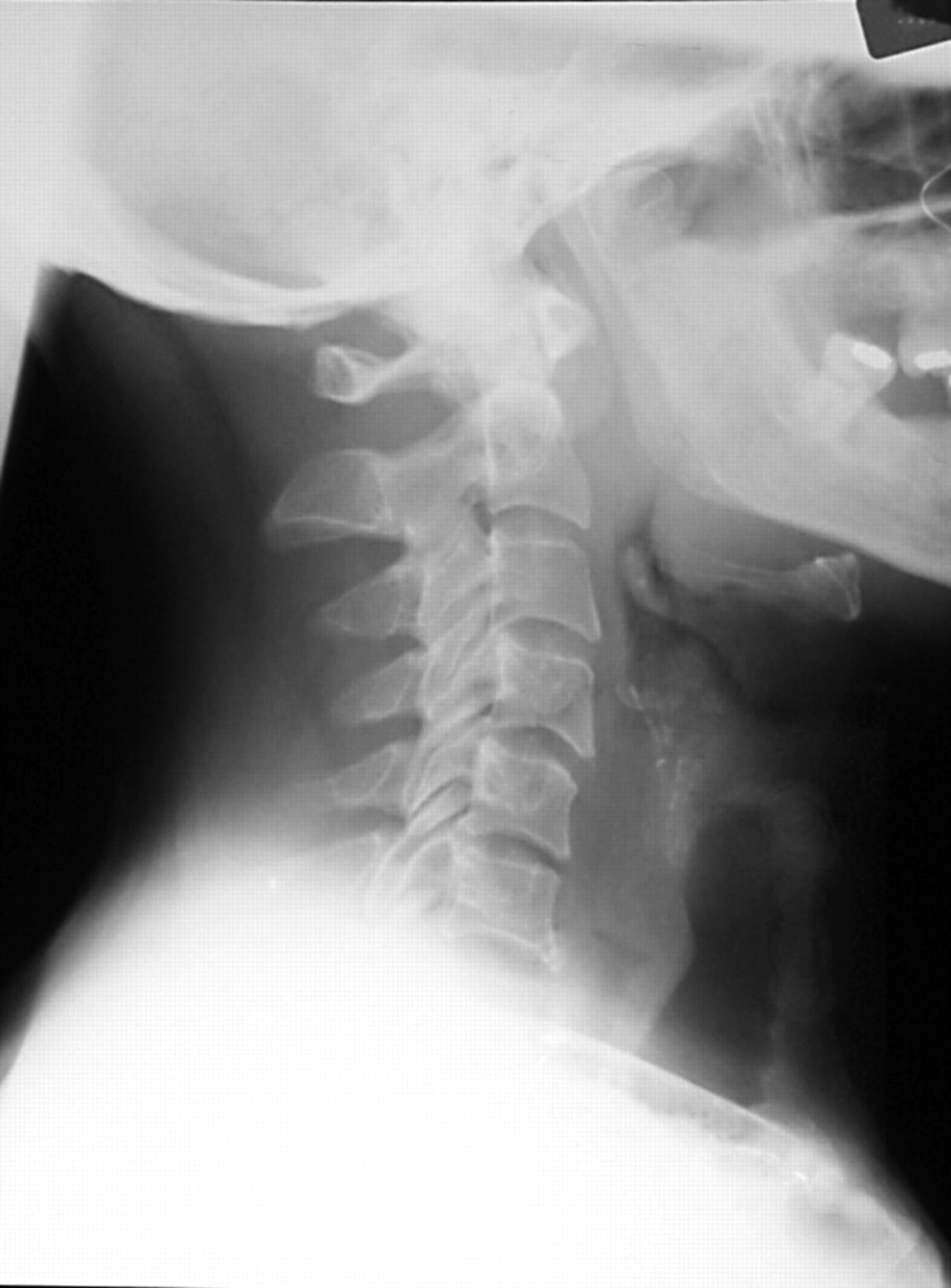
A 55 year old man presented to accident and emergency reporting that he had swallowed his dentures. He fell asleep with his dentures in situ and awoke adentulous with dysphagia and dysphonia. There was mild tenderness on palpation of the cricoid cartilage but no surgical emphysema. Lateral soft tissue neck radiograph revealed no radio-opaque foreign body, however the patient was referred to the ear, nose, and throat department for assessment. The radiograph demonstrated a column of air visible in the proximal oesophagus and widening of the prevertebral fascia (fig 1). Flexible nasopharyngolaryngoscopy showed evidence of the dental plate within the hypopharynx, lodged within the lumen of the crocopharyngeus.
Lateral neck radiograph demonstrates air within the proximal oesophagus and widening of prevertebral fascia.
Emergency pharyngoscopy was therefore performed. The dentures were impacted within the cricopharyngeus and there were surrounding mucosal abrasions. The dentures were removed intact and the patient was discharged the following morning symptom free and was well at follow up two weeks later.
DISCUSSION
Impacted foreign bodies in the trachea and oesophagus are a common problem. Impacted dentures, usually broken or partial dentures, accounted for 11.5% of foreign bodies in a series by Abdullah et al.1 Ingestion usually occurs after trauma, intoxication, loss of consciousness or sleep, so there may not be a definite history of ingestion. Prompt management is vital if significant morbidity, and mortality is to be avoided.1,2
Soft tissue lateral radiographs of the neck are routinely performed. There is, however, debate as to the value of the use of plain radiographs in such cases as dental plates are radiolucent (in contrast with natural teeth, which can be seen on plain radiography). This has been the case since the 1940s when radiolucent acrylic materials replaced radio-opaque vulcanite as the main denture material.2–4 It is no surprise therefore that in Abdullah’s series only 22% of dental prostheses impacted in the oesophagus showed evidence of a foreign body on the lateral neck radiograph. All of these had radiolucent wires attached.
Jones et al5 found that lateral soft tissue films of the neck changed the management in only 1.4% cases. Signs commonly associated with the ingestion of a foreign body include foreign body shadow, air in the oesophagus, increased pre-vertebral shadow, salivary pooling/level, and loss of cervical lordosis.6 According to Marais et al none of these commonly recognised radiological features are associated with a high positive prediction rate.6
It should be noted that foreign bodies in the oesophagus can result in significant morbidity and mortality if the duration of impaction is prolonged. The foreign body may become buried in the progressive mucosal oedema resulting in possible fistulisation and perforation. Cases of tracheo-oesophageal fistulas, aortic erosion, aorto-oesophageal fistulas, and even oesophago-broncho-aortic fistula after perforation have been reported.7–10 Not surprisingly complications have been shown to be more likely if there is a delay in treatment with the rate of complications rising from 3.2% after 24 hours to 23.5% after 48 hours.11,12 The radiolucent nature of dentures on neck radiographs, particularly if not appreciated by the treating emergency physician, may contribute to such a delay in diagnosis and definitive treatment.
The use of a radio-opaque material in the manufacture of dental plates may reduce the incidence of missed or delayed diagnoses when such plates are ingested. As yet none of the methods for making dentures visible on plain radiograph meet all of the required needs of a denture base material.13 It is therefore important, in order to prevent accidental ingestion, that dentures should be made to fit properly and damaged or malfitting dentures should be discarded and replaced. In addition patients should be strongly advised against wearing them in bed.12
CONCLUSION
In cases of dental plate ingestion the history may be vague and few clinical signs may be present. In addition dentures may be radiolucent, therefore accident and emergency physicians must maintain a high index of suspicion in such cases.
Lateral soft tissue radiographs do have a role in confirming pathology if they are abnormal, eliciting signs such as air in the oesophagus and increased prevertebral shadow. However, a normal radiograph does not exclude pathology. Therefore, even in the absence of radiological abnormalities, urgent referral to ENT is still indicated in the presence of the following signs/symptoms:
-
total or partial dysphagia (difficulty swallowing)
-
discomfort or pain in the throat or neck
-
pooling or excessive production of saliva.