Article Text

Statistics from Altmetric.com
CLINICAL HISTORY
A 55 year old man was admitted to the rheumatology department owing to inflammatory back pain (occurring at 4 00 am, not aggravated by movement) without sciatica, increasingly severe over two years and associated with asthenia and weight loss (10 kg). No abnormalities were found on physical examination, except for tenderness on palpation of the spinous processes of L5 and S1. There was no significant past medical history. Laboratory tests showed a mild inflammatory picture: erythrocyte sedimentation rate 25 mm/1st h (normal 5–15), C reactive protein 12 mg/l (normal 0–10), fibrinogen 5.1 g/l (normal 2–4). Protein electrophoresis, red and white blood cell count, glucose, renal, and liver function tests were normal. Chemical bone markers (urinary pyridinoline, alkaline phosphatase, osteocalcin, parathormone, 25-hydroxyvitamin D, serum calcium and phosphorus, and urinary calcium and phosphorus) were normal.
RADIOLOGICAL FINDINGS
Standard lumbar spine and pelvic radiographs were normal.
Technetium-99m bone scintigraphy showed no abnormal uptake in the spine or skull but disclosed an increased peripheral uptake.
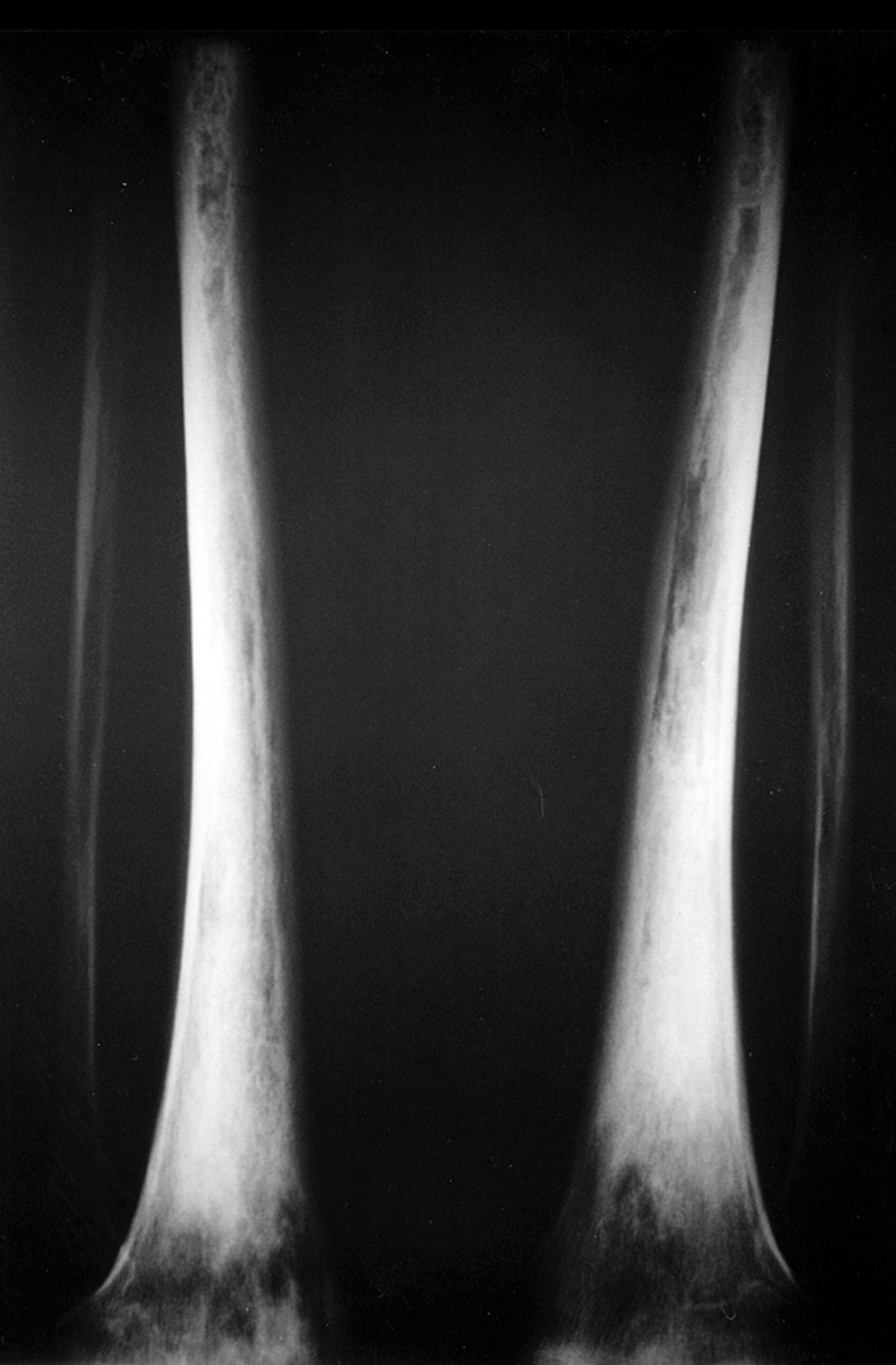
A skeletal survey showed multiple mixed bone lesions with sclerotic areas surrounding smaller lytic foci. These roughly bilateral, symmetrical lesions involved the metaphyses and diaphyses of some long bones but spared the epiphyses. Bones affected were the femora, tibiae, radii, wrist, and tarsal bones (fig 1⇓).

Skeletal bone radiographs showed bilateral and symmetrical cortical osteosclerosis of the diaphysial and metaphysial regions of the legs, corresponding to the increased uptake in bone scan, but clinically asymptomatic.
With magnetic resonance imaging (MRI), these lesions appeared in low signal on T1 weighted sequences (fig 2⇓) and in high heterogeneous signal on T2 weighted sequences with fat annulation. The computed tomography (CT) scan showed central osteosclerosis of the medullary canal with no evidence of osteolysis or periostosis.

In T1 spin echo weighted sequences the histiocytosis infiltration appeared as metaphyso-diaphysial low signal lesions, corresponding to the osteosclerotic lesions in standard radiographs.
An abdominal CT scan disclosed tissue infiltration surrounding the kidneys.
Biopsy of the tibial area displayed foamy lipid laden histiocytes, confirming the diagnosis of Erdheim-Chester disease.
TREATMENT
Because of the systemic involvement, prednisone (20 mg daily) treatment was started and, after one month, significant clinical and laboratory improvement was seen. At nine months the patient's general condition had returned to normal, his back pain had completely disappeared, and all laboratory values had returned to normal. Moreover, the retroperitoneal infiltration seen on abdominal CT scan had significantly decreased. Steroid treatment was progressively decreased, but at 18 months, with a daily regimen of 6 mg prednisone by mouth, the inflammatory lumbar pain reappeared, without any increase of the inflammatory markers (erythrocyte sedimentation rate and C reactive protein). Prednisone was increased to 7 mg daily and the pain disappeared.
DISCUSSION
Erdheim-Chester disease is a rare, non-Langerhans form of histiocytosis characterised by infiltrates of foamy, lipid laden histiocytes and bilateral symmetrical foci of sclerosis in appendicular long bones.1,2 It usually affects adults over the age of 40 years and there is a slight male predominance.3
In a review of 59 cases,2 bone pain was the most common symptom (28 patients), and was sometimes isolated (seven patients). It mainly affected the legs, especially the knees and the ankles, and is described as a juxta-articular, mild but permanent, and non-inflammatory pain. In the same study, general symptoms such as fever, weight loss, and weakness were present in 11 patients, as was so in our case. Retroperitoneal fibrosis, regardless of the cause, can lead to dull back pain as in our patient, or more commonly to abdominal pain.4–,6 However, even if the retroperitoneum and kidneys are commonly affected in Erdheim-Chester disease (17/59 patients),2 it is rarely symptomatic (dysuria, abdominal pain, obstructive renal impairment, and enlarged palpable kidneys), and lumbar pain is not especially reported in this disease. Reports of about 23 patients with retroperitoneal Erdheim-Chester disease, including 13 with only focal xanthogranulomatosis in the retroperitoneum, have been published.2,7–,9
Each clinical sign in Erdheim-Chester disease can lead to misdiagnosis or to the diagnosis of another histiocytic disorder. The only specific signs are the radiological findings of osteosclerosis and the histological features. Symmetrical sclerosis of the metaphysis and diaphysis of the long tubular bone are pathognomonic radiological changes.2,10 It is not reported in Langerhans' cell histiocytosis, in which skeletal lesions are osteolytic and very rarely located on the long bone.11 This widespread sclerosis of the appendicular skeleton is rarely associated with lytic lesions and spares the epiphysis and axial flat bones. Because clinical bone symptoms can be mild or absent, bone scintigraphy may be valuable, as it discloses all sites of bone affected in one diagnostic procedure.12 Differential diagnosis includes mastocytosis, fluoride intoxication, myeloid metaplasia, lymphoma, metastatic disease, toxic osteoarthropathy, and adult progressive diaphysial dysplasia (Engelmann disease).
Extraskeletal manifestations can occur in almost all organs, including lungs, pericardium, skin, orbit, and retroperitoneum. The retroperitoneal deposits tend to be mainly in the upper retroperitoneum in the perirenal fat, as in our case, sometimes encasing the adrenal glands and even extending into the mediastinum.13,14 Retroperitoneal xanthogranuloma is distinguished from inflammatory fibrosarcoma by its numerous foamy histiocytes, relative lack of plasma cells, and lack of nuclear atypia; Erdheim-Chester infiltration is distinguished from retroperitoneal fibrosis principally by its many foamy histiocytes, lack of plasma cells, and lack of vasculitis.7
Clinical trials for treatment of Erdheim-Chester disease have not been conducted; thus therapeutic options are based on anecdotal experience, and generally do not provide information about the effects on disease outcome. Systemic corticosteroids, chemotherapy (for example, vinblastine, vincristine, cyclosphosphamide, doxorubidicin), and radiation treatment have been used.2,15 In this case, we obtained a good clinical and laboratory response with a partial regression of the retroperitoneal infiltration; the patient's disease was stabilised with a small dose of steroids, but he remains dependent on it. The prognosis depends to a large part on the extent and distribution of extraosseous disease. In a review of 59 patients, Veyssier-Belot et al reported death related to the disease in 59% of cases, including 36% in less than six months2: the most commonly reported causes of death are respiratory and heart failure. The efficacy of the various treatments is difficult to evaluate because of the rarity of the disease.
THE LESSONS
Erdheim-Chester is a rare non-Langerhans histiocytosis showing characteristic radiological and histological features.
Extraskeletal manifestations can occur in almost all organs, leading to a poor prognosis.
As bone pain is the first revealing symptom, rheumatologists should be able to recognise the disease.
REFERENCES
Footnotes
Series editor: Anthony D Woolf