Advertisement

Main menu

  • Home
  • Content
    • Current Issue
    • Publication Preview--Ahead of Print
    • Past Issue Archive
    • Case of the Week Archive
    • Classic Case Archive
    • Case of the Month Archive
    • COVID-19 Content and Resources
  • For Authors
  • About Us
    • About AJNR
    • Editors
    • American Society of Neuroradiology
  • Submit a Manuscript
  • Podcasts
    • Subscribe on iTunes
    • Subscribe on Stitcher
  • More
    • Subscribers
    • Permissions
    • Advertisers
    • Alerts
    • Feedback
  • Other Publications
    • ajnr

User menu

  • Subscribe
  • Alerts
  • Log in

Search

  • Advanced search
American Journal of Neuroradiology

American Journal of Neuroradiology

  • Subscribe
  • Alerts
  • Log in

Advanced Search

  • Home
  • Content
    • Current Issue
    • Publication Preview--Ahead of Print
    • Past Issue Archive
    • Case of the Week Archive
    • Classic Case Archive
    • Case of the Month Archive
    • COVID-19 Content and Resources
  • For Authors
  • About Us
    • About AJNR
    • Editors
    • American Society of Neuroradiology
  • Submit a Manuscript
  • Podcasts
    • Subscribe on iTunes
    • Subscribe on Stitcher
  • More
    • Subscribers
    • Permissions
    • Advertisers
    • Alerts
    • Feedback
  • Follow AJNR on Twitter
  • Visit AJNR on Facebook
  • Follow AJNR on Instagram
  • Join AJNR on LinkedIn
  • RSS Feeds
Research ArticleBRAIN

Complete Duplication or Extreme Fenestration of the Basilar Artery

Jonas H. Goldstein, Richard Woodcock, Huy M. Do, C. Douglas Phillips and Jacques E. Dion
American Journal of Neuroradiology January 1999, 20 (1) 149-150;
Jonas H. Goldstein
aFrom the Department of Radiology, Box 170, University of Virginia Health Sciences Center, Charlottesville, VA 22908.
  • Find this author on Google Scholar
  • Find this author on PubMed
  • Search for this author on this site
Richard Woodcock
aFrom the Department of Radiology, Box 170, University of Virginia Health Sciences Center, Charlottesville, VA 22908.
  • Find this author on Google Scholar
  • Find this author on PubMed
  • Search for this author on this site
Huy M. Do
aFrom the Department of Radiology, Box 170, University of Virginia Health Sciences Center, Charlottesville, VA 22908.
  • Find this author on Google Scholar
  • Find this author on PubMed
  • Search for this author on this site
C. Douglas Phillips
aFrom the Department of Radiology, Box 170, University of Virginia Health Sciences Center, Charlottesville, VA 22908.
  • Find this author on Google Scholar
  • Find this author on PubMed
  • Search for this author on this site
Jacques E. Dion
aFrom the Department of Radiology, Box 170, University of Virginia Health Sciences Center, Charlottesville, VA 22908.
  • Find this author on Google Scholar
  • Find this author on PubMed
  • Search for this author on this site
  • Article
  • Figures & Data
  • Info & Metrics
  • References
  • PDF
Loading

Abstract

Summary: We describe a 42-year-old man with complete duplication or extreme fenestration of the basilar artery. We review the developmental anatomy and embryology and discuss the possible clinical implications and associated findings of this anomaly.

The term fenestration refers to a localized duplication of a vessel. While fenestration of the basilar artery has been reported to be as high as 6% in postmortem studies, its angiographic prevalence has been described as ranging from 0.04% to 0.6% (1). Basilar artery fenestration most commonly occurs in the lower half of the vessel (2), extending for a length of less than 5 mm (3, 4). Complete duplication of the basilar artery, with each vertebral artery continuing separately to form a posterior cerebral artery, has been described previously (5, 6). We present a case of complete duplication or extreme fenestration of the basilar artery demonstrated by catheter digital subtraction angiography (DSA).

Case Report

A 42-year-old obese man with rhizomelia and mild mental retardation was admitted to the emergency department with abrupt onset of a left temporal headache accompanied by nausea and vomiting. A noncontrast head CT scan was obtained and showed no evidence of subarachnoid hemorrhage. Lumbar puncture revealed equivocal findings for subarachnoid hemorrhage. Diagnostic cerebral angiography was requested to exclude an aneurysm or vascular malformation.

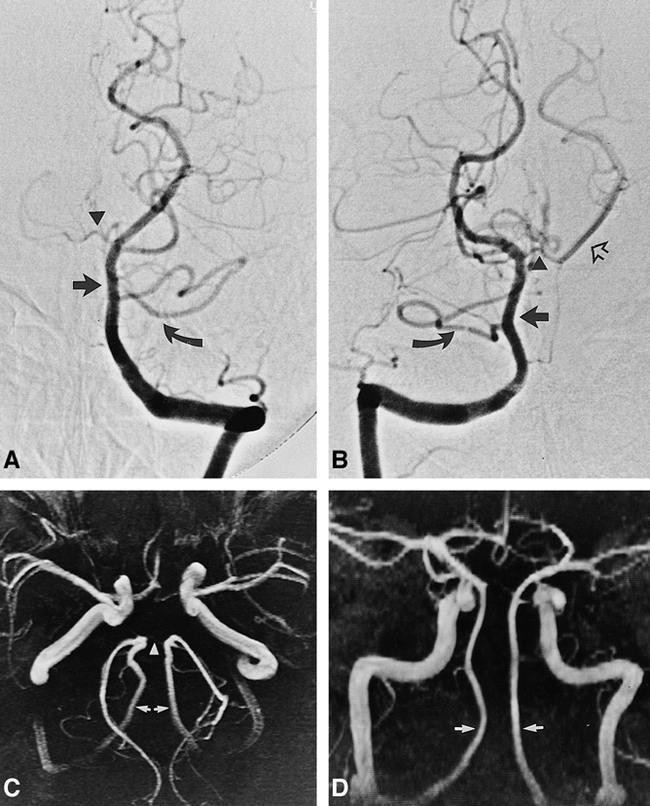
Cerebral angiography showed the left vertebral artery as arising from the aortic arch and giving origin to the thyrocervical trunk in its cervical course. Likewise, the thyrocervical trunk on the right arose from the vertebral artery. The left vertebral artery injection did not show reflux into the contralateral vertebral artery. Although the ipsilateral posterior cerebral, superior, anterior inferior, and posterior inferior cerebellar arteries were present and normal, their contralateral counterparts were not identified (Fig 1A), with the exception of transient opacification of the right superior cerebellar artery via a small communicating branch. The right vertebral artery injection revealed similar findings (Fig 1B), with the addition of two small bridging vessels to the contralateral vertebral artery, one at the proximal and one at the distal segment. Additional vascular anomalies included the left pericallosal artery arising from the right anterior cerebral artery, and the right ophthalmic artery arising from the internal carotid artery cavernous segment. The posterior communicating arteries were not identified and the remainder of the examination failed to reveal a cause of subarachnoid hemorrhage. Subsequently, an MR imaging/MR angiographic examination was performed, with the MR angiogram confirming findings on the conventional angiogram (Fig 1C and D). The patient was clinically stable for 48 hours and was discharged.

fig 1.
  • Download figure
  • Open in new tab
  • Download powerpoint
fig 1.

Left and right vertebral artery angiograms (A and B) and MR angiograms (C and D) in a 42-year-old man with rhizomelia and mild mental retardation.

A, Anteroposterior projection of left vertebral angiogram shows filling of basilar artery to the left of midline (straight arrow). There is opacification of a common trunk for the AICA and PICA (curved arrow), with no filling of the right AICA/PICA trunk. A small branch is seen from the distal basilar artery (arrowhead).

B, Anteroposterior projection of right vertebral angiogram shows filling of basilar artery to the right of midline (straight solid arrow). There is opacification of a common trunk for the AICA and PICA on the right (curved arrow), with no filling of the left AICA. A branch from the distal basilar artery (arrowhead) opacifies left PCA (open arrow).

C, Angled anteroposterior projection of MR angiogram shows two vertebrobasilar systems (arrows), each filling an ipsilateral PCA. The distal basilar cross-filling branch faintly opacifies (arrowhead).

D, Anteroposterior projection of MR angiogram shows proximal portion of the two vertebrobasilar systems (arrows).

Discussion

The embryology of the basilar artery has been well described by Padget (7). Fusion of the embryonic longitudinal neural arteries into a single basilar artery occurs in a craniocaudal direction (8) by approximately the fifth fetal week (7). Fenestrations or “windows” within the basilar artery occur as a result of failure of fusion of the neural arteries and of regression of the bridging arteries that connect the longitudinal arteries. The term fenestration has been used interchangeably with partial duplication (9) or segmental duplication (10–12). Although partial or segmental duplications (fenestrations) have a reported rate of occurrence of up to 5% to 6% (11), true or complete duplication is extremely rare (4–6).

This case illustrates complete duplication or extreme fenestration of the basilar artery, with each vertebral artery terminating in a posterior cerebral artery. Given the patient's rhizomelia and mental retardation, we were curious as to whether this vascular anomaly was part of a chromosomal syndrome. A normal karyotype result had been obtained several years earlier, however. This nonfusion of the embryologic longitudinal neural arches was thought to be an incidental finding. It was interesting to note both a small superior and inferior connecting artery (Fig 1B); we hypothesized that the superior connecting artery was composed of the P1 segments of the posterior cerebral arteries and the terminal basilar artery, which together may represent a posterior cerebral vessel analog to the anterior communicating artery. These two small bridging arteries were not thought to be subjected to the same type of hemodynamic alterations as true fenestrations, and therefore did not indicate an increased risk of aneurysmal formation sufficient to justify further surveillance.

Footnotes

  • ↵1 Address reprint requests to Jonas H. Goldstein, MD.

References

  1. ↵
    Sanders WP, Sorek PA, Mehta BA. Fenestration of intracranial arteries with special attention to associated aneurysms and other anomalies. AJNR Am J Neuroradiol 1993;14:675-680
    Abstract/FREE Full Text
  2. ↵
    Takahashi M. Atlas of Vertebral Angiography. Tokyo: Bunkyo-ku; 1974;22-24
  3. ↵
    Finlay HM, Canham PB. The layered fabric of cerebral artery fenestrations. Stroke 1994;25:1799-1806
    Abstract/FREE Full Text
  4. ↵
    Newton TH, Potts DG. Radiology of the Skull and Brain: Angiography. St Louis: Mosby; 1974;69:1775-1795
  5. ↵
    Adachi B. Das Arteriensystem der Japaner. Kyoto, Japan: Kaiserlich-Japanischen Univerität zu Kyoto; 1928
  6. Krayenbühl H, Yasargil G. Die vaskulären Erkrankungen im Gebiet der Arteria vertebralis und Arteria basialis; eine anatomische und pathologische, klinische und neuroradiologische Studie. Stuttgart: Thieme; 1957
  7. ↵
    Padget DH. The development of the cranial arteries in the human embryo. Contrib Embryol 1948;32:205-261
  8. ↵
    Lasjaunias P, Berenstein A. Surgical Neuroangiography: Functional Vascular Anatomy of Brain, Spinal Cord, and Spine. New York: Springer; 1990;3:199-207
  9. ↵
    Tran-Dinh HD, Soo YS, Jayasinghe LS. Duplication of the vertebrobasilar system. Australas Radiol 1991;35:220-224
    CrossRefPubMed
  10. ↵
    De Caro R, Serafini MT, Galli S, et al. Anatomy of segmental duplication in the human basilar artery: possible site of aneurysm formation. Clin Neuropathol 1995;14:303-309
    PubMed
  11. ↵
    Berry AD, Kepes JJ, Wetzel NM. Segmental duplication of the basilar artery with thrombosis. Stroke 1988;119:256-260
  12. Black SP, Ansbacher LE. Saccular aneurysm associated with segmental duplication of the basilar artery: a morphologic study. J Neurosurg 1984;61:1005-1008
    CrossRefPubMed
  • Received September 18, 1997.
  • Accepted after revision April 13, 1998.
  • Copyright © American Society of Neuroradiology
View Abstract
PreviousNext
Back to top

In this issue

American Journal of Neuroradiology
Vol. 20, Issue 1
1 Jan 1999
  • Table of Contents
  • Index by author
Advertisement
Print
Download PDF
Email Article

Thank you for your interest in spreading the word on American Journal of Neuroradiology.

NOTE: We only request your email address so that the person you are recommending the page to knows that you wanted them to see it, and that it is not junk mail. We do not capture any email address.

Enter multiple addresses on separate lines or separate them with commas.
Complete Duplication or Extreme Fenestration of the Basilar Artery
(Your Name) has sent you a message from American Journal of Neuroradiology
(Your Name) thought you would like to see the American Journal of Neuroradiology web site.
CAPTCHA
This question is for testing whether or not you are a human visitor and to prevent automated spam submissions.
Citation Tools
Complete Duplication or Extreme Fenestration of the Basilar Artery
Jonas H. Goldstein, Richard Woodcock, Huy M. Do, C. Douglas Phillips, Jacques E. Dion
American Journal of Neuroradiology Jan 1999, 20 (1) 149-150;

Citation Manager Formats

  • BibTeX
  • Bookends
  • EasyBib
  • EndNote (tagged)
  • EndNote 8 (xml)
  • Medlars
  • Mendeley
  • Papers
  • RefWorks Tagged
  • Ref Manager
  • RIS
  • Zotero
Share
Complete Duplication or Extreme Fenestration of the Basilar Artery
Jonas H. Goldstein, Richard Woodcock, Huy M. Do, C. Douglas Phillips, Jacques E. Dion
American Journal of Neuroradiology Jan 1999, 20 (1) 149-150;
del.icio.us logo Digg logo Reddit logo Twitter logo CiteULike logo Facebook logo Google logo Mendeley logo
  • Tweet Widget
  • Facebook Like
  • Google Plus One

Jump to section

  • Article
    • Abstract
    • Case Report
    • Discussion
    • Footnotes
    • References
  • Figures & Data
  • Info & Metrics
  • References
  • PDF

Related Articles

  • No related articles found.
  • PubMed
  • Google Scholar

Cited By...

  • Republished: A rare case of absent left common carotid artery with bovine origin of the left external carotid artery
  • A rare case of absent left common carotid artery with bovine origin of the left external carotid artery
  • Crossref
  • Google Scholar

This article has not yet been cited by articles in journals that are participating in Crossref Cited-by Linking.

More in this TOC Section

  • Enhanced Axonal Metabolism during Early Natalizumab Treatment in Relapsing-Remitting Multiple Sclerosis
  • Progression of Microstructural Damage in Spinocerebellar Ataxia Type 2: A Longitudinal DTI Study
  • SWI or T2*: Which MRI Sequence to Use in the Detection of Cerebral Microbleeds? The Karolinska Imaging Dementia Study
Show more BRAIN

Similar Articles

Advertisement

News and Updates

  • Lucien Levy Best Research Article Award
  • Thanks to our 2022 Distinguished Reviewers
  • Press Releases

Resources

  • Evidence-Based Medicine Level Guide
  • How to Participate in a Tweet Chat
  • AJNR Podcast Archive
  • Ideas for Publicizing Your Research
  • Librarian Resources
  • Terms and Conditions

Opportunities

  • Share Your Art in Perspectives
  • Get Peer Review Credit from Publons
  • Moderate a Tweet Chat

American Society of Neuroradiology

  • Neurographics
  • ASNR Annual Meeting
  • Fellowship Portal
  • Position Statements

© 2023 by the American Society of Neuroradiology | Print ISSN: 0195-6108 Online ISSN: 1936-959X

Powered by HighWire