
Abstract
Summary: Transcranial Doppler sonography shows potential as a noninvasive technique for long-term follow-up of treated intracranial saccular aneurysms. This technical note describes a color Doppler artifact related to microcoil architecture that might represent a potential pitfall in transcranial Doppler sonographic evaluation of aneurysmal cavity thrombosis, since it may be wrongly interpreted as residual flow or aneurysmal cavity recanalization.
Transcranial Doppler sonography is a promising noninvasive technique for the detection and posttherapeutic follow-up of intracranial saccular aneurysms. This technical note describes a color Doppler artifact related to microcoil architecture that might represent a potential pitfall in transcranial Doppler sonographic evaluation of aneurysmal cavity thrombosis.
Case Report
Cerebral digital subtraction angiography was performed in a 45-year-old patient with subarachnoid hemorrhage proved at CT. Three saccular aneurysms were discovered: a broad-based, irregularly shaped aneurysm of the anterior communicating artery (ACoA), a 3-mm aneurysm of the right anterior choroidal artery (AChA) (Fig 1), and a 1-mm aneurysm of the right middle cerebral artery (MCA) bifurcation (Fig 1). The ACoA aneurysm was considered the source of the hemorrhage, and surgical clipping was performed; it was decided to treat the AChA aneurysm through an endovascular approach. The very small MCA aneurysm was left untreated. The AChA aneurysm was successfully embolized with a single Guglielmi detachable coil (diameter, 3 mm; length, 40 mm) (GDC-10 soft, Target Therapeutics, Fremont, CA) (Fig 2). Transcranial Doppler sonography was performed through a right temporal window before and after endovascular treatment as part of the embolization protocol adopted in our institution (Figs 3 and 4). At posttherapeutic transcranial Doppler sonography, the coil appeared as a hyperechoic structure with shadowing, and a color signal was noted as a rapidly changing mixture of red and blue behind the coil (Fig 4). No flow was noted close to or in the aneurysm, whereas a digital subtraction angiogram obtained immediately before transcranial Doppler sonography showed that the aneurysmal cavity was completely excluded from the cerebral circulation. As a complement to this clinical observation, an in vitro essay consisting of insonation of a Guglielmi detachable coil enclosed between two hydrated agar sheets (Sonar-Aid, Geistlich-Pharma, Switzerland) was performed, allowing us to reproduce the artifactual color signal (Fig 5).

Digital subtraction angiogram, superselective study of the right internal carotid artery, arterial phase, lateral projection, displays the MCA bifurcation.
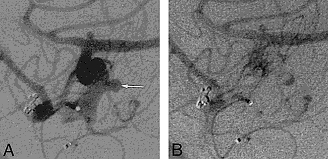
A, A small saccular aneurysm (arrow) is seen at the right anterior choroidal artery origin. Note also the minute aneurysm at the MCA bifurcation.
B, On late arterial phase image, a characteristic aneurysmal inflow-outflow pattern (“yin-yang”) may be observed.
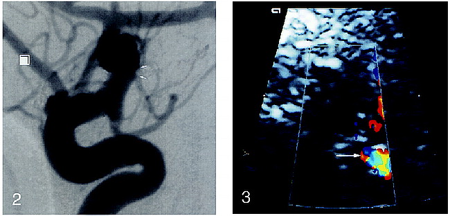
Digital subtraction angiogram, selective study of the right internal carotid artery, arterial phase, lateral projection. After treatment with a single detachable coil, the aneurysmal cavity is totally excluded from the cerebral circulation (arrows).
fig 3. Transcranial Doppler sonogram, right transtemporal approach. Before treatment, a color Doppler signal is seen within the aneurysmal cavity (arrow). It also shows the typical “yin- yang” appearance, corresponding to the inflow and outflow regions of the aneurysm.

Transcranial Doppler sonogram, right transtemporal approach. After treatment, a twinkling artifact (arrow) is observed immediately behind the hyperechoic signal of the microcoil.
fig 5. In vitro insonation of a Guglielmi detachable coil enclosed within two hydrated agar sheets. Color Doppler image shows the hyperechoic structure of the coil (arrow) and the twinkling artifact (arrowhead).
Discussion
The color artifact reported herein corresponds to the color Doppler twinkling artifact described by Rahmouni et al (1). These authors showed that artifactual color appears to be generated at random, strongly reflecting a medium composed of individual reflectors. Color is not always restricted to blood flow and can be seen adjacent to areas of severe vascular stenosis (2) or in echo-free regions without flow (3). In the reported observation, no flow spectrum could be recorded in the color areas, and the spectrum was composed of close vertical bands with no outer wrapping. This artifact has been observed in the abdominal field, particularly in the detection of renal or ureteral calculi, but its potential interest in locating intracerebral coils has, to our knowledge, not been reported previously.
Conclusion
Transcranial Doppler sonography shows potential as a noninvasive technique for long-term follow-up of treated intracranial saccular aneurysms. The artifact described herein thus seems important to recognize, as it could be mistaken for residual flow or aneurysmal cavity recanalization.
Footnotes
↵1 Address reprint requests to Haleem G. Khan, MD.
- Received January 22, 1998.
- Copyright © American Society of Neuroradiology
In this issue


Jump to section
Related Articles
Cited By...
- A Novel Approach for Quantification and Analysis of the Color Doppler Twinkling Artifact With Application in Noninvasive Surface Roughness Characterization: An In Vitro Phantom Study
- Sonographic Twinkling Artifact in a Renal Graft With Prolene Mesh
- Twinkling Artifact in Gallbladder Adenomyomatosis
- Twinkling Artifact in Color Doppler Imaging of the Orbit