Advertisement

Main menu

  • Home
  • Content
    • Current Issue
    • Publication Preview--Ahead of Print
    • Past Issue Archive
    • Case of the Week Archive
    • Classic Case Archive
    • Case of the Month Archive
    • COVID-19 Content and Resources
  • For Authors
  • About Us
    • About AJNR
    • Editors
    • American Society of Neuroradiology
  • Submit a Manuscript
  • Podcasts
    • Subscribe on iTunes
    • Subscribe on Stitcher
  • More
    • Subscribers
    • Permissions
    • Advertisers
    • Alerts
    • Feedback
  • Other Publications
    • ajnr

User menu

  • Subscribe
  • Alerts
  • Log in

Search

  • Advanced search
American Journal of Neuroradiology

American Journal of Neuroradiology

  • Subscribe
  • Alerts
  • Log in

Advanced Search

  • Home
  • Content
    • Current Issue
    • Publication Preview--Ahead of Print
    • Past Issue Archive
    • Case of the Week Archive
    • Classic Case Archive
    • Case of the Month Archive
    • COVID-19 Content and Resources
  • For Authors
  • About Us
    • About AJNR
    • Editors
    • American Society of Neuroradiology
  • Submit a Manuscript
  • Podcasts
    • Subscribe on iTunes
    • Subscribe on Stitcher
  • More
    • Subscribers
    • Permissions
    • Advertisers
    • Alerts
    • Feedback
  • Follow AJNR on Twitter
  • Visit AJNR on Facebook
  • Follow AJNR on Instagram
  • Join AJNR on LinkedIn
  • RSS Feeds
Research ArticleHEAD AND NECK

CT and MR Findings of Michel Anomaly: Inner Ear Aplasia

Kathlyn Marsot-Dupuch, Alessandro Dominguez-Brito, Karim Ghasli and Claude-Henri Chouard
American Journal of Neuroradiology February 1999, 20 (2) 281-284;
Kathlyn Marsot-Dupuch
  • Find this author on Google Scholar
  • Find this author on PubMed
  • Search for this author on this site
Alessandro Dominguez-Brito
  • Find this author on Google Scholar
  • Find this author on PubMed
  • Search for this author on this site
Karim Ghasli
  • Find this author on Google Scholar
  • Find this author on PubMed
  • Search for this author on this site
Claude-Henri Chouard
  • Find this author on Google Scholar
  • Find this author on PubMed
  • Search for this author on this site
  • Article
  • Figures & Data
  • Info & Metrics
  • References
  • PDF
Loading

Article Figures & Data

Figures

  • fig 1.
    • Download figure
    • Open in new tab
    • Download powerpoint
    fig 1.

    Case 1: 9-year-old girl with profound bilateral congenital sensorineural hearing loss.

    A, Axial CT scan shows right petrous bone aplasia with absence of inner ear structures. The medial wall of the middle ear is flattened (arrow), being in close contact with the infratentorial nervous structures. Note normal differentiation of the malleus.

    B, Coronal CT scan shows a normally developed external and middle right ear. The long process of the incus (arrow) leans against the medial middle ear wall. The oval window, the stapes, and the second portion of the facial nerve are absent. A small dehiscence of the medial wall of the middle ear (arrowhead), located at the IAM, probably corresponds to the entrance of the facial nerve.
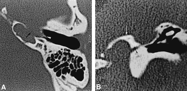
  • fig 2.
    • Download figure
    • Open in new tab
    • Download powerpoint
    fig 2.

    Case 2: 16-year-old boy, brother of girl in case 1, also with profound bilateral congenital sensorineural hearing loss.

    A, Axial CT scan shows an aberrant course of the left internal carotid artery, which bulges into the middle ear (white arrow). Dehiscence of the lateral wall of the petrous bone adjacent to the horizontal carotid artery is present (black arrow). A small lateral tract corresponds to a persistent stapediohyoid artery (white arrowhead). The facial nerve passes behind the internal carotid artery (black arrowheads).

    B, Coronal CT scan shows enlargement of inferior tympanic canaliculus (arrowhead). Arrow indicates jugular foramen.

PreviousNext
Back to top

In this issue

American Journal of Neuroradiology
Vol. 20, Issue 2
1 Feb 1999
  • Table of Contents
  • Index by author
Advertisement
Print
Download PDF
Email Article

Thank you for your interest in spreading the word on American Journal of Neuroradiology.

NOTE: We only request your email address so that the person you are recommending the page to knows that you wanted them to see it, and that it is not junk mail. We do not capture any email address.

Enter multiple addresses on separate lines or separate them with commas.
CT and MR Findings of Michel Anomaly: Inner Ear Aplasia
(Your Name) has sent you a message from American Journal of Neuroradiology
(Your Name) thought you would like to see the American Journal of Neuroradiology web site.
CAPTCHA
This question is for testing whether or not you are a human visitor and to prevent automated spam submissions.
Citation Tools
CT and MR Findings of Michel Anomaly: Inner Ear Aplasia
Kathlyn Marsot-Dupuch, Alessandro Dominguez-Brito, Karim Ghasli, Claude-Henri Chouard
American Journal of Neuroradiology Feb 1999, 20 (2) 281-284;

Citation Manager Formats

  • BibTeX
  • Bookends
  • EasyBib
  • EndNote (tagged)
  • EndNote 8 (xml)
  • Medlars
  • Mendeley
  • Papers
  • RefWorks Tagged
  • Ref Manager
  • RIS
  • Zotero
Share
CT and MR Findings of Michel Anomaly: Inner Ear Aplasia
Kathlyn Marsot-Dupuch, Alessandro Dominguez-Brito, Karim Ghasli, Claude-Henri Chouard
American Journal of Neuroradiology Feb 1999, 20 (2) 281-284;
Reddit logo Twitter logo Facebook logo Mendeley logo
  • Tweet Widget
  • Facebook Like
  • Google Plus One

Jump to section

  • Article
    • Abstract
    • Case Reports
    • Discussion
    • Conclusion
    • Acknowledgments
    • Footnotes
    • References
  • Figures & Data
  • Info & Metrics
  • References
  • PDF

Related Articles

  • No related articles found.
  • PubMed
  • Google Scholar

Cited By...

  • Pediatric Sensorineural Hearing Loss, Part 2: Syndromic and Acquired Causes
  • Complete Labyrinthine Aplasia: Clinical and Radiologic Findings with Review of the Literature
  • Imaging appearances of unusual conditions of the middle and inner ear
  • Crossref
  • Google Scholar

This article has not yet been cited by articles in journals that are participating in Crossref Cited-by Linking.

More in this TOC Section

  • Correlation of Apparent Diffusion Coefficient at 3T with Prognostic Parameters of Retinoblastoma
  • Parathyroid Lesions: Characterization with Dual-Phase Arterial and Venous Enhanced CT of the Neck
  • Comparison of Contrast Effect on the Cochlear Perilymph after Intratympanic and Intravenous Gadolinium Injection
Show more HEAD AND NECK

Similar Articles

Advertisement

News and Updates

  • Lucien Levy Best Research Article Award
  • Thanks to our 2022 Distinguished Reviewers
  • Press Releases

Resources

  • Evidence-Based Medicine Level Guide
  • How to Participate in a Tweet Chat
  • AJNR Podcast Archive
  • Ideas for Publicizing Your Research
  • Librarian Resources
  • Terms and Conditions

Opportunities

  • Share Your Art in Perspectives
  • Get Peer Review Credit from Publons
  • Moderate a Tweet Chat

American Society of Neuroradiology

  • Neurographics
  • ASNR Annual Meeting
  • Fellowship Portal
  • Position Statements

© 2023 by the American Society of Neuroradiology | Print ISSN: 0195-6108 Online ISSN: 1936-959X

Powered by HighWire