
Abstract
Summary: We describe a cervical congenital paraspinal arteriovenous malformation (AVM) drained by paraspinal and epidural ectatic veins, which caused massive erosion of the C6 and C7 vertebral bodies, threatening the cervical stability and necessitating treatment. During the first session, six arterial embolizations were performed to reduce the size and the flow of the AVM. Two months later, a venous approach was used to occlude the remnant venous exit of the AVM and achieve a complete cure. All embolizations were performed using N-butylcyanoacrylate.
Spinal vascular malformations can be classified on the basis of their location and angioarchitecture (1, 2): intramedullary arteriovenous malformations (AVMs), intradural AVFs, dural AVFs, epidural AVFs and AVMs, paravertebral AVFs and AVMs, intraosseous vertebral AVMs, and Cobb syndrome.
Paravertebral malformations are rare, but the cervical location is reported to be the most common (3). In most of the cases, paravertebral malformations are congenital (3). We report a congenital cervical paravertebral AVM consisting of several AVFs associated with Klippel-Trenaunay syndrome of the left arm. The lesion drained into multiple ectatic veins, some of which were inside the spinal canal, causing C5 and C6 vertebral body erosion and shifting of the spinal cord.
Case Report
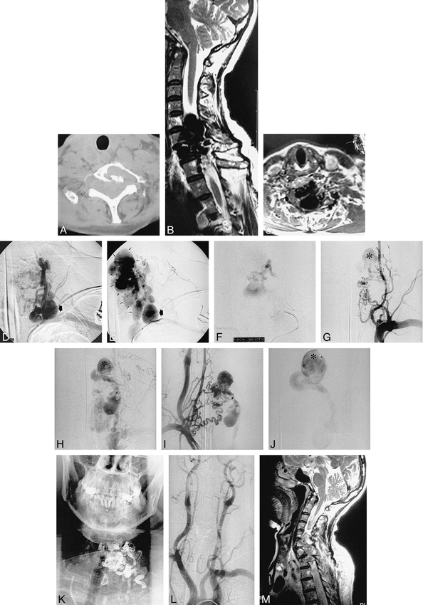
In 1997, a 48-year-old woman with a congenital Klippel-Trenaunay syndrome of the left arm with cutaneous and subcutaneous angiodysplasia of the left hemithorax associated with a paravertebral cervical AVM was referred to our institution. The appearance was in keeping with a metameric vascular malformation. No neurologic deficit was ever noted. the Klippel-Trenaunay syndrome was diagnosed at birth. The paraspinal AVM was angiographically documented in 1990 after a thrill was palpated in the neck. Treatment was not proposed to the patient at that time. In December 1996, plain X-rays of the cervical spine showed progressive erosion of C6 and C7 vertebral bodies, which threatened the stability of the cervical spine and indicated treatment, although the patient remained asymptomatic. CT confirmed the severity of bone destruction (Fig 1A). MR imaging showed a large AVM located inside and outside the left part of the spinal canal between C4 and T1, shifting the spinal cord to the right (Fig 1B and C). No abnormal signal of the cord was noted.

48-year-old woman with a congenital Klippel-Trenaunay syndrome of the left arm with cutaneous and subcutaneous angiodysplasia of left hemithorax associated with a paravertebral cervical AVM.
A, CT scan, obtained at the level of C7 before treatment, shows a massive erosion of the C7 vertebral body.
B, Sagittal T2-weighted spin-echo 2610/115 (TR/TE) MR image, obtained before treatment, shows the AVM with large epidural high-flow pouches.
C, Axial T1-weighted 500/18 MR image, obtained after contrast enhancement at the level of C6 before treatment, shows that the cord is shifted to the right (large arrow).
D, Anteroposterior view subtracted angiogram of the left subclavian injection, obtained during the arterial phase, shows several AVFs supplied by collaterals of the costocervical trunk and of the thyrocervical trunk. Note a large aneurysm at the origin of the left thyrobicervicoscapular artery (large arrow).
E, Anteroposterior view subtracted angiogram of the left subclavian injection, obtained during the venous phase, shows several AVFs supplied by collaterals of the costocervical trunk and of the thyrocervical trunk. Note a large aneurysm at the origin of the left thyrobicervicoscapular artery (large arrow) and the ectatic epidural and paraspinal veins draining the vascular malformation (small arrows).
F, Anteroposterior view subtracted hyperselective angiogram of a collateral of the left costocervical trunk, obtained before NBCA injection, shows an AVF.
G, Anteroposterior view subtracted angiogram of the left subclavian injection, obtained during the arterial phase 2 months after the first session of treatment, shows that the remnant AVM drained through a single venous pouch (asterisk).
H, Anteroposterior view subtracted angiogram of the left subclavian injection, obtained during the venous phase 2 months after the first session of treatment, shows that the remnant AVM drained through a single venous pouch (asterisk).
I, Anteroposterior view subtracted angiogram of the right subclavian injection, obtained 2 months after the first session of treatment, shows that the remnant AVM drained through a single venous pouch (asterisk).
J, Angiogram shows direct opacification of the same venous pouch as that shown in G through I, by retrograde venous catheterization, just before glue injection.
K, Anteroposterior view X-ray of the final cast of glue (NBCA mixed with lipiodol).
L, Anteroposterior view subtracted angiogram of the aortic arch, obtained 2 days after the end of treatment, shows a complete occlusion of the AVM.
M, Sagittal T2-weighted spin-echo 2610/115 MR image, obtained 6 months after the last session of treatment, shows no area of signal void, suggesting persistence of complete thrombosis.
Angiography showed several AVFs supplied by the collaterals of the thyrocervical trunk and by the collaterals of the costocervical trunk, predominantly on the left side (Fig 1D). Additionally, at the origin of the thyrocervical trunk, there was a large aneurysm. The cervical cord was supplied by anterior spinal arteries coming from the vertebral arteries; none of the vertebral arteries were involved in the process. The AVM drained into ectatic paraspinal and epidural veins, causing the erosion of the vertebral bodies (Fig 1E). The exact locations of the fistulas were difficult to assess, but we think that they were predominantly in the paraspinal muscles. The decision to administer endovascular treatment was made to arrest the progression of the vertebral bodies' erosion.
During the first session of treatment, via an arterial approach and using microcatheters (Ultralight 10, MIS; no longer available), multiple embolizations of AVFs (Fig 1F), as well as embolization of the aneurysm at the origin of the left thyrocervical trunk, were performed. All embolizations were conducted with a mixture of N-butylcyanoacrylate (NBCA) and lipiodol (one-third NBCA, two-thirds lipiodol). A control angiogram showed a significant decrease of the AVM volume and flow. The patient remained asymptomatic.
Two months later, a control angiogram showed that the remnant AVM drained into a single vein (Fig 1G–I). At that point, to achieve a complete cure, we decided to occlude the venous drainage. The right femoral vein was punctured, and a 6-French introducer was inserted. Through a 6-French guiding catheter (Envoi, Cordis, Miami, FL) positioned in the left innominate trunk, a microcatheter (Starfast 18, Nycomed, Paris, France) was advanced into the draining vein for glue injection (one-quarter NBCA, three-quarters lipiodol) (Fig 1J). The control series showed total occlusion of the AVM. Complete cure was confirmed by angiograms obtained 2 days later (Fig 1K and L). The postoperative course was unremarkable. A regimen of steroids was administered for 4 days, and the patient was discharged. Six months later, MR imaging was performed and showed no area of void signal inside the AVM (Fig 1M), suggesting persistence of complete thrombosis.
Discussion
Because paravertebral AVMs are not progressive, as are tumors, and because they are not known to hemorrhage, the indications for treatment should be based on clinical findings (ie, high output cardiac failure, neurologic symptoms induced by either spinal cord compression, adjacent dilated vessels, or venous hypertension [congestive myelopathy], clinical evidence of compression of adjacent structure [eg, oesophagus/dysphagia], and, finally, bone erosion threatening the stability of the spine, as in this case, or major spinal deformity). Surgical treatment of this type of AVM is difficult if not impossible. Embolization by a venous approach alone is usually not feasible because the exact anatomy of the lesion and of its venous drainage cannot be well shown by angiography because of the excessively rapid flow. Furthermore, occlusion of the draining veins of a nonreduced AVM may present a high risk of hemorrhage. On the other hand, arterial approach alone is rarely entirely effective because catheterization of AVFs is progressively more difficult with successive embolization. Like Willinsky et al (4), who reported the first case of spinal epidural AVFs treated first by an arterial and then by a venous approach, we think that it is mandatory to occlude the venous side of the shunt(s) when multiple fistulas or a complex arterial network is present and when the cord is not draining through the vein to be occluded.
In our opinion and based on our experience, although a single-hole fistula is best treated using a detachable balloon, fluid embolic material, such as glue, is the most appropriate material for arterial embolization of complex AVMs. A combination of coils and liquid adhesives may be another approach. For venous occlusion, coils can be used, giving the theoretical advantage of progressive occlusion. Direct puncture of the venous component of a paraspinal AVM might be easier than retrograde catheterization. The venous pouch in this case, however, was epidural, and we avoided the attendant risk of epidural hematoma by first attempting retrograde catheterization.
Conclusion
Complex paraspinal AVMs may coexist with extensive vascular malformations involving metameric distribution or an entire limb. The high-flow malformations may be curable with carefully planned and staged embolization of arterial inflow combined with venous occlusion.
Acknowledgments
We thank Nelly Elbaz-Weill for a review of the English.
Footnotes
↵1 Address reprint requests to Prof. Moret, Service de Neuroradiologie Interventionnelle, Fondation Ophtalmologique Adolphe De Rothschild, 27 rue Manin, 75019 Paris, France.
- Received August 14, 1998.
- Copyright © American Society of Neuroradiology


