Advertisement

Main menu

  • Home
  • Content
    • Current Issue
    • Publication Preview--Ahead of Print
    • Past Issue Archive
    • Case of the Week Archive
    • Classic Case Archive
    • Case of the Month Archive
    • COVID-19 Content and Resources
  • For Authors
  • About Us
    • About AJNR
    • Editors
    • American Society of Neuroradiology
  • Submit a Manuscript
  • Podcasts
    • Subscribe on iTunes
    • Subscribe on Stitcher
  • More
    • Subscribers
    • Permissions
    • Advertisers
    • Alerts
    • Feedback
  • Other Publications
    • ajnr

User menu

  • Subscribe
  • Alerts
  • Log in

Search

  • Advanced search
American Journal of Neuroradiology

American Journal of Neuroradiology

  • Subscribe
  • Alerts
  • Log in

Advanced Search

  • Home
  • Content
    • Current Issue
    • Publication Preview--Ahead of Print
    • Past Issue Archive
    • Case of the Week Archive
    • Classic Case Archive
    • Case of the Month Archive
    • COVID-19 Content and Resources
  • For Authors
  • About Us
    • About AJNR
    • Editors
    • American Society of Neuroradiology
  • Submit a Manuscript
  • Podcasts
    • Subscribe on iTunes
    • Subscribe on Stitcher
  • More
    • Subscribers
    • Permissions
    • Advertisers
    • Alerts
    • Feedback
  • Follow AJNR on Twitter
  • Visit AJNR on Facebook
  • Follow AJNR on Instagram
  • Join AJNR on LinkedIn
  • RSS Feeds
Research ArticlePEDIATRICS

Relationship between MR Imaging and Histopathologic Findings of the Brain in Extremely Sick Preterm Infants

Ursula Felderhoff-Mueser, Mary A. Rutherford, Waney V. Squier, Philip Cox, Elia F. Maalouf, Serena J. Counsell, Graeme M. Bydder and A. David Edwards
American Journal of Neuroradiology July 1999, 20 (7) 1349-1357;
Ursula Felderhoff-Mueser
  • Find this author on Google Scholar
  • Find this author on PubMed
  • Search for this author on this site
Mary A. Rutherford
  • Find this author on Google Scholar
  • Find this author on PubMed
  • Search for this author on this site
Waney V. Squier
  • Find this author on Google Scholar
  • Find this author on PubMed
  • Search for this author on this site
Philip Cox
  • Find this author on Google Scholar
  • Find this author on PubMed
  • Search for this author on this site
Elia F. Maalouf
  • Find this author on Google Scholar
  • Find this author on PubMed
  • Search for this author on this site
Serena J. Counsell
  • Find this author on Google Scholar
  • Find this author on PubMed
  • Search for this author on this site
Graeme M. Bydder
  • Find this author on Google Scholar
  • Find this author on PubMed
  • Search for this author on this site
A. David Edwards
  • Find this author on Google Scholar
  • Find this author on PubMed
  • Search for this author on this site
  • Article
  • Figures & Data
  • Info & Metrics
  • References
  • PDF
Loading

Article Figures & Data

Figures

  • Tables
  • fig 1.
    • Download figure
    • Open in new tab
    • Download powerpoint
    fig 1.

    Preterm infant of 26 weeks' (+3 days) gestation (case 6) at age 3 weeks.

    A, T2-weighted (3500/208) FSE MR sequence shows the anterior cap within the periventricular white matter as high signal intensity (arrow) with a medial low signal component. There are abnormal low signal intensities within the globus pallidus and thalamus.

    B, T2-weighted (3500/32/950) IR FSE sequence shows the anterior cap within the periventricular white matter as an area of low signal intensity (arrow) with a high-signal medial component. There are abnormal high signal intensities within the globus pallidus, putamen, and thalamus. The posterior limb of the internal capsule is seen as very low signal intensity.
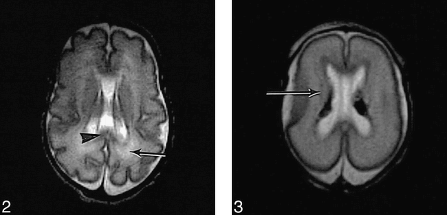
  • fig 2.
    • Download figure
    • Open in new tab
    • Download powerpoint
    fig 2.

    T2-weighted (3500/208) FSE sequence in preterm infant (not in study) of 26 weeks' gestation at age 5 weeks shows normal appearance of the corpus callosum (arrowhead) in the posterior periventricular white matter (arrow). fig 3. T2-weighted (3500/208) FSE sequence in preterm infant of 24 weeks' (+4 days) gestation (case 7) at age 2 days shows the intermediate layer of white matter as low signal intensity (arrow). There are bilateral low signal intensity germinal layer hemorrhages
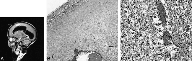
  • fig 4.
    • Download figure
    • Open in new tab
    • Download powerpoint
    fig 4.

    Preterm infant of 23 weeks' (+6 days) gestation (case 3) at age 3 weeks.

    A, T2-weighted (3500/208) FSE sequence shows a large intraventricular hemorrhage with fan-shaped parenchymal involvement. The perihemorrhagic white matter is of very high signal intensity (arrow).

    B, Histologic section shows intraventricular hemorrhage. A zone of venous infarction radiates out into the white matter from the site of bleeding (arrow in center of infarct). Relatively normal white matter is seen to the left of the picture (arrowhead). The infarcted area is very pale owing to tissue loss (hematoxylin-eosin, original magnification ×22.5).

    C, Higher-magnification histologic section of the venous infarction shows very congested capillaries. The adjacent tissue contains many large macrophages with ingested red cells (arrows). There are few free red cells. The tissue shows early cystic breakdown (hematoxylin-eosin, original magnification ×360).
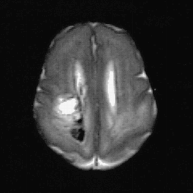
  • fig 5.
    • Download figure
    • Open in new tab
    • Download powerpoint
    fig 5.

    Postmortem T2-weighted (3500/208) FSE sequence in preterm infant of 2 weeks' (+5 days) gestation (case 4) shows bilateral abnormal signal intensity within the posterior periventricular white matter and a low signal intensity hemorrhagic component on the right
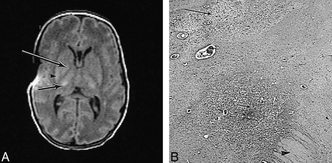
  • fig 6.
    • Download figure
    • Open in new tab
    • Download powerpoint
    fig 6.

    Preterm infant of 28 weeks' gestation (case 2) at age 4 weeks.

    A, T1-weighted (600/20) SE sequence shows abnormal high signal intensity within the posterior putamen (arrowhead), globus pallidus (long arrow) on the right, and lateral thalamus (short arrow). The posterior limb of the internal capsule is of uniform low signal intensity. There is an artifact of uncertain origin over the right insula cortex.

    B, High-power histologic section of damaged deep gray matter shows that the posterior limb of the internal capsule is edematous (arrowhead). The adjacent globus pallidus is darkly stained owing to capillary proliferation and mineralization of dead neurons (short arrow). The posterior part of the putamen is pale as a result of extensive tissue loss (long arrow) (hematoxylin-eosin, original magnification × 22.5).

Tables

  • Figures
  • Table1

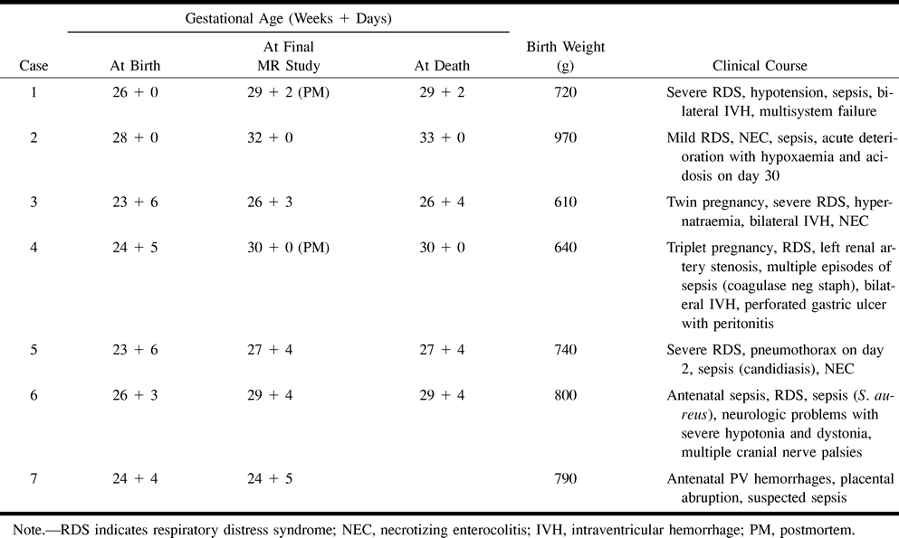
    Clinical data of premature infants enrolled in this study

PreviousNext
Back to top

In this issue

American Journal of Neuroradiology
Vol. 20, Issue 7
1 Jul 1999
  • Table of Contents
  • Index by author
Advertisement
Print
Download PDF
Email Article

Thank you for your interest in spreading the word on American Journal of Neuroradiology.

NOTE: We only request your email address so that the person you are recommending the page to knows that you wanted them to see it, and that it is not junk mail. We do not capture any email address.

Enter multiple addresses on separate lines or separate them with commas.
Relationship between MR Imaging and Histopathologic Findings of the Brain in Extremely Sick Preterm Infants
(Your Name) has sent you a message from American Journal of Neuroradiology
(Your Name) thought you would like to see the American Journal of Neuroradiology web site.
CAPTCHA
This question is for testing whether or not you are a human visitor and to prevent automated spam submissions.
Citation Tools
Relationship between MR Imaging and Histopathologic Findings of the Brain in Extremely Sick Preterm Infants
Ursula Felderhoff-Mueser, Mary A. Rutherford, Waney V. Squier, Philip Cox, Elia F. Maalouf, Serena J. Counsell, Graeme M. Bydder, A. David Edwards
American Journal of Neuroradiology Jul 1999, 20 (7) 1349-1357;

Citation Manager Formats

  • BibTeX
  • Bookends
  • EasyBib
  • EndNote (tagged)
  • EndNote 8 (xml)
  • Medlars
  • Mendeley
  • Papers
  • RefWorks Tagged
  • Ref Manager
  • RIS
  • Zotero
Share
Relationship between MR Imaging and Histopathologic Findings of the Brain in Extremely Sick Preterm Infants
Ursula Felderhoff-Mueser, Mary A. Rutherford, Waney V. Squier, Philip Cox, Elia F. Maalouf, Serena J. Counsell, Graeme M. Bydder, A. David Edwards
American Journal of Neuroradiology Jul 1999, 20 (7) 1349-1357;
Reddit logo Twitter logo Facebook logo Mendeley logo
  • Tweet Widget
  • Facebook Like
  • Google Plus One

Jump to section

  • Article
    • Abstract
    • Methods
    • Results
    • Discussion
    • Conclusion
    • Acknowledgments
    • Footnotes
    • References
  • Figures & Data
  • Info & Metrics
  • References
  • PDF

Related Articles

  • No related articles found.
  • PubMed
  • Google Scholar

Cited By...

  • Preoperative Brain Injury in Transposition of the Great Arteries Is Associated With Oxygenation and Time to Surgery, Not Balloon Atrial Septostomy
  • Preventing Brain Injury in Newborns With Congenital Heart Disease: Brain Imaging and Innovative Trial Designs
  • Magnetic resonance imaging of preterm brain injury
  • Cerebral Maturation in Premature Infants: Quantitative Assessment Using MR Imaging
  • Comparison of Findings on Cranial Ultrasound and Magnetic Resonance Imaging in Preterm Infants
  • Crossref
  • Google Scholar

This article has not yet been cited by articles in journals that are participating in Crossref Cited-by Linking.

More in this TOC Section

  • Neuroimaging Features of Biotinidase Deficiency
  • Medullary Tegmental Cap Dysplasia: Fetal and Postnatal Presentations of a Unique Brainstem Malformation
  • Diagnostic Utility of 3D Gradient-Echo MR Imaging Sequences through the Filum Compared with Spin-Echo T1 in Children with Concern for Tethered Cord
Show more PEDIATRICS

Similar Articles

Advertisement

News and Updates

  • Lucien Levy Best Research Article Award
  • Thanks to our 2022 Distinguished Reviewers
  • Press Releases

Resources

  • Evidence-Based Medicine Level Guide
  • How to Participate in a Tweet Chat
  • AJNR Podcast Archive
  • Ideas for Publicizing Your Research
  • Librarian Resources
  • Terms and Conditions

Opportunities

  • Share Your Art in Perspectives
  • Get Peer Review Credit from Publons
  • Moderate a Tweet Chat

American Society of Neuroradiology

  • Neurographics
  • ASNR Annual Meeting
  • Fellowship Portal
  • Position Statements

© 2023 by the American Society of Neuroradiology | Print ISSN: 0195-6108 Online ISSN: 1936-959X

Powered by HighWire