
Article Figures & Data
Figures
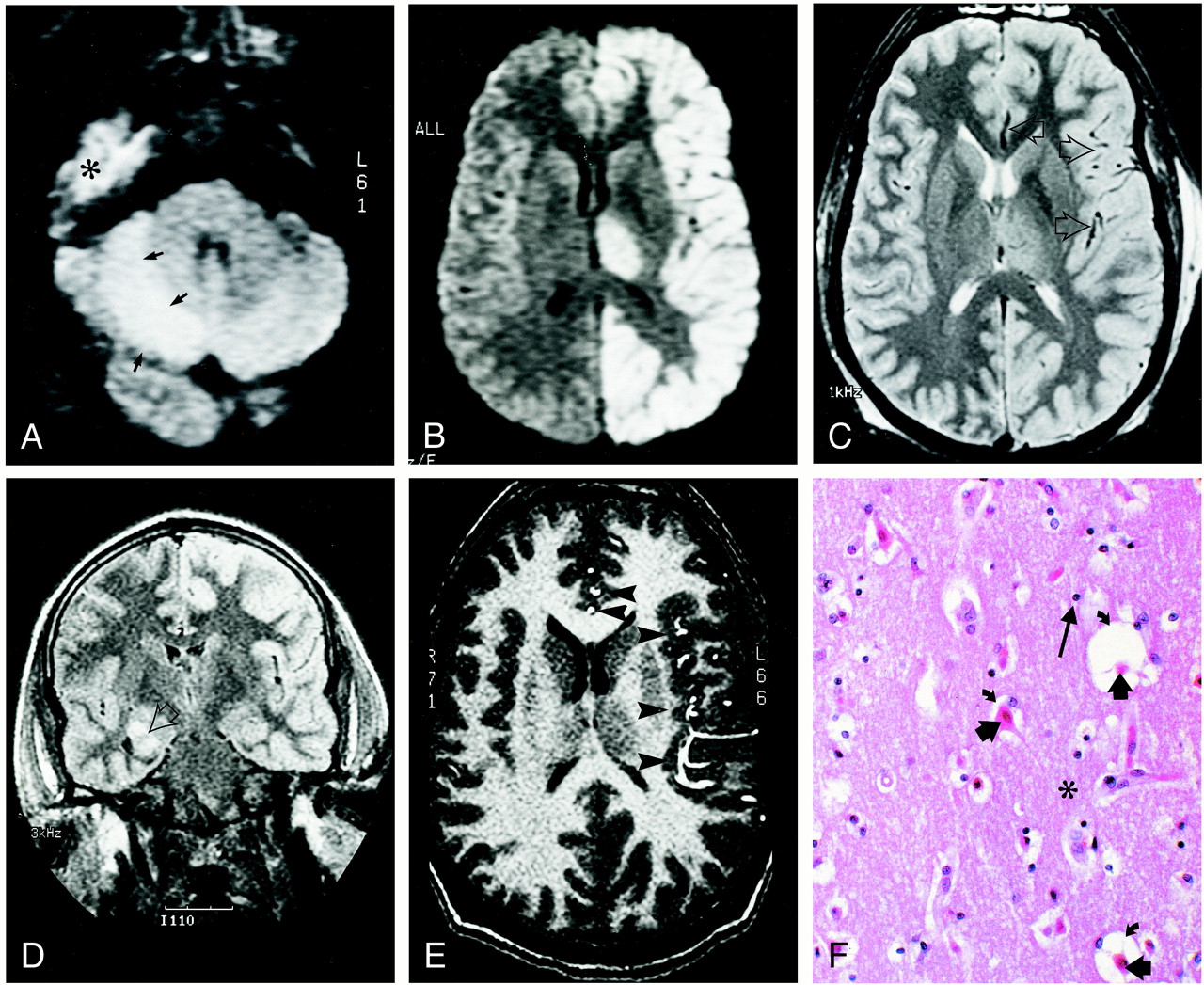
- fig 1.
24-year-old man who experienced status epilepticus 4 days prior to imaging.
A and B, Diffusion-weighted trace images (9999/91.4 [TR/TE], B = 1000 s/mm2) through the levels of cerebellum (A) and thalami (B) show diffusion abnormality involving the right cerebellar cortex (arrows in A), the entire left hemispheric cortex, and the left thalamus (B). The high signal in the right middle fossa (indicated with * in A) is artifactual.
C, Spin-echo T2-weighted (2800/80/1 [TR/TE/excitations]) image through the same level as B shows mildly increased intensity in the diffusely thickened left hemispheric cortex and the left thalamus. Note the relative prominence of signal void in middle and anterior cerebral artery branches (arrows) on the left side.
D, Fluid-attenuated inversion recovery coronal image (9002/145/2200 [TR/TE/TI]) reveals the right hippocampal ganglioglioma (arrow). Note increased intensity in the left hemispheric cortex.
E, Flow-sensitive gradient-echo T1-weighted image (9.3/2.1/90 degrees [TR/TE/flip angle]). Prominent anterior and middle cerebral artery branches (arrowheads) over the left hemispheric cortex suggest enhanced arterial flow in the left hemisphere.
F, Histologic section through the left frontal cortex stained with hemotoxylin and eosin shows eosinophilic neurons (short, thick arrows). The surrounding neuropil (indicated with *) is edematous, as demonstrated by expanded perineural spaces (curved arrows), but there is no fragmentation of the neuropil, and glial cells (long arrow) showed no evidence of necrosis or apoptosis. These findings correspond to selective neuronal necrosis.

In this issue


Jump to section
Related Articles
Cited By...
- Status Epilepticus in Adults: A Review of Diagnosis and Treatment
- Isolated Reversible Thalamic Vasogenic Edema Following a Generalized Seizure
- Diffusion MRI abnormalities after prolonged febrile seizures with encephalopathy
- MRI volumetry of the thalamus in temporal, extratemporal, and idiopathic generalized epilepsy
- Visualization of Evolving Status Epilepticus with Diffusion and Perfusion MR Imaging