Advertisement

Main menu

  • Home
  • Content
    • Current Issue
    • Publication Preview--Ahead of Print
    • Past Issue Archive
    • Case of the Week Archive
    • Classic Case Archive
    • Case of the Month Archive
    • COVID-19 Content and Resources
  • For Authors
  • About Us
    • About AJNR
    • Editors
    • American Society of Neuroradiology
  • Submit a Manuscript
  • Podcasts
    • Subscribe on iTunes
    • Subscribe on Stitcher
  • More
    • Subscribers
    • Permissions
    • Advertisers
    • Alerts
    • Feedback
  • Other Publications
    • ajnr

User menu

  • Subscribe
  • Alerts
  • Log in

Search

  • Advanced search
American Journal of Neuroradiology

American Journal of Neuroradiology

  • Subscribe
  • Alerts
  • Log in

Advanced Search

  • Home
  • Content
    • Current Issue
    • Publication Preview--Ahead of Print
    • Past Issue Archive
    • Case of the Week Archive
    • Classic Case Archive
    • Case of the Month Archive
    • COVID-19 Content and Resources
  • For Authors
  • About Us
    • About AJNR
    • Editors
    • American Society of Neuroradiology
  • Submit a Manuscript
  • Podcasts
    • Subscribe on iTunes
    • Subscribe on Stitcher
  • More
    • Subscribers
    • Permissions
    • Advertisers
    • Alerts
    • Feedback
  • Follow AJNR on Twitter
  • Visit AJNR on Facebook
  • Follow AJNR on Instagram
  • Join AJNR on LinkedIn
  • RSS Feeds
Research ArticleBRAIN

An Adult Case of Congenital External Carotid–Jugular Arteriovenous Fistula with Reversible Circulatory Insufficiency in the Cerebellum and Lower Brain Stem

Masahiro Horiuchi, Tsutomu Kamo, Hiroshi Sugihara, Koshiro Fujisawa, Yoichi Takahashi, Ritsuko Ikeda, Hiroyuki Morishima and Yoshitaro Yamaguchi
American Journal of Neuroradiology February 2001, 22 (2) 273-276;
Masahiro Horiuchi
  • Find this author on Google Scholar
  • Find this author on PubMed
  • Search for this author on this site
Tsutomu Kamo
  • Find this author on Google Scholar
  • Find this author on PubMed
  • Search for this author on this site
Hiroshi Sugihara
  • Find this author on Google Scholar
  • Find this author on PubMed
  • Search for this author on this site
Koshiro Fujisawa
  • Find this author on Google Scholar
  • Find this author on PubMed
  • Search for this author on this site
Yoichi Takahashi
  • Find this author on Google Scholar
  • Find this author on PubMed
  • Search for this author on this site
Ritsuko Ikeda
  • Find this author on Google Scholar
  • Find this author on PubMed
  • Search for this author on this site
Hiroyuki Morishima
  • Find this author on Google Scholar
  • Find this author on PubMed
  • Search for this author on this site
Yoshitaro Yamaguchi
  • Find this author on Google Scholar
  • Find this author on PubMed
  • Search for this author on this site
  • Article
  • Figures & Data
  • Info & Metrics
  • References
  • PDF
Loading

Abstract

Summary: A 27-year-old man with congenital external carotid–jugular arteriovenous fistula presented with a diminished level of consciousness and an ataxic gait. Axial fluid-attenuated inversion-recovery (FLAIR) MR imaging revealed venous congestion, a dilated right jugular vein, and an area of high signal intensity in the brain stem and cerebellum. Angiography showed a dilated right external carotid artery and jugular vein and the presence of a fistula. After coil embolization of the fistula, axial MR FLAIR images showed only a few areas of high signal intensity in the brain stem and cerebellum. The causal factor was venous congestion in the inferior petrosal sinus and basilar plexus due to high blood pressure in the jugular vein. This case is presented for its unusual clinical and radiologic findings.

Arteriovenous malformations (AVMs) arising between the external carotid artery and the jugular vein are rare: a review of the literature revealed only nine cases that presented with symptoms other than a pulsatile mass (1–8). Only three of these patients, including two with vertigo, had neurologic findings attributable to the brain stem or cerebellum. We describe our findings in a patient with symptoms of congenital external carotid–jugular arteriovenous fistula.

Case Report

A 27-year-old man presented with various clinical symptoms, including ataxia, in April 1999. He had suffered from an attention deficit disorder since childhood, and his school records had been consistently poor. The patient had no history of trauma. A pulsatile swelling beneath his right ear caused it to move with each pulse, a finding that had been clearly visible since birth. He had experienced a gait disturbance for the 2 years prior to admission and often fell down. In addition, he had suffered from headache since April 1998 and pollakiuria since February 1999, at which time he had consulted a neurosurgeon, who diagnosed congenital external carotid–jugular arteriovenous fistula; however, the cause of the ataxia could not be explained.

Upon admission, the patient's blood pressure was 106/76 mm Hg and clinical findings included headache, nausea, and loss of appetite. No edema or cardiac failure was observed. Bruit was detectable above the pulsatile swelling beneath his right ear. His consciousness level was found to be impaired and he was absent-minded with poor recent memory. Bilateral choked disk, horizontal nystagmus, and truncal ataxia were present. Serum histochemical levels were all found to be within normal limits.

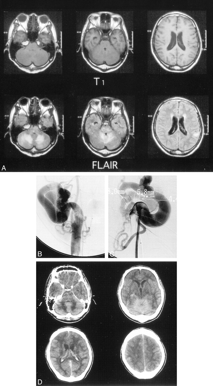
Axial fluid-attenuated inversion-recovery (FLAIR) MR imaging on the day of admission revealed venous congestion, a dilated right jugular vein, and an area of high signal intensity in the brain stem and cerebellum. A small infarction was detected in the temporal lobe (Fig 1A). Angiography showed a dilated right external carotid artery and internal jugular vein connected by a fistula (Fig 1B and C).

fig 1.
  • Download figure
  • Open in new tab
  • Download powerpoint
fig 1.

27-year-old man with congenital external carotid–jugular arteriovenous fistula who presented with a diminished level of consciousness and an ataxic gait.

A, Axial FLAIR MR images (6000/120) on the day of hospital admission show venous congestion, a dilated right jugular vein, and an area of high signal intensity in the brain stem and cerebellum. A small infarction is visible in the temporal lobe.

B and C, Angiograms, frontal (B) and lateral (C) views, 1 day after admission show a dilated right external carotid artery and internal jugular vein and the presence of a fistula.

D, Brain CT studies 5 hours after angiography show congestion and edema.

fig 1.
  • Download figure
  • Open in new tab
  • Download powerpoint
fig 1.

fig. 1 Continued.

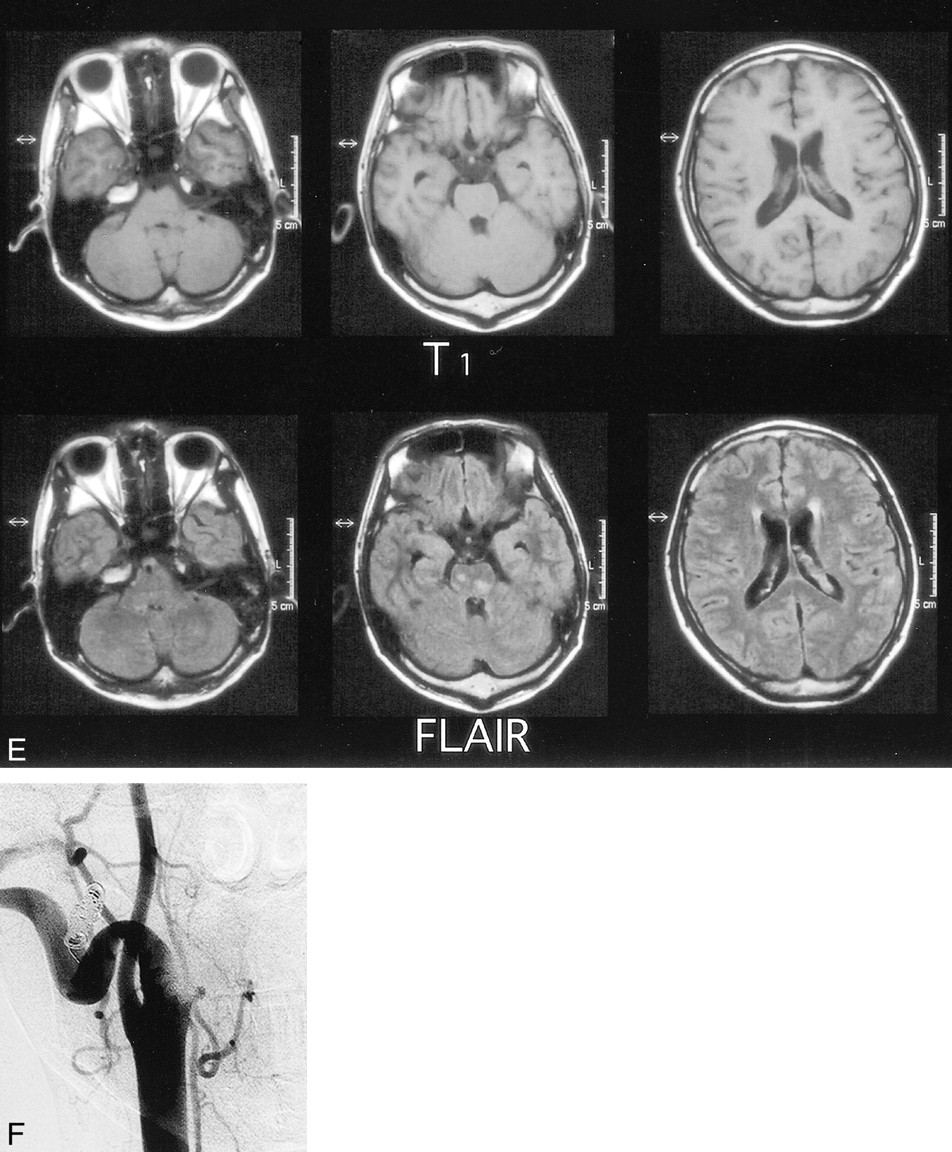
E, Axial FLAIR MR images (6000/120) after embolization show only a few areas of high signal intensity in the brain stem and cerebellum.

F, Angiogram 1 month after admission shows that the external carotid artery and jugular vein are almost equivalent in size, and no arteriovenous communication is detected.

After the angiography, the patient's consciousness level worsened to a state of somnolence. A brain CT study 5 hours after angiography showed congestion and edema (Fig 1D). The patient was intubated and the fistula was embolized with coils 2 days after admission.

Following embolization, the pollakiuria and nausea disappeared, and the headache and ataxia diminished. Eighteen days postoperatively, axial MR FLAIR images revealed only a few areas of high signal intensity in the brain stem and cerebellum (Fig 1E). The patient had retrograde amnesia of the 2-month period prior to recovery.

Before embolization, angiography depicted venous congestion, and both the artery phase and delayed venous phase were observed. After embolization, however, no congestion was detected and only the artery phase was observed. The external carotid artery and internal jugular vein were almost equivalent in size, and no arteriovenous communication was observed (Fig 1F). The patient's right ear no longer moved with each pulse. Before surgical intervention, oxygen saturation values in the superior vena cava were 93.8%, PCO2 was 24.0 mm Hg, and PO2 was 87.9 mm Hg. After the procedure, the equivalent values were 86.2%, 31.7 mm Hg, and 47.6 mm Hg, respectively.

Discussion

The universality of the response of peripheral vessels to fistula formation warrants recognition of the hemodynamic principle that states that flowing blood, like flowing water, seeks the path of least resistance (9). The most common symptoms among patients with an arteriovenous fistula are a pulsatile mass, swelling, and bruit accompanied by a partial increase in blood flow (10, 11). In our case, low oxygen saturation in the superior vena cava and reversible venous congestion may have resulted from the steal phenomenon (12).

Axial MR FLAIR imaging in our patient showed that a preexisting small infarction with high signal intensity in the temporal lobe either decreased or disappeared after surgical embolization of the fistula. This finding indicated that venous congestion represented a reversible circulatory insufficiency. Willinsky et al (13) proposed the term venous congestive encephalopathy to describe neurologic deficits caused by venous hypertension. These authors reported two patients with ataxia caused by dural AVMs. In both cases, MR imaging revealed edema in the cerebellum while angiography showed dural AVMs at the torcular herophili near the inferior petrosal sinus with reflux into cerebellar veins. The cause of ataxia in the present case was venous congestion in the inferior petrosal sinus and basilar plexus due to high blood pressure in the jugular vein.

In some cases involving direct surgical correction of a fistula, segmental parotidectomy is performed; however, this is associated with a risk of injury to the facial nerve (14, 15). Endovascular treatment does not leave facial scars nor does it carry the risk of bleeding during surgical exposure (14). For these reasons, we elected to perform endovascular treatment in our case.

Footnotes

  • ↵1 Address reprint requests to Masahiro Horiuchi, MD, Department of Internal Medicine, Division of Neurology, St. Marianna University School of Medicine, 2-16-1 Sugao, Miyamae-ku, Kawasaki, 216-8511, Japan.

References

  1. ↵
    Halsted WS. Congenital arteriovenous and lymphaticovenous fistula-unique clinical and experimental observations. Trans Am Surg Assoc 1919;37:262-272
  2. Rienhoff WF. Congenital arteriovenous fistula: an embryologic study, with the report of a case. Bull Johns Hopkins Hosp 1924;35:271-284
  3. Gage M. Congenital arteriovenous aneurysm of right external carotid artery and posterior facial vein with absence of branches of external carotid artery: new method of obliterating inaccessible venous sac and fistula. Ann Surg 1951;134:444-450
  4. Svolos D, Nomikos N, Tzouliadis V. Congenital arteriovenous aneurysm in the neck: a case report. J Neurosurg 1965;23:68-71
    PubMed
  5. Tingaud R, Videau J, Masse CI, Magendie J, Boissieras P, Mayeux CI. Fistule arterio-veineuse congenitale entre le tronc de la carotide externe et la veine jugulaire externe (un cas). J Chir (Paris) 1970;99:391-400
    PubMed
  6. Scialfa G, Valsecchi F, Toncon C. Treatment of external carotid arteriovenous fistula with detachable balloon. Neuroradiology 1979;17:265-267
    PubMed
  7. Kuss JJ, Karli A, Fischbach M, et al. Aneurysme carotido-jugulaire congenital. Arch Fr Pediatr 1979;36:502-507
    PubMed
  8. Tekkok IH, Akkurt C, Suzer T, Ozcan OE. Congenital external carotid-jugular fistula: report of two cases and review of literature. Neurosurgery 1992;30:272-276
    CrossRefPubMed
  9. ↵
    Holman E. Reflections on arteriovenous fistulas. Ann Thorac Surg 1971;11:176-186
    CrossRefPubMed
  10. ↵
    Szilagyi DE, Elliott JP, Derusso FJ, Smith RF. Peripheral congenital arteriovenous fistulas. Surgery 1965;57:61-81
  11. Kawakami K, Takahashi A, Sugawara T, Nakamura N, Yoshimoto T, Suzuki J. Spontaneous arteriovenous fistula of external carotid artery treated by detachable balloon: a case report. Neurol Surg 1986;15:549-553
  12. ↵
    Costantino A, Vinters HV. A pathologic correlate of the “steal” phenomenon in a patient with cerebral arteriovenous malformation. Stroke 1986;17:103-106
    Abstract/FREE Full Text
  13. ↵
    Willinsky R, Terbrugge K, Montanera W, Mikulis D, Wallace C. Venous congestion: MR findings in dural arteriovenous malformations with cortical venous drainage. AJNR Am J Neuroradiol 1994;15:1501-1507
    Abstract/FREE Full Text
  14. ↵
    Prevot J, Babut JM. Congenital cervical jugulo-carotid fistula. J Pediatr Surg 1970;5:431-436
    CrossRefPubMed
  15. Gobin YP, de la Fuente AG, Herbreteau D, Houdart E, Merland JJ. Endovascular treatment of external carotid-jugular fistula in the parotid region. Neurosurgery 1993;33:812-816
    CrossRefPubMed
  • Received May 12, 2000.
  • Copyright © American Society of Neuroradiology
View Abstract
PreviousNext
Back to top

In this issue

American Journal of Neuroradiology
Vol. 22, Issue 2
1 Feb 2001
  • Table of Contents
  • Index by author
Advertisement
Print
Download PDF
Email Article

Thank you for your interest in spreading the word on American Journal of Neuroradiology.

NOTE: We only request your email address so that the person you are recommending the page to knows that you wanted them to see it, and that it is not junk mail. We do not capture any email address.

Enter multiple addresses on separate lines or separate them with commas.
An Adult Case of Congenital External Carotid–Jugular Arteriovenous Fistula with Reversible Circulatory Insufficiency in the Cerebellum and Lower Brain Stem
(Your Name) has sent you a message from American Journal of Neuroradiology
(Your Name) thought you would like to see the American Journal of Neuroradiology web site.
CAPTCHA
This question is for testing whether or not you are a human visitor and to prevent automated spam submissions.
Citation Tools
An Adult Case of Congenital External Carotid–Jugular Arteriovenous Fistula with Reversible Circulatory Insufficiency in the Cerebellum and Lower Brain Stem
Masahiro Horiuchi, Tsutomu Kamo, Hiroshi Sugihara, Koshiro Fujisawa, Yoichi Takahashi, Ritsuko Ikeda, Hiroyuki Morishima, Yoshitaro Yamaguchi
American Journal of Neuroradiology Feb 2001, 22 (2) 273-276;

Citation Manager Formats

  • BibTeX
  • Bookends
  • EasyBib
  • EndNote (tagged)
  • EndNote 8 (xml)
  • Medlars
  • Mendeley
  • Papers
  • RefWorks Tagged
  • Ref Manager
  • RIS
  • Zotero
Share
An Adult Case of Congenital External Carotid–Jugular Arteriovenous Fistula with Reversible Circulatory Insufficiency in the Cerebellum and Lower Brain Stem
Masahiro Horiuchi, Tsutomu Kamo, Hiroshi Sugihara, Koshiro Fujisawa, Yoichi Takahashi, Ritsuko Ikeda, Hiroyuki Morishima, Yoshitaro Yamaguchi
American Journal of Neuroradiology Feb 2001, 22 (2) 273-276;
del.icio.us logo Digg logo Reddit logo Twitter logo CiteULike logo Facebook logo Google logo Mendeley logo
  • Tweet Widget
  • Facebook Like
  • Google Plus One

Jump to section

  • Article
    • Abstract
    • Case Report
    • Discussion
    • Footnotes
    • References
  • Figures & Data
  • Info & Metrics
  • References
  • PDF

Related Articles

  • No related articles found.
  • PubMed
  • Google Scholar

Cited By...

  • No citing articles found.
  • Crossref
  • Google Scholar

This article has not yet been cited by articles in journals that are participating in Crossref Cited-by Linking.

More in this TOC Section

  • Enhanced Axonal Metabolism during Early Natalizumab Treatment in Relapsing-Remitting Multiple Sclerosis
  • Progression of Microstructural Damage in Spinocerebellar Ataxia Type 2: A Longitudinal DTI Study
  • SWI or T2*: Which MRI Sequence to Use in the Detection of Cerebral Microbleeds? The Karolinska Imaging Dementia Study
Show more BRAIN

Similar Articles

Advertisement

News and Updates

  • Lucien Levy Best Research Article Award
  • Thanks to our 2022 Distinguished Reviewers
  • Press Releases

Resources

  • Evidence-Based Medicine Level Guide
  • How to Participate in a Tweet Chat
  • AJNR Podcast Archive
  • Ideas for Publicizing Your Research
  • Librarian Resources
  • Terms and Conditions

Opportunities

  • Share Your Art in Perspectives
  • Get Peer Review Credit from Publons
  • Moderate a Tweet Chat

American Society of Neuroradiology

  • Neurographics
  • ASNR Annual Meeting
  • Fellowship Portal
  • Position Statements

© 2023 by the American Society of Neuroradiology | Print ISSN: 0195-6108 Online ISSN: 1936-959X

Powered by HighWire