
Article Figures & Data
Figures
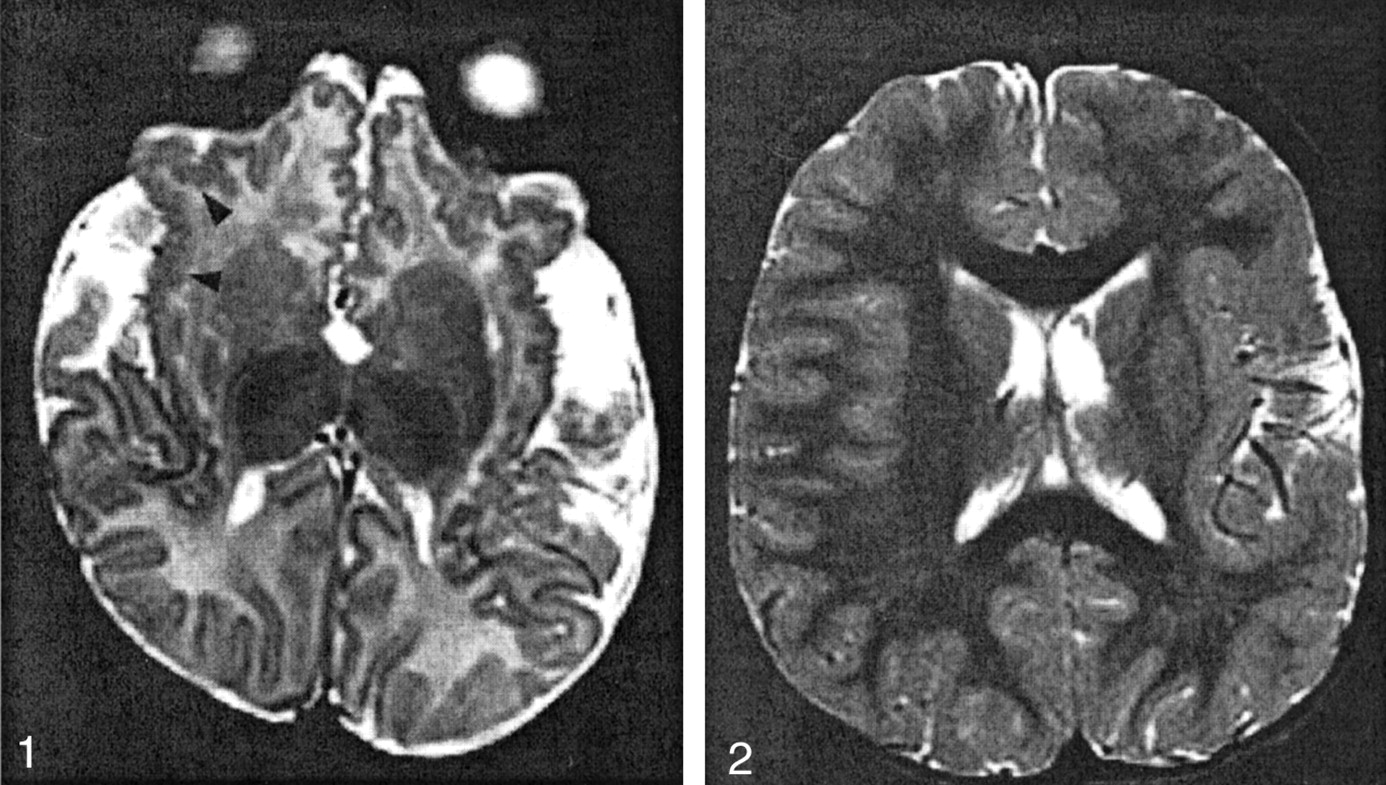
- Fig 1.
Patient 2 (aged 2 months), with bilateral frontal and sylvian PMG.
T2-weighted image shows pattern 1: abnormal cortex with a small, fine, and undulating appearance in the insulae and orbital surfaces of the frontal lobes (arrowheads). The cortical thickness was 4 mm.
- Fig 2.
Patient 12 (aged 3 years), with left hemispheric PMG.
T2-weighted image shows pattern 2: abnormally thick sylvian and perisylvian cortex (7 mm at the left sylvian cortex) with a bumpy appearance at the surface and the cortical-white matter junction.
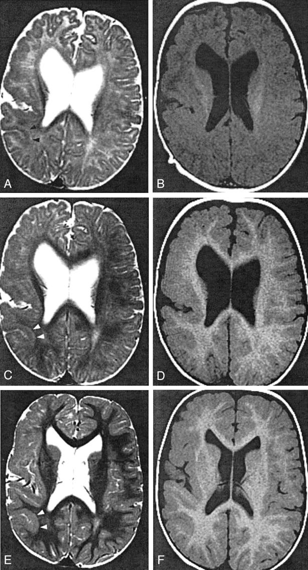
- Fig 3.
Patient 4, with right hemispheric PMG.
A and B, Images obtained when the patient was 3 months old. T2-weighted image (A) shows pattern 1: 4-mm-thick cortex in the parietal lobe (arrowheads). T1-weighted image (B) shows pattern 2: 5-mm thickness in the same region.
C and D, Images obtained when the patient was 11 months old. T2-weighted image (C) at the parietal lobe shows pattern 1: 4-mm thickness and a 2-mm-thick layer of T2 prolongation between the cortex and myelinated white matter (arrowheads). T1-weighted image (D) shows pattern 2: 6-mm thickness of the same cortex.
E and F, Images obtained when the patient was 2 years old. In the parietal lobe (arrowheads), T2- (E) and T1-weighted (F) images show pattern 2: 6-mm thickness. The T1-weighted image (F) shows pattern 2 in the frontal lobe: 5-mm thickness. Conversely, the T2- weighted image (E) reveals pattern 1 in the frontal lobe: 3-mm thickness with subjacent layer of T2 prolongation.
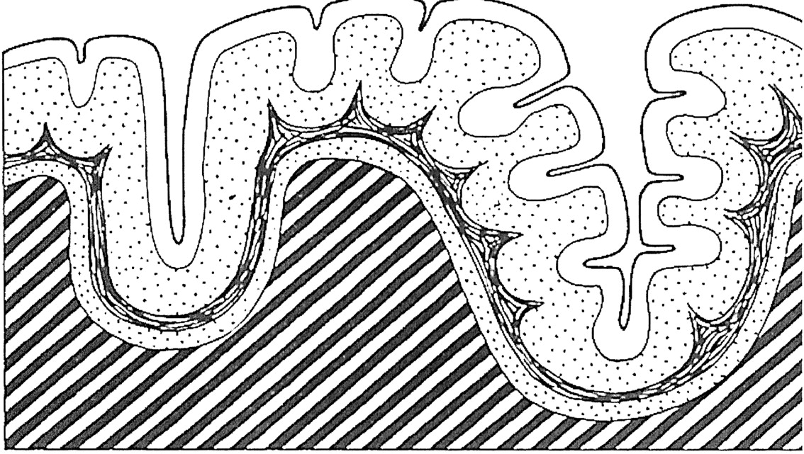
- Fig 4.
Diagrammatic representation of the cerebral cortex in four-layered PMG. (Reprinted from Fig 210, R. Excourolle and J. Poirier, Manual of Basic Neuropathology. 2nd ed. [1978] with permission of Elsevier Science.)


Tables
Clinical and MR imaging findings in PMG patients
Patient Age Sex Clinical Manifestation Other MR Findings PMG Location Evaluated Lesion T1 Pattern Thickness (mm) Axial (Coronal) T2 Pattern Thickness (mm) Axial (Coronal) Layer between Cortex and White Matter 1 3D F Zellweger syndrome Germinolytic cysts bil. Syl Syl 2 4 (4) 1 4 2 2M M Epilepsy bil. F, Syr, P Syl 2 4 (3) 1 4 (3) 3 3M F Infantile spasmus Microcephaly, hypoplastic CC bil. Syl, P Syl Undifferentiated 1 3 (4) 4 3M M Large head, retardation rt. hemi P 2 5 (5) 1 4 7M P 2 6 (6) 1 4 11M P 2 6 1 4 Present 2Y F 2 5 1 3 Present P 2 6 2 6 5 5M M Retardation bil. T T Undifferentiated 1 3 6 7M M Infantile spasmus diffuse Syl 2 6 1 3 (3) 7 12M F In utero drug exposure lt. F, Syl Syl 2 7 (6) 1 4 8 15M F rt. hemiparesis lt. F, P, bil. Syr F 2 6 (5) 1 3 Present P 2 6 (6) 2 6 9 15M M lt. hemiparesis rt. hemi F 2 7 (7) 1 4 Present P 2 7 (6) 2 7 10 18M F Retardation, focal seizure small rt. Th, peducle rt. hemi F 2 6 (6) 1 3 Present P 2 6 (6) 2 6 11 18M M Seizure heterotopia bil. Syl, P Syr 2 6 (6) 2 6 12 3Y M rt. hemiparesis lt. hemi Syr 2 7 (6) 2 7 13 8Y M Speech delay rt. hemi, lt. F Ins 2 8 (8) 2 8 14 8Y M Hydrocephalus heterotopia, Agenesis of CC bil. F F 2 6 (6) 2 6 15 9Y F Retardation heterotopia, hypoplastic CC bil. F F 2 8 (7) 2 8 16 11Y M Retardation heterotopia, agenesis of CC lt. F F 2 7 (6) 2 7 17 43Y F Epilepsy bil. Syr Syr 2 6 (6) 2 6 Note.—PMG, polymicrogyria; D, day; M, month; Y, year; CC, corpus callosum; Th, thalamus; bil, bilateral; rt, right; lt, left; F, frontal; Syl, Sylvian; P, parietal; hemi, hemispheric.


