
Article Figures & Data
Figures
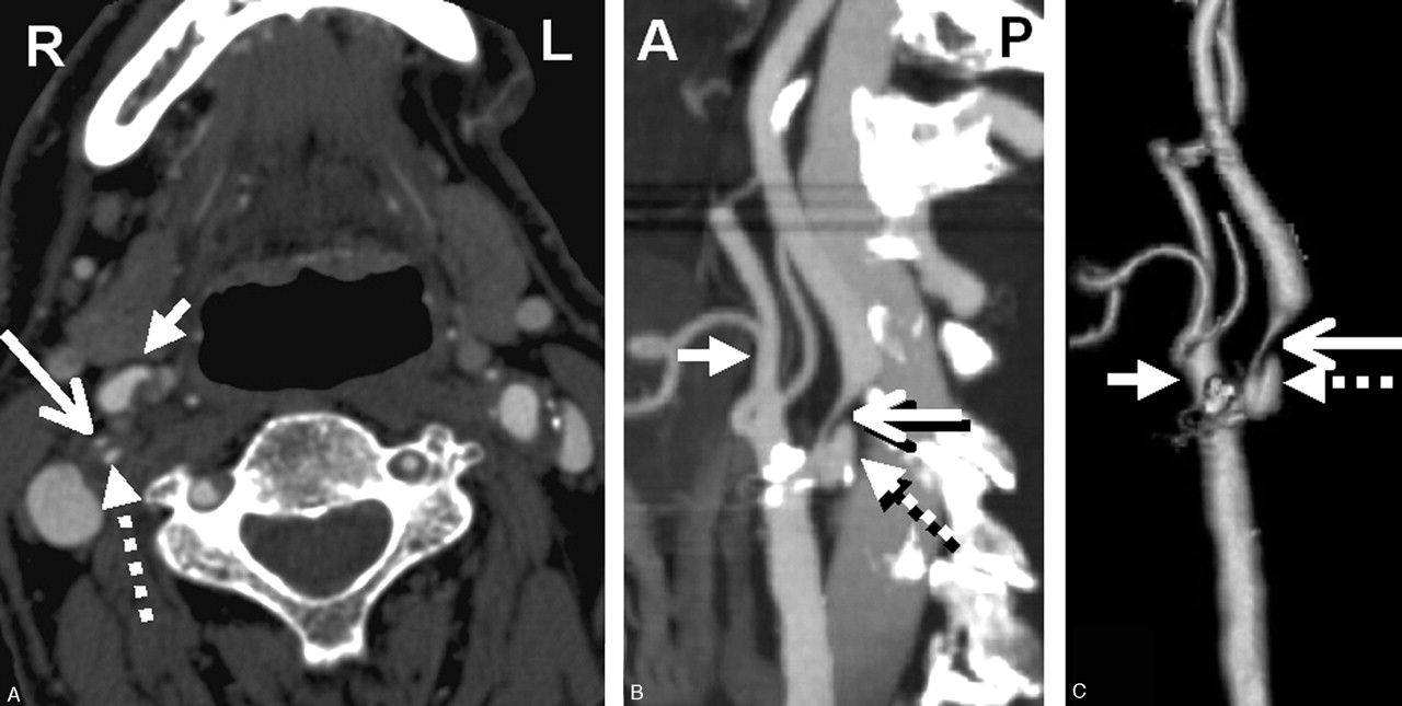
- Fig 1.
CTA showing right severe carotid bulb stenosis with deep plaque ulceration. A, Axial source image. B, Sagittal reformat. C, 3D rendered image (large arrow, stenotic ICA; dashed arrow, bulb plaque ulcer; small arrow, proximal ECA).
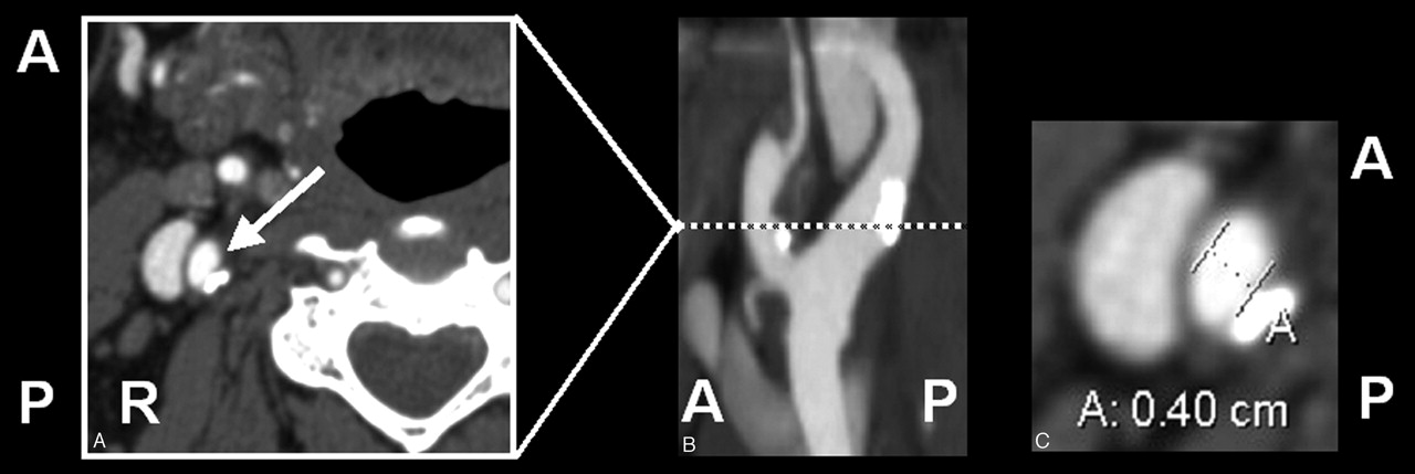
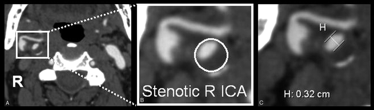
- Fig 2.
Carotid stenosis measurements were obtained from axial source data at the narrowest portion of the carotid bulb. A, Source image. B, Magnified right carotid portion. C, Submillimeter measuring tool determining 3.2 mm (0.32 cm).
- Fig 3.
Oblique artery measure. A, Axial source image with arrow toward ICA. B, Sagittal reformat showing oblique ICA axis of a slightly tortuous ICA. C, Axial measurement, perpendicular to oblique axis. This demonstrates a need for viewing reformats in addition to axial source images.
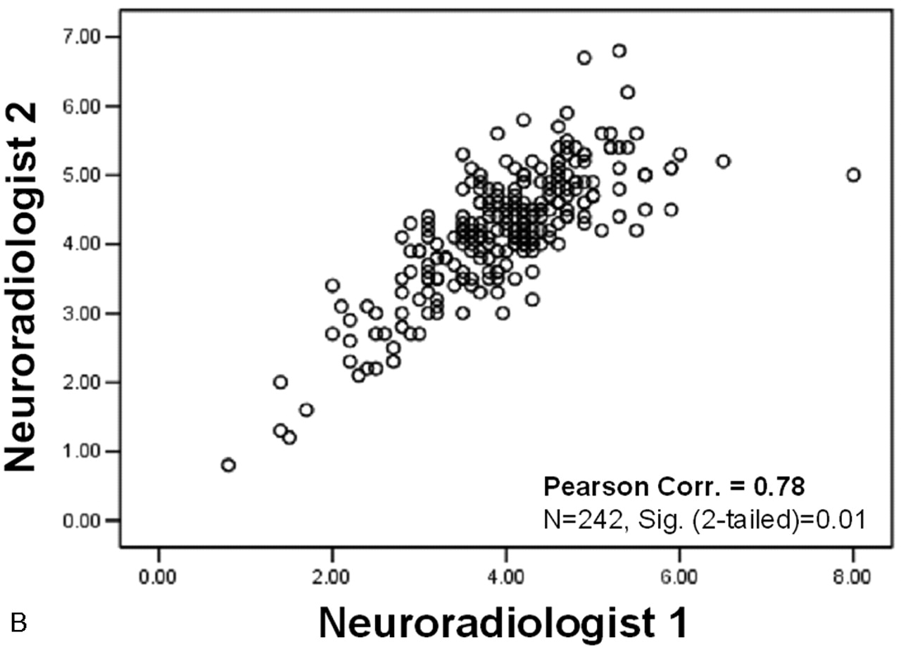
- Fig 4.
Correlation scatter plots. Interobserver agreement between reader measurements. A, Maximum stenosis (mm). B, Distal ICA (mm). C, Derived NASCET-style percent stenosis. (Correlations: 2-tailed significance = 0.01. n values reflect exclusion of pairwise missing data; excludes “near-occlusion” cases).
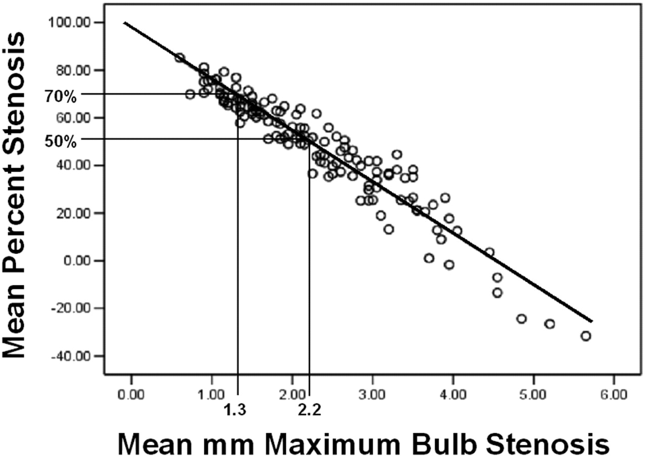
- Fig 5.
Linear regression. Mean percent stenosis to mean millimeter maximum carotid bulb stenosis. (Pearson correlation = −0.95; n = 136; 2-tailed significance = 0.01; R2 linear = 0.895; SE of estimate = 7.63; B value = −21.539).
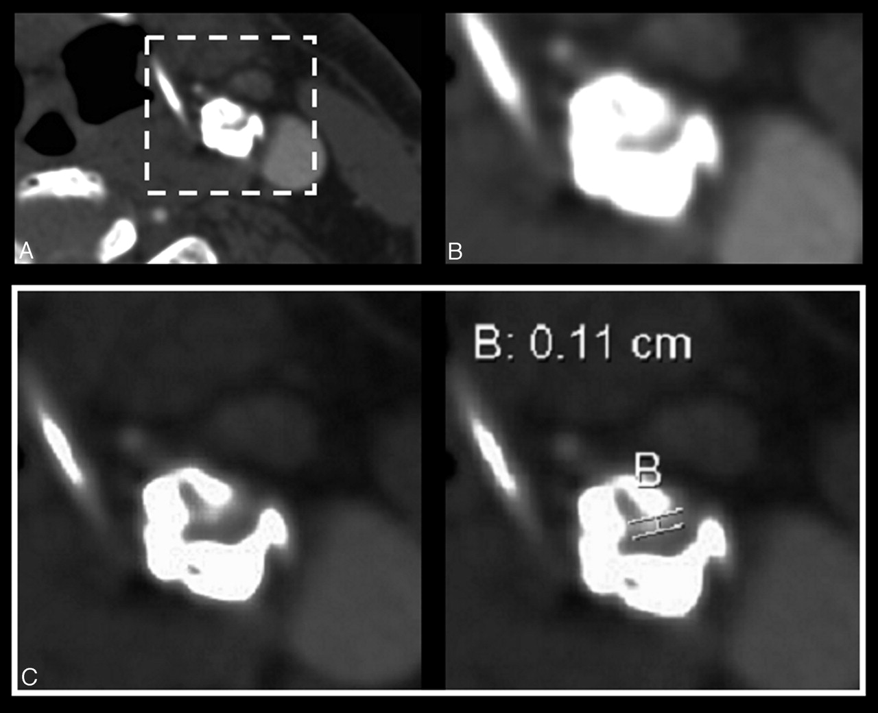
- Fig 6.
Axial source images of left severe ICA stenosis; small residual lumen with vessel wall plaque and thick calcification on the sides. A, Source axial image with “standard” W : L settings for CTA evaluation (W 750 : L 200). B, Magnified left ICA (W 750 : L 200). C, Magnified with wide window for appropriate visualization and measurement of residual carotid lumen (W 900 : L 275). A wider W : L setting allows conspicuity between the different attenuations of contrast in lumen and calcification along the sides.


Tables
- Table 1:
Estimate of NASCET-style derived percent stenosis from the millimeter carotid bulb stenosis measurements
mm Stenosis % Stenosis (95% PI) 2.2 50–55 2.1 52–57 2.0 54–59 1.9 57–62 1.8 59–64 1.7 61–66 1.6 64–68 1.5 66–70 1.4 68–72 1.3 70–74 1.2 73–76 1.1 75–78 1.0 77–80 0.9 80–82 0.8 82–84 0.7 84–86 0.6 86–88 0.5 89–90 0.4 91–92 0.3 93–94 0.2 95–96 Note:— Percent stenosis is expressed as a range with 95% prediction intervals (PI).
- Table 2:
Contingency table with the NASCET-style ratios defining the presence of disease in the final carotid stenosis group analyzed (n = 136) and the millimeter stenosis measurements as the test variable
Stenosis Measurements <50% 50–69% 70%+ >2.2 mm 60 5 0 65 1.4 to 2.2 mm 3 42 2 47 1.3 mm or less 0 9 15 24 63 56 17 n = 136
In this issue


Jump to section
Related Articles
Cited By...
- Association of Carotid Artery Disease with Collateralization and Infarct Growth in Patients with Acute Middle Cerebral Artery Occlusion
- Comparison of 30-Day Outcomes after Carotid Artery Stenting in Patients with Near-Occlusion and Severe Stenosis: A Propensity Score Matching Analysis
- Cumulative incidence of restenosis in the endovascular treatment of extracranial carotid artery stenosis: a meta-analysis
- Carotid CTA at the Lowest Tube Voltage (70 kV) in Comparison with Automated Tube Voltage Adaption
- Prolonged cerebral circulation time is more associated with symptomatic carotid stenosis than stenosis degree or collateral circulation
- Quantifying Intracranial Internal Carotid Artery Stenosis on MR Angiography
- Vitamin D and Vulnerable Carotid Plaque
- Nonstenotic carotid plaque on CT angiography in patients with cryptogenic stroke
- Prediction of Carotid Intraplaque Hemorrhage Using Adventitial Calcification and Plaque Thickness on CTA
- Carotid Near-Occlusion: A Comprehensive Review, Part 1--Definition, Terminology, and Diagnosis
- Optimal Prediction of Carotid Intraplaque Hemorrhage Using Clinical and Lumen Imaging Markers
- Diagnostic Accuracy of 4 Commercially Available Semiautomatic Packages for Carotid Artery Stenosis Measurement on CTA
- Intraoperative Sonography During Carotid Endarterectomy: Normal Appearance and Spectrum of Complications
- Intraluminal Thrombus, Intraplaque Hemorrhage, Plaque Thickness, and Current Smoking Optimally Predict Carotid Stroke
- Correlation between carotid bifurcation calcium burden on non-enhanced CT and percentage stenosis, as confirmed by digital subtraction angiography
- Performance of Semiautomatic Assessment of Carotid Artery Stenosis on CT Angiography: Clarification of Differences with Manual Assessment
- Carotid Artery Stenosis: Making Complex Assessments of a Simple Problem or Simplifying Approach to a Complex Disease?
- Interpretation Errors in CT Angiography of the Head and Neck and the Benefit of Double Reading
- Reporting standards for angioplasty and stent-assisted angioplasty for intracranial atherosclerosis
- Characterization of Carotid Plaque Hemorrhage: A CT Angiography and MR Intraplaque Hemorrhage Study
- Role of CT Angiographic Plaque Morphologic Characteristics in Addition to Stenosis in Predicting the Symptomatic Side in Carotid Artery Disease
- Should Modeling Methodology Suppress Anatomic Excellence?
- Reporting Standards for Angioplasty and Stent-Assisted Angioplasty for Intracranial Atherosclerosis
- Contrast-Enhanced MR Angiography Is Not More Accurate Than Unenhanced 2D Time-of-Flight MR Angiography for Determining >=70% Internal Carotid Artery Stenosis
- Dangerous Advances in Measurements from Digital Subtraction Angiography: When Is a Millimeter Not a Millimeter?
- High-Resolution 3T MR Angiography of the Carotid Arteries: Comparison of Manual and Semiautomated Quantification of Stenosis
- Response to Letter by Bladin et al
- Simplification of the Residual Lumen Geometry in Measuring Carotid Stenosis
- Carotid Stenosis Index Revisited With Direct CT Angiography Measurement of Carotid Arteries to Quantify Carotid Stenosis