
Article Figures & Data
Figures
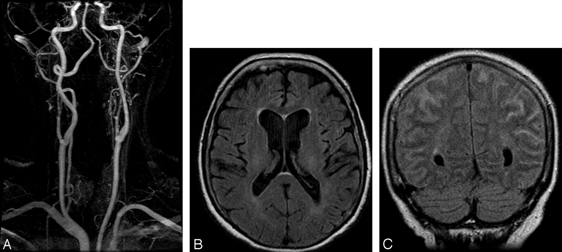
- Fig 1.
A 74-year-old man with normal renal function who underwent a gadolinium-enhanced MR imaging of the head and MR angiography (MRA) of the neck because of a transient ischemic attack and who had a follow-up head MR imaging the next day. A, Normal MRA findings of the neck. B, Precontrast axial FLAIR image shows no signal intensity in the SAS on the date of the gadolinium administration. C, Repeat noncontrast coronal FLAIR image 24 hours later shows increased signal intensity in the SAS. The patient had an emergent lumbar puncture, which was negative for subarachnoid hemorrhage, infection, or malignant cells.
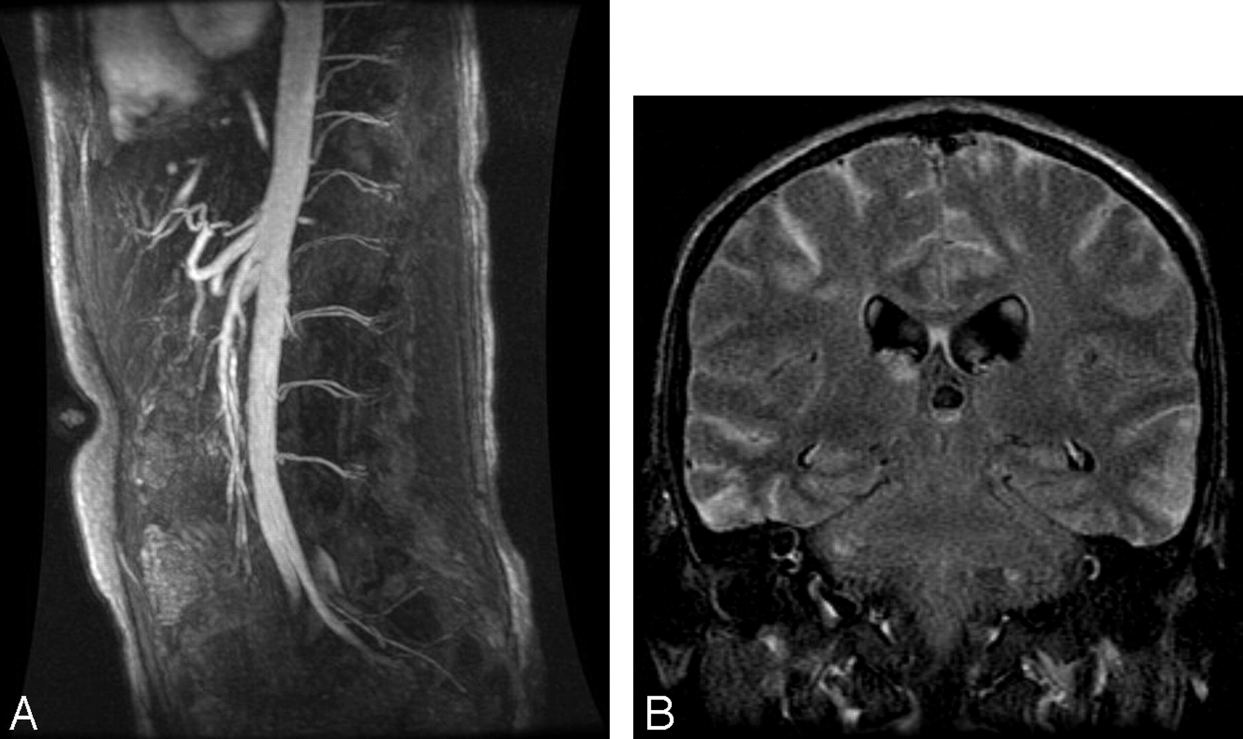
- Fig 2.
A 21-year-old man with chronic renal insufficiency who underwent gadolinium-enhanced MRA of the abdomen followed by MR imaging of the brain 6 days later to rule out causes of syncope. A, Normal findings on abdominal MRA. B, Precontrast FLAIR image shows diffuse increased signal intensity in the SAS 6 days after the gadolinium injection. The patient had an emergent lumbar puncture, which was as negative for subarachnoid hemorrhage, infection, or malignant cells.
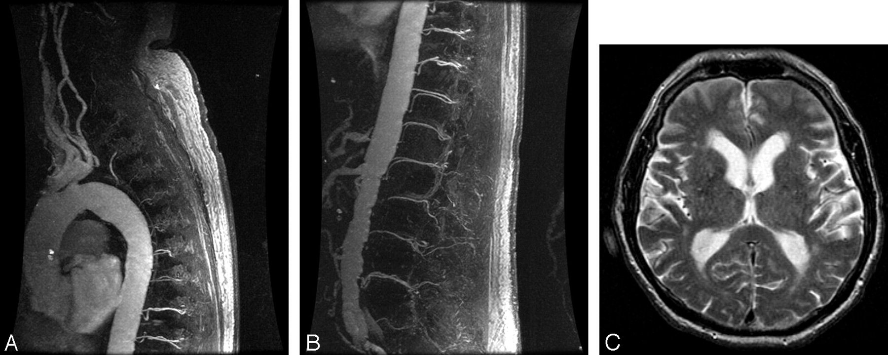
- Fig 3.
An 81-year-old man with normal renal function who underwent a triple-dose (60 mL) 3D MRA of the spine to exclude a dural arteriovenous fistula, followed by MR imaging of the brain 24 hours later for a syncopal episode. Upper thoracic (A) and thoracolumbar (B) spinal MRA images demonstrate no evidence of a dural arteriovenous fistula. C, Precontrast FLAIR image shows increased signal intensity in the SAS and in the lateral ventricles 24 hours after gadolinium injection. The patient returned to the department and was imaged a second time due to the T2 spin-echo appearance to ensure that there was not a technical error in the initial FLAIR sequence and was found to have similar results.
- Fig 4.
A 79-year-old man with normal renal function evaluated for a transient ischemic attack. A, Precontrast axial FLAIR image shows no abnormal signal intensity in the SAS. B, Forty-eight hours postgadolinium injection, repeat MR imaging shows diffuse increased signal intensity in the subarachnoid space (SAS) on coronal FLAIR images due to delayed clearance of the gadolinium chelate. C, Repeat MR imaging 72 hours postgadolinium injection shows resolution of the increased signal intensity in the SAS.


Tables
Patient characteristics and time interval between gadolinium administration and increased signal in the subarachnoid space on FLAIR
Patient No. Age (yr) Intervala (days) Gadolinium (mL/kg) Creatinine Level (mg/dL) Lumbar Puncture Other Pertinent Findings 1 74 2 0.82 4.4 Negativeb Minimal leukoaraiosis 2 21 6 0.42 4.1 Negative None 3 73 6 0.27 4.4 CSF protein 50 mg/dL, otherwise negative Acute occipital infarct 4 70 1 0.35 2.5 CSF protein 154 mg/dL, otherwise negative Old infarction frontal lobe, tiny anterior meningioma without mass effect 5 79 1 0.24 2.4 No CSF collected Right PCA infarct, prostate metastasis to the clivus 6 81 1 0.61 1.5 CSF protein 108 mg/dL Minimal leukoaraiosis 7 74 1 0.31 1.0 Negative None 8 68 1 0.43 0.9 No CSF collected Right carotid occlusion with chronic watershed infarct 9 79 1 0.63 1.0 Negative Old lacunar infarcts 10 65 2 0.52 1.2 Negative None 11 56 2 0.40 2.3 Negative None Note:—PCA indicates posterior cerebral artery.
a Between gadolinium administration and increased signal in the subarachnoid space on FLAIR.
b For subarachnoid hemorrhage, infection, or malignant cells.
In this issue


Jump to section
Related Articles
Cited By...
- HARMless: Transient Cortical and Sulcal Hyperintensity on Gadolinium-Enhanced FLAIR after Elective Endovascular Coiling of Intracranial Aneurysms
- Flat Detector Angio-CT following Intra-Arterial Therapy of Acute Ischemic Stroke: Identification of Hemorrhage and Distinction from Contrast Accumulation due to Blood-Brain Barrier Disruption
- Elevated Cerebral Blood Volume Contributes to Increased FLAIR Signal in the Cerebral Sulci of Propofol-Sedated Children
- Isolated Acute Nontraumatic Cortical Subarachnoid Hemorrhage