Advertisement

Main menu

  • Home
  • Content
    • Current Issue
    • Publication Preview--Ahead of Print
    • Past Issue Archive
    • Case of the Week Archive
    • Classic Case Archive
    • Case of the Month Archive
    • COVID-19 Content and Resources
  • For Authors
  • About Us
    • About AJNR
    • Editors
    • American Society of Neuroradiology
  • Submit a Manuscript
  • Podcasts
    • Subscribe on iTunes
    • Subscribe on Stitcher
  • More
    • Subscribers
    • Permissions
    • Advertisers
    • Alerts
    • Feedback
  • Other Publications
    • ajnr

User menu

  • Subscribe
  • Alerts
  • Log in

Search

  • Advanced search
American Journal of Neuroradiology

American Journal of Neuroradiology

  • Subscribe
  • Alerts
  • Log in

Advanced Search

  • Home
  • Content
    • Current Issue
    • Publication Preview--Ahead of Print
    • Past Issue Archive
    • Case of the Week Archive
    • Classic Case Archive
    • Case of the Month Archive
    • COVID-19 Content and Resources
  • For Authors
  • About Us
    • About AJNR
    • Editors
    • American Society of Neuroradiology
  • Submit a Manuscript
  • Podcasts
    • Subscribe on iTunes
    • Subscribe on Stitcher
  • More
    • Subscribers
    • Permissions
    • Advertisers
    • Alerts
    • Feedback
  • Follow AJNR on Twitter
  • Visit AJNR on Facebook
  • Follow AJNR on Instagram
  • Join AJNR on LinkedIn
  • RSS Feeds
OtherHEAD & NECK

Aberrant Middle Turbinate Masquerading as a Nasopharyngeal Mass

P.H. Lee, A.S. Lee and R.A. Bhadelia
American Journal of Neuroradiology September 2008, 29 (8) 1564-1565; DOI: https://doi.org/10.3174/ajnr.A1116
P.H. Lee
  • Find this author on Google Scholar
  • Find this author on PubMed
  • Search for this author on this site
A.S. Lee
  • Find this author on Google Scholar
  • Find this author on PubMed
  • Search for this author on this site
R.A. Bhadelia
  • Find this author on Google Scholar
  • Find this author on PubMed
  • Search for this author on this site
  • Article
  • Figures & Data
  • Info & Metrics
  • References
  • PDF
Loading

Abstract

SUMMARY: We present a unique variant of the middle turbinate that extends posteriorly through the choana to wrap around the posterior free edge of the nasal septum. The embryology and anatomy of the lateral nasal wall and the nasal septum are reviewed.

The anatomy of the nasal cavity and the lateral nasal wall is complex, with many variants. We present a unique variant of the middle turbinate that was initially thought clinically to be a mass.

Case Report

A 43-year-old man presented with approximately 2 weeks’ history of bilateral epistaxes. The medical history was significant only for seasonal rhinitis. There was no history of any facial traumatic injury. On examination of the nasal cavity, dry mucous membranes along the anterior nasal septum bilaterally were noted. Nasal endoscopic examination revealed a well-circumscribed pedunculated mass apparently extending from the left posterior nasal septum. No blood or blood clots were associated with this lesion. A CT scan was performed for further evaluation.

CT examination demonstrated that the right middle turbinate assumed an unusual configuration and location. The turbinate was straightened anteriorly (Fig 1), more posteriorly located than the contralateral normal side, curved posteromedially around the posterior free edge of the nasal septum, and extended into the left nasopharynx (Figs 2,3). It was determined to be a middle turbinate in an unusual location because it had a typical bony attachment to the lateral nasal wall, and there was no normal middle turbinate on that side. No other lesions were identified.

Fig 1.
  • Download figure
  • Open in new tab
  • Download powerpoint
Fig 1.

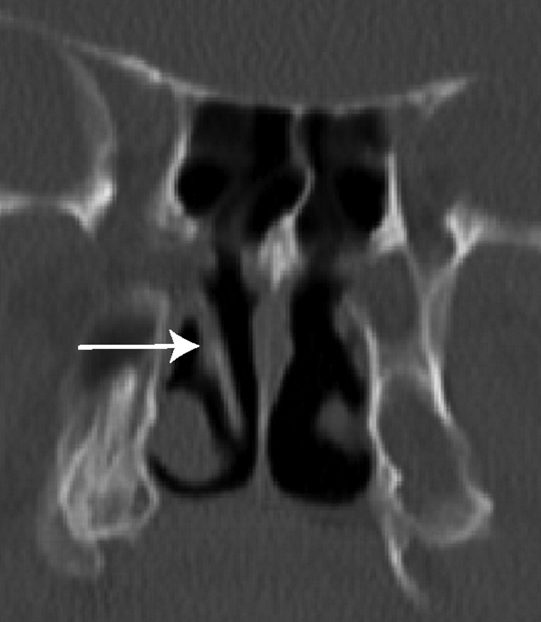
A coronal CT image showing the straightened right middle turbinate (arrow).

Fig 2.
  • Download figure
  • Open in new tab
  • Download powerpoint
Fig 2.

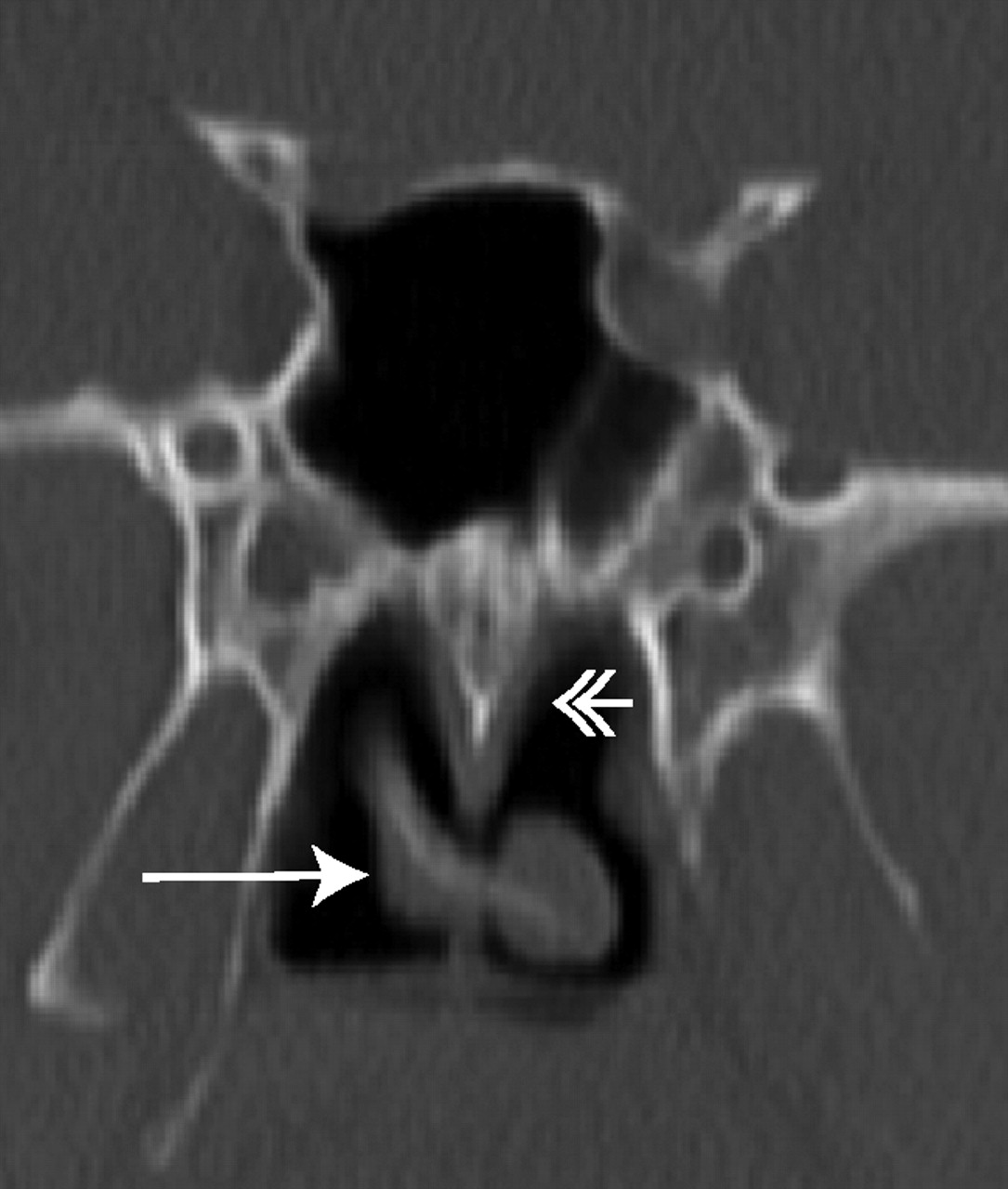
Axial CT image showing the right middle turbinate (arrow) extending through the right choana to wrap around the posterior free edge of the nasal septum (double-headed arrow).

Fig 3.
  • Download figure
  • Open in new tab
  • Download powerpoint
Fig 3.

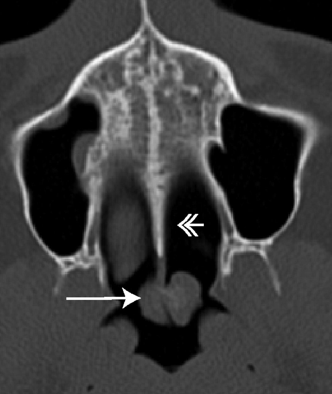
Coronal CT image showing the right middle turbinate (arrow) extending through the right choana to wrap around the posterior free edge of the nasal septum (double-headed arrow).

The epistaxis was thought to be from the dry mucous membranes. Because the aberrant middle turbinate was considered to be an incidental finding, no treatment was pursued.

Discussion

There are many anatomic variations of the middle turbinate including paradoxical curvature, pneumatization (concha bullosa and lamella concha), and secondary or accessory middle turbinates.1 A secondary middle turbinate is described as a bony projection from the lateral nasal wall that has a similar appearance to a normally present middle turbinate. This is thought to be either an incomplete anterior wall of the ethmoid bulla or a supernumerary turbinate.2 An accessory middle turbinate is described as an uncinate process that is medially bent with an anterior fold.3 However, to the best of our knowledge, an aberrantly configured middle turbinate, as described in this case report, has not been reported previously.

The embryology and anatomy of the paranasal sinuses and the other nasal structures are complex.4,5 Three facial projections develop in the embryo—the frontonasal, maxillary, and mandibular processes—that join to form the central face. Arising from the frontonasal process is the nasal capsule, which, in turn, forms the nasal cartilaginous structures. The nasal capsule divides into the mesethmoid and the ectethmoid. The mesethmoid joins with the tectoseptal extension of the maxillary process to form the nasal septum. The ectethmoid forms the lateral nasal wall, from which multiple ridges arise and coalesce to 3 or 4 ridges. The inferior turbinate is formed from the maxilloturbinal ridge and is considered to be an independent bone. The other turbinates are part of the ethmoid bone, and the middle turbinate forms from the first ethmoturbinal ridge, the superior turbinate from the second ethmoturbinal ridge, and the supreme turbinate (variably present) from the third ethmoturbinal ridge.

The supporting bony attachments for the free-hanging portions of the turbinates are called lamellae.6 Five lamellae start anteriorly: 1) the lamella of the uncinate process; 2) the lamella of the ethmoid bulla; 3) the basal lamella of the middle turbinate; 4) the basal lamella of the superior turbinate; and 5) the lamella of the supreme turbinate, if present.

The middle turbinate is a part of the ethmoid bone. Its anterior portion inserts into the ascending process of the maxilla and the posteromedial margin of the agger nasi cells. Its superior insertion is to the lateral edge of the cribriform plate. The posterior portion is oriented horizontally and attaches to the ethmoid crest of the perpendicular plate of the palatine bone.

The nasal septum is composed anteriorly by the quadrilateral cartilage, posterosuperiorly by the perpendicular plate of the ethmoid bone, and posteroinferiorly by the vomer. The vomer articulates inferiorly with the nasal crest of the maxillae and the palatine bones. The developments of the lateral nasal wall and the nasal septum occur concurrently, but the exact timing can be variable.7

The case of our patient is unique in that the right middle turbinate does not have the usual curvature, extends posteriorly through the right choana, and wraps around the posterior free edge of the nasal septum to cross over to the contralateral nasopharynx. This finding is likely not a posttraumatic deformity because there is no history of a traumatic injury and no evidence of deformity or fractures of the adjacent bones. We speculate that the anatomic features of this patient may have developed from the middle turbinate having a medial course or a medial projection that extended posterior to the developing nasal septum and therefore was subsequently pulled to its current configuration as the nasal septum grew.

To the best of our knowledge, a variant middle turbinate as described in this case report has not been described previously. It is important to document anatomic variations in this region because they may initially be mistaken for masses, as in the case of this patient, and may affect surgical planning.

References

  1. ↵
    Laine FJ, Smoker WR. The ostiomeatal unit and endoscopic surgery: anatomy, variations, and imaging findings in inflammatory diseases. AJR Am J Roentgenol 1992;159:849–57
    CrossRefPubMed
  2. ↵
    Apaydin FD, Duce MN, Yildiz A, et al. Inferomedially projecting pneumatised secondary middle turbinate. Eur J Radiol 2002;43:42–44
    CrossRefPubMed
  3. ↵
    Aksungur EH, Biçcakçci K, Inal M, et al. CT demonstration of accessory nasal turbinates: secondary middle turbinate and bifid inferior turbinate. Eur J Radiol 1999;31:174–76
    CrossRefPubMed
  4. ↵
    Arredondo de Arreola G, López Serna N, de Hoyos Parra R, et al. Morphogenesis of the lateral nasal wall from 6 to 36 weeks. Otolaryngol Head Neck Surg 1996;114:54–60
    CrossRefPubMed
  5. ↵
    Rice D. Embryology. In: Donald PJ, Gluckman JL, Rice DH, ed. The Sinuses. New York: Raven Press;1995 :15–24
  6. ↵
    Stammberger HR, Kennedy DW. Paranasal sinuses: anatomic terminology and nomenclature. The Anatomic Terminology Group. Ann Otol Rhinol Laryngol Suppl 1995;167:7–16
    PubMed
  7. ↵
    Levine HW, Clemente MP. Sinus Surgery: Endoscopic and Microscopic Approaches. New York: Thieme;2005
  • Received December 24, 2007.
  • Accepted after revision February 6, 2008.
  • Copyright © American Society of Neuroradiology
View Abstract
PreviousNext
Back to top

In this issue

American Journal of Neuroradiology: 29 (8)
American Journal of Neuroradiology
Vol. 29, Issue 8
September 2008
  • Table of Contents
  • Index by author
Advertisement
Print
Download PDF
Email Article

Thank you for your interest in spreading the word on American Journal of Neuroradiology.

NOTE: We only request your email address so that the person you are recommending the page to knows that you wanted them to see it, and that it is not junk mail. We do not capture any email address.

Enter multiple addresses on separate lines or separate them with commas.
Aberrant Middle Turbinate Masquerading as a Nasopharyngeal Mass
(Your Name) has sent you a message from American Journal of Neuroradiology
(Your Name) thought you would like to see the American Journal of Neuroradiology web site.
CAPTCHA
This question is for testing whether or not you are a human visitor and to prevent automated spam submissions.
Citation Tools
Aberrant Middle Turbinate Masquerading as a Nasopharyngeal Mass
P.H. Lee, A.S. Lee, R.A. Bhadelia
American Journal of Neuroradiology Sep 2008, 29 (8) 1564-1565; DOI: 10.3174/ajnr.A1116

Citation Manager Formats

  • BibTeX
  • Bookends
  • EasyBib
  • EndNote (tagged)
  • EndNote 8 (xml)
  • Medlars
  • Mendeley
  • Papers
  • RefWorks Tagged
  • Ref Manager
  • RIS
  • Zotero
Share
Aberrant Middle Turbinate Masquerading as a Nasopharyngeal Mass
P.H. Lee, A.S. Lee, R.A. Bhadelia
American Journal of Neuroradiology Sep 2008, 29 (8) 1564-1565; DOI: 10.3174/ajnr.A1116
Reddit logo Twitter logo Facebook logo Mendeley logo
  • Tweet Widget
  • Facebook Like
  • Google Plus One
Purchase

Jump to section

  • Article
    • Abstract
    • Case Report
    • Discussion
    • References
  • Figures & Data
  • Info & Metrics
  • References
  • PDF

Related Articles

  • No related articles found.
  • PubMed
  • Google Scholar

Cited By...

  • No citing articles found.
  • Crossref
  • Google Scholar

This article has not yet been cited by articles in journals that are participating in Crossref Cited-by Linking.

More in this TOC Section

  • Imaging the Tight Orbit: Radiologic Manifestations of Orbital Compartment Syndrome
  • CT and MR Imaging Appearance of the Pedicled Submandibular Gland Flap: A Potential Imaging Pitfall in the Posttreatment Head and Neck
  • Increased Labyrinthine T1 Postgadolinium Signal Intensity Is Associated with the Degree of Ipsilateral Sensorineural Hearing Loss in Patients with Sporadic Vestibular Schwannoma
Show more Head & Neck

Similar Articles

Advertisement

News and Updates

  • Lucien Levy Best Research Article Award
  • Thanks to our 2022 Distinguished Reviewers
  • Press Releases

Resources

  • Evidence-Based Medicine Level Guide
  • How to Participate in a Tweet Chat
  • AJNR Podcast Archive
  • Ideas for Publicizing Your Research
  • Librarian Resources
  • Terms and Conditions

Opportunities

  • Share Your Art in Perspectives
  • Get Peer Review Credit from Publons
  • Moderate a Tweet Chat

American Society of Neuroradiology

  • Neurographics
  • ASNR Annual Meeting
  • Fellowship Portal
  • Position Statements

© 2023 by the American Society of Neuroradiology | Print ISSN: 0195-6108 Online ISSN: 1936-959X

Powered by HighWire