
Article Figures & Data
Figures
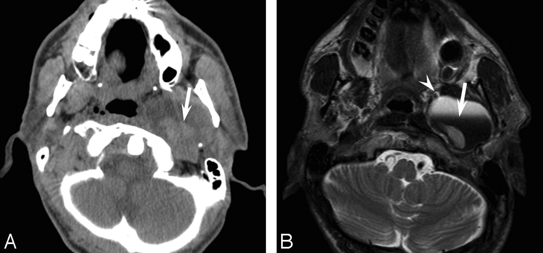
- Fig 1.
Case 1. An 85-year-old woman with a vagus nerve schwannoma in the right neck. A, Unenhanced CT scan shows a well-demarcated, hypoattenuated lesion, with a fluid-fluid level (arrow). B, Axial T2-weighted fast spin-echo MR image (TR, 4102 ms; TE, 90 ms) shows a cystic tumor with a fluid-fluid level (arrow) and a deviation of the right common carotid artery (arrowhead).
- Fig 2.
Case 2. A 44-year-old man with a vagus nerve schwannoma in the left parapharyngeal space. A, Unenhanced CT scan shows a well-demarcated, hypoattenuated lesion, with a fluid-fluid level (arrow). B, Axial T2-weighted fast spin-echo MR image (TR, 5710 ms; TE, 90 ms) shows a unilocular cystic tumor with a fluid-fluid level (arrow) and a deviation of the left internal carotid artery (arrowhead).
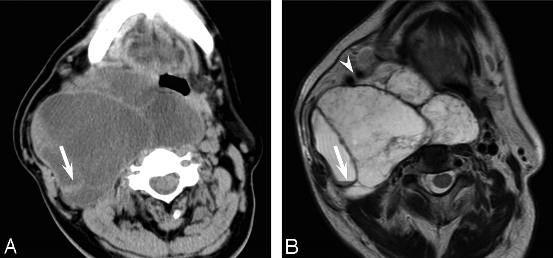
- Fig 3.
Case 3. A 59-year-old man with a large schwannoma in the right neck, extending to the posterior cranial fossa. A, Unenhanced CT scan shows a well-demarcated, hypoattenuated lesion, with a fluid-fluid level (arrow). B, Axial T2-weighted fast spin-echo MR image (TR, 4102 ms; TE, 90 ms) shows a multicystic tumor with a fluid-fluid level (arrow) and a deviation of the right internal and external carotid arteries (arrowhead).


