Advertisement

Main menu

  • Home
  • Content
    • Current Issue
    • Publication Preview--Ahead of Print
    • Past Issue Archive
    • Case of the Week Archive
    • Classic Case Archive
    • Case of the Month Archive
    • COVID-19 Content and Resources
  • For Authors
  • About Us
    • About AJNR
    • Editors
    • American Society of Neuroradiology
  • Submit a Manuscript
  • Podcasts
    • Subscribe on iTunes
    • Subscribe on Stitcher
  • More
    • Subscribers
    • Permissions
    • Advertisers
    • Alerts
    • Feedback
  • Other Publications
    • ajnr

User menu

  • Subscribe
  • Alerts
  • Log in

Search

  • Advanced search
American Journal of Neuroradiology

American Journal of Neuroradiology

  • Subscribe
  • Alerts
  • Log in

Advanced Search

  • Home
  • Content
    • Current Issue
    • Publication Preview--Ahead of Print
    • Past Issue Archive
    • Case of the Week Archive
    • Classic Case Archive
    • Case of the Month Archive
    • COVID-19 Content and Resources
  • For Authors
  • About Us
    • About AJNR
    • Editors
    • American Society of Neuroradiology
  • Submit a Manuscript
  • Podcasts
    • Subscribe on iTunes
    • Subscribe on Stitcher
  • More
    • Subscribers
    • Permissions
    • Advertisers
    • Alerts
    • Feedback
  • Follow AJNR on Twitter
  • Visit AJNR on Facebook
  • Follow AJNR on Instagram
  • Join AJNR on LinkedIn
  • RSS Feeds
Case ReportBrain

White Matter Lesions Suggestive of Amyotrophic Lateral Sclerosis Attributed to Celiac Disease

K.J. Brown, V. Jewells, H. Herfarth and M. Castillo
American Journal of Neuroradiology May 2010, 31 (5) 880-881; DOI: https://doi.org/10.3174/ajnr.A1826
K.J. Brown
  • Find this author on Google Scholar
  • Find this author on PubMed
  • Search for this author on this site
V. Jewells
  • Find this author on Google Scholar
  • Find this author on PubMed
  • Search for this author on this site
H. Herfarth
  • Find this author on Google Scholar
  • Find this author on PubMed
  • Search for this author on this site
M. Castillo
  • Find this author on Google Scholar
  • Find this author on PubMed
  • Search for this author on this site
  • Article
  • Figures & Data
  • Info & Metrics
  • References
  • PDF
Loading

Abstract

SUMMARY: CD is an autoimmune-mediated disorder of the gastrointestinal tract. Initial symptom presentation is variable and can include neurologic manifestations that may comprise ataxia, neuropathy, dizziness, epilepsy, and cortical calcifications rather than gastrointestinal-hindering diagnosis and management. We present a case of a young man with progressive neurologic symptoms and brain MR imaging findings worrisome for ALS. During the diagnostic work-up, endomysium antibodies were discovered, and CD was confirmed by upper gastrointestinal endoscopy with duodenal biopsies. MR imaging findings suggestive of ALS improved after gluten-free diet institution.

Abbreviations

ALS
amyotrophic lateral sclerosis
CD
celiac disease
FLAIR
fluid-attenuated inversion recovery
IgA
immunoglobulin A

CD is an inflammatory condition of the small intestine also called celiac sprue, nontropical sprue, and gluten-sensitive enteropathy. The most common presenting symptoms include diarrhea, steatorrhea, weight loss, iron deficiency anemia, and abdominal distension. Imaging findings specific to the brain have been reported primarily involving the periventricular white matter.

Case Report

A 32-year-old man with a 1-year history of balance difficulties presented with 1 week of worsening symptoms, including hand tremors and gait disturbance. Admission review of systems revealed a 60-pound (27.22 kg) weight loss during the past year, which he attributed to diarrhea consisting of 3–4 loose stools per day. Physical examination showed a broad-based gait leaning to the right, dysmetria with right finger-to-nose, hyperreflexia, an upgoing right toe, right lower extremity weakness, and right foot drop. Sensation was normal. Laboratory studies revealed a slightly elevated phosphorus concentration (4.8 mg/dL), but findings were otherwise normal. Brain MR imaging showed abnormal increased signal intensity on T2 and FLAIR in the bilateral corona radiata, extending inferiorly into the corticospinal tracts without contrast enhancement (Fig 1).

Fig 1.
  • Download figure
  • Open in new tab
  • Download powerpoint
Fig 1.

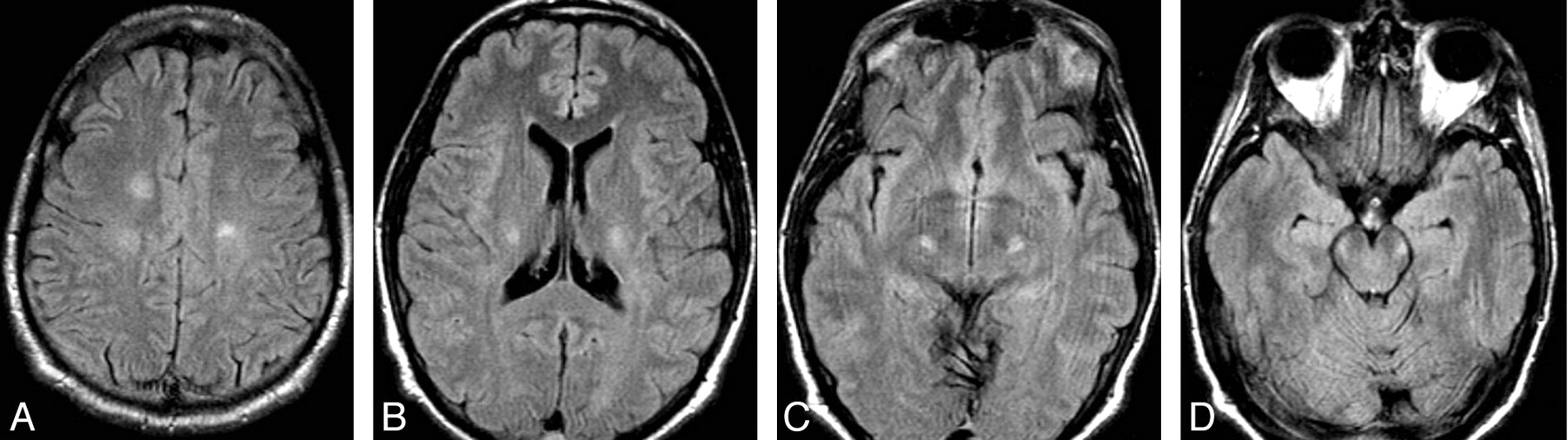
FLAIR images from initial brain MR imaging demonstrate abnormal increased signal intensity in the white matter of the centra semiovale and bilateral corticospinal tracts.

Initial diagnostic considerations included ALS and Friedrich ataxia. Electromyelography findings of the right upper and lower extremities were normal. Total spine MR imaging and CT findings of the chest, abdomen, and pelvis were normal. CSF findings for herpes simplex virus, human herpesvirus 6 cytomegalovirus, varicella-zoster virus, Epstein-Barr virus, and Venereal Disease Research Laboratory test were negative, but +4 oligoclonal bands were noted. Findings were negative for paraneoplastic antibodies. Ataxia work-up included a positive antiendomysial antibody titer of 1:160. Duodenal biopsies yielded crypt hyperplastic villous atrophy with numerous intraepithelial lymphocytes, most consistent with gluten-sensitive enteropathy.

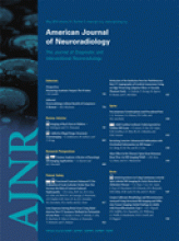
The patient started a strict gluten-free diet with improvement of symptoms during the next several months. Repeat brain MR imaging (23 months after initial presentation) revealed resolution of initial abnormalities (Fig 2 ). Continued physical therapy and a gluten-free diet have been prescribed.

Fig 2.
  • Download figure
  • Open in new tab
  • Download powerpoint
Fig 2.

Follow-up FLAIR images after a gluten-free diet show complete resolution of signal-intensity abnormalities.

Discussion

CD is an inflammatory condition of the small intestine also called celiac sprue, nontropical sprue, and gluten-sensitive enteropathy.1 The disease appears in early childhood and has a second peak in the third-to-fourth decades. Males and females are equally affected, and its incidence has increased in Northern Europe and the United States.2,3 Multiple other disorders have been associated with CD, including Down syndrome, insulin-dependent diabetes mellitus, IgA deficiency, and rheumatoid arthritis.1

Symptoms are precipitated in genetically predisposed individuals by ingestion of foods containing gluten, such as wheat, rye, and barley. Consumption of triggering foods leads to an autoimmune-mediated inflammatory response in the small intestine, resulting in villous atrophy and malabsorption.3 The most common presenting symptoms include diarrhea, steatorrhea, weight loss, iron deficiency anemia, and abdominal distension.

The diagnosis is sometimes difficult to make given that not all patients present in the same manner and that symptoms overlap other gastrointestinal disorders. Half of patients have iron deficiency anemia or osteoporosis, without diarrhea. Diagnostic evaluation for CD consists of antibody testing and duodenal biopsy.4 The most sensitive and specific antibodies for its confirmation are tissue transglutaminase IgA and endomysial IgA antibodies. Due to the low specificity compared with tissue transglutaminase and endomysium antibodies, gliadin antibodies should no longer be used in the diagnostic work-up for CD. Duodenal biopsies are still the criterion standard for tissue diagnosis and reveal villous atrophy5 and increased intraepithelial lymphocytes.6

Neurologic findings associated with CD disease were initially reported in 1966 by Cooke and Smith.7 Most common symptoms are ataxia and neuropathy, followed by epilepsy, ALS and multiple sclerosis-like symptoms, isolated motor neuron disorders, sensory ataxia, and dizziness. These complications may arise as consequences of vitamin and mineral deficiencies. Other authors discuss a potential pathophysiologic role of antigliadin antibodies in a neurotoxic autoimmune process.8

MR imaging is used primarily in the diagnostic work-up of CD-associated conditions. Nonspecific periventricular white matter lesions of various sizes with increased T2/FLAIR signal intensity and without enhancement have been described.9,10 The lesions may be unilateral and bilateral.11 Occipital calcifications have also been reported. In 2007, Turner et al12 reported a case of CD mimicking ALS with T2/FLAIR signal-intensity abnormalities in the left corticospinal tract, which improved on a gluten-free diet. Our patient also showed abnormal signal intensity involving both corticospinal tracts on MR imaging. His symptoms and MR imaging lesions improved and resolved, respectively, on appropriate treatment of CD. Ours is the second patient reported with this group of findings. Because ALS is a progressive and untreatable disease while CD is easily treatable, considering the latter as a cause of neurologic disorders in patients with ALS-like symptoms may be indicated.

References

  1. 1.↵
    1. Green PH,
    2. Cellier C
    . Celiac disease. N Engl J Med 2007;357:1731–43
    CrossRefPubMed
  2. 2.↵
    1. Collin P,
    2. Kaukinen K,
    3. Vogelsang H,
    4. et al
    . Antiendomysial and antihuman recombinant tissue transglutaminase antibodies in the diagnosis of coeliac disease: a biopsy-proven European multicentre study. Eur J Gastroenterol Hepatol Jan 2005;17:85–91
    CrossRef
  3. 3.↵
    1. Hadjivassiliou M,
    2. Williamson CA,
    3. Woodroofe N,
    4. et al
    . The immunology of gluten sensitivity: beyond the gut. Trends Immunol 2004;25:278–82
  4. 4.↵
    1. Mandal A,
    2. Mehdi I,
    3. Munshi S,
    4. et al
    . Value of routine duodenal biopsy in diagnosing coeliac disease in patients with iron deficiency anaemia. Postgrad Med J 2004;80:475–77
    Abstract/FREE Full Text
  5. 5.↵
    1. Brocchi E,
    2. Corazza GR,
    3. Caletti G,
    4. et al
    . Endoscopic demonstration of loss of duodenal folds in the diagnosis of celiac disease. N Engl J Med 1988;319:741–44
    PubMed
  6. 6.↵
    1. Sollid L
    . Intraepithelial lymphocytes in celiac disease: license to kill revealed. Immunity 2004;21:303–04
    PubMed
  7. 7.↵
    1. Cooke WT,
    2. Smith WT
    . Neurological disorders associated with adult celiac disease. Brain 1966;89:683–722
    FREE Full Text
  8. 8.↵
    1. Hadjivassiliou M,
    2. Gibson A,
    3. Davies-Jones CA,
    4. et al
    . Does cryptic gluten sensitivity play a part in neurological illness? Lancet 1996;347:369–71
    CrossRefPubMed
  9. 9.↵
    1. Hadjivassiliou M,
    2. Grünewald RA,
    3. Lawden M,
    4. et al
    . Headache and CNS white matter abnormalities associated with gluten sensitivity. Neurology 2001;56:385–88
    Abstract/FREE Full Text
  10. 10.↵
    1. Kieslich M,
    2. Errázuriz G,
    3. Posselt HG,
    4. et al
    . Brain white-matter lesions in celiac disease: a prospective study of 75 diet-treated patients. Pediatrics 2001;108:E21
    CrossRefPubMed
  11. 11.↵
    1. Lea ME,
    2. Harbord M,
    3. Sage MR
    . Bilateral occipital calcification associated with celiac disease, folate deficiency, and epilepsy. AJNR Am J of Neuroradiology 1995;16:1498–500
  12. 12.↵
    1. Turner M,
    2. Chocan G,
    3. Quaghebeur G,
    4. et al
    . A case of celiac disease mimicking amyotrophic lateral sclerosis. Nat Clin Pract Neurol 2007;3:581–84
    CrossRefPubMed
  • Received June 23, 2009.
  • Accepted after revision July 3, 2009.
  • Copyright © American Society of Neuroradiology
View Abstract
PreviousNext
Back to top

In this issue

American Journal of Neuroradiology: 31 (5)
American Journal of Neuroradiology
Vol. 31, Issue 5
1 May 2010
  • Table of Contents
  • Index by author
Advertisement
Print
Download PDF
Email Article

Thank you for your interest in spreading the word on American Journal of Neuroradiology.

NOTE: We only request your email address so that the person you are recommending the page to knows that you wanted them to see it, and that it is not junk mail. We do not capture any email address.

Enter multiple addresses on separate lines or separate them with commas.
White Matter Lesions Suggestive of Amyotrophic Lateral Sclerosis Attributed to Celiac Disease
(Your Name) has sent you a message from American Journal of Neuroradiology
(Your Name) thought you would like to see the American Journal of Neuroradiology web site.
CAPTCHA
This question is for testing whether or not you are a human visitor and to prevent automated spam submissions.
Citation Tools
White Matter Lesions Suggestive of Amyotrophic Lateral Sclerosis Attributed to Celiac Disease
K.J. Brown, V. Jewells, H. Herfarth, M. Castillo
American Journal of Neuroradiology May 2010, 31 (5) 880-881; DOI: 10.3174/ajnr.A1826

Citation Manager Formats

  • BibTeX
  • Bookends
  • EasyBib
  • EndNote (tagged)
  • EndNote 8 (xml)
  • Medlars
  • Mendeley
  • Papers
  • RefWorks Tagged
  • Ref Manager
  • RIS
  • Zotero
Share
White Matter Lesions Suggestive of Amyotrophic Lateral Sclerosis Attributed to Celiac Disease
K.J. Brown, V. Jewells, H. Herfarth, M. Castillo
American Journal of Neuroradiology May 2010, 31 (5) 880-881; DOI: 10.3174/ajnr.A1826
del.icio.us logo Digg logo Reddit logo Twitter logo CiteULike logo Facebook logo Google logo Mendeley logo
  • Tweet Widget
  • Facebook Like
  • Google Plus One
Purchase

Jump to section

  • Article
    • Abstract
    • Abbreviations
    • Case Report
    • Discussion
    • References
  • Figures & Data
  • Info & Metrics
  • References
  • PDF

Related Articles

  • No related articles found.
  • PubMed
  • Google Scholar

Cited By...

  • Aberrant enteric neuromuscular system and dysbiosis in amyotrophic lateral sclerosis
  • Crossref (15)
  • Google Scholar

This article has been cited by the following articles in journals that are participating in Crossref Cited-by Linking.

  • Neurophysiology of the “Celiac Brain”: Disentangling Gut-Brain Connections
    Manuela Pennisi, Alessia Bramanti, Mariagiovanna Cantone, Giovanni Pennisi, Rita Bella, Giuseppe Lanza
    Frontiers in Neuroscience 2017 11
  • Cortical involvement in celiac disease before and after long-term gluten-free diet: A Transcranial Magnetic Stimulation study
    Manuela Pennisi, Giuseppe Lanza, Mariagiovanna Cantone, Riccardo Ricceri, Raffaele Ferri, Carmela Cinzia D’Agate, Giovanni Pennisi, Vincenzo Di Lazzaro, Rita Bella, François Tremblay
    PLOS ONE 2017 12 5
  • The Role of the Microbiota–Gut–Brain Axis and Antibiotics in ALS and Neurodegenerative Diseases
    Mark Obrenovich, Hayden Jaworski, Tara Tadimalla, Adil Mistry, Lorraine Sykes, George Perry, Robert Bonomo
    Microorganisms 2020 8 5
  • Peripheral Neuropathic Symptoms in Celiac Disease and Inflammatory Bowel Disease
    Ting-Chin David Shen, Benjamin Lebwohl, Himanshu Verma, Nikhil Kumta, Christina Tennyson, Suzanne Lewis, Ellen Scherl, Arun Swaminath, Kristina M. Capiak, Dan DiGiacomo, Brian P. Bosworth, H. Brannagan Thomas, Peter H. R. Green
    Journal of Clinical Neuromuscular Disease 2012 13 3
  • Aberrant enteric neuromuscular system and dysbiosis in amyotrophic lateral sclerosis
    Yongguo Zhang, Destiny Ogbu, Shari Garrett, Yinglin Xia, Jun Sun
    Gut Microbes 2021 13 1
  • No association between biopsy-verified celiac disease and subsequent amyotrophic lateral sclerosis - a population-based cohort study
    J. F. Ludvigsson, D. Mariosa, B. Lebwohl, F. Fang
    European Journal of Neurology 2014 21 7
  • A Gut Feeling in Amyotrophic Lateral Sclerosis: Microbiome of Mice and Men
    Sarah Martin, Carolina Battistini, Jun Sun
    Frontiers in Cellular and Infection Microbiology 2022 12
  • Central Pontine Myelinolysis Presenting With Tremor in a Child With Celiac Disease
    Preeti Sharma, Suvasini Sharma, Neha Panwar, Deonath Mahto, Praveen Kumar, Atin Kumar, Satinder Aneja
    Journal of Child Neurology 2014 29 3
  • Coeliac disease mimicking Amyotrophic Lateral Sclerosis
    Enrica Bersano, Alessandro Stecco, Sandra D’Alfonso, Lucia Corrado, Maria Francesca Sarnelli, Valentina Solara, Roberto Cantello, Letizia Mazzini
    Amyotrophic Lateral Sclerosis and Frontotemporal Degeneration 2015 16 3-4
  • No association between gluten sensitivity and amyotrophic lateral sclerosis
    Anne E. Visser, Raha Pazoki, Sara L. Pulit, Wouter van Rheenen, Joost Raaphorst, Anneke J. van der Kooi, Isis Ricaño-Ponce, Cisca Wijmenga, Henny G. Otten, Jan H. Veldink, Leonard H. van den Berg
    Journal of Neurology 2017 264 4

More in this TOC Section

  • Statin Therapy Does Not Affect the Radiographic and Clinical Profile of Patients with TIA and Minor Stroke
  • Usefulness of Quantitative Susceptibility Mapping for the Diagnosis of Parkinson Disease
  • White Matter Alterations in the Brains of Patients with Active, Remitted, and Cured Cushing Syndrome: A DTI Study
Show more BRAIN

Similar Articles

Advertisement

News and Updates

  • Lucien Levy Best Research Article Award
  • Thanks to our 2022 Distinguished Reviewers
  • Press Releases

Resources

  • Evidence-Based Medicine Level Guide
  • How to Participate in a Tweet Chat
  • AJNR Podcast Archive
  • Ideas for Publicizing Your Research
  • Librarian Resources
  • Terms and Conditions

Opportunities

  • Share Your Art in Perspectives
  • Get Peer Review Credit from Publons
  • Moderate a Tweet Chat

American Society of Neuroradiology

  • Neurographics
  • ASNR Annual Meeting
  • Fellowship Portal
  • Position Statements

© 2023 by the American Society of Neuroradiology | Print ISSN: 0195-6108 Online ISSN: 1936-959X

Powered by HighWire