Advertisement

Main menu

  • Home
  • Content
    • Current Issue
    • Publication Preview--Ahead of Print
    • Past Issue Archive
    • Case of the Week Archive
    • Classic Case Archive
    • Case of the Month Archive
    • COVID-19 Content and Resources
  • For Authors
  • About Us
    • About AJNR
    • Editors
    • American Society of Neuroradiology
  • Submit a Manuscript
  • Podcasts
    • Subscribe on iTunes
    • Subscribe on Stitcher
  • More
    • Subscribers
    • Permissions
    • Advertisers
    • Alerts
    • Feedback
  • Other Publications
    • ajnr

User menu

  • Subscribe
  • Alerts
  • Log in

Search

  • Advanced search
American Journal of Neuroradiology

American Journal of Neuroradiology

  • Subscribe
  • Alerts
  • Log in

Advanced Search

  • Home
  • Content
    • Current Issue
    • Publication Preview--Ahead of Print
    • Past Issue Archive
    • Case of the Week Archive
    • Classic Case Archive
    • Case of the Month Archive
    • COVID-19 Content and Resources
  • For Authors
  • About Us
    • About AJNR
    • Editors
    • American Society of Neuroradiology
  • Submit a Manuscript
  • Podcasts
    • Subscribe on iTunes
    • Subscribe on Stitcher
  • More
    • Subscribers
    • Permissions
    • Advertisers
    • Alerts
    • Feedback
  • Follow AJNR on Twitter
  • Visit AJNR on Facebook
  • Follow AJNR on Instagram
  • Join AJNR on LinkedIn
  • RSS Feeds
Research ArticleTechnical Note

High-Resolution Double Inversion Recovery Black-Blood Imaging of Cervical Artery Dissection Using 3T MR Imaging

M.A. Hunter, C. Santosh, E. Teasdale and K.P. Forbes
American Journal of Neuroradiology December 2012, 33 (11) E133-E137; DOI: https://doi.org/10.3174/ajnr.A2599
M.A. Hunter
aFrom the Department of Radiology (M.A.H.), Royal Alexander Hospital, Paisley, United Kingdom
  • Find this author on Google Scholar
  • Find this author on PubMed
  • Search for this author on this site
C. Santosh
bDepartment of Neuroradiology (C.S., E.T., K.P.F.), Institute of Neurological Sciences, Glasgow, United Kingdom
  • Find this author on Google Scholar
  • Find this author on PubMed
  • Search for this author on this site
E. Teasdale
bDepartment of Neuroradiology (C.S., E.T., K.P.F.), Institute of Neurological Sciences, Glasgow, United Kingdom
  • Find this author on Google Scholar
  • Find this author on PubMed
  • Search for this author on this site
K.P. Forbes
bDepartment of Neuroradiology (C.S., E.T., K.P.F.), Institute of Neurological Sciences, Glasgow, United Kingdom
  • Find this author on Google Scholar
  • Find this author on PubMed
  • Search for this author on this site
  • Article
  • Figures & Data
  • Info & Metrics
  • References
  • PDF
Loading

Abstract

SUMMARY: We performed high-resolution DIR-BBI of the cervical arteries at 3T in 19 subjects with cervical dissection. It offered excellent visualization of both the lumen and arterial wall, allowing detection of the primary and secondary features of dissection. We suggest that this is a highly useful technique for diagnosis of cervical dissection, either routinely or in equivocal cases of suspected dissection. It also offers further insight into the pathogenesis of this disorder.

ABBREVIATIONS:

BBI
black-blood imaging
CA
carotid artery
CTA
CT angiography
DIR
double inversion recovery
MCA
middle cerebral artery
PCA
posterior cerebral artery
PICA
posterior inferior cerebellar artery
VA
vertebral artery

DIR-BBI has proved to be an excellent technique for structural assessment of the vascular system, nulling signal intensity from flowing blood and perivascular fat. When performed with a surface coil at 3T, it offers a high-resolution assessment of the cervical artery wall and lumen. The technique has been widely used for assessment of carotid atheromatous plaque, allowing plaque subcomponents and substructural changes to be identified.1

We were interested in applying this technique to cervical artery dissection, a pathology that arises in the arterial wall, to see if DIR-BBI might either assist in the diagnosis and/or add to the known pathogenesis and imaging appearances of this condition. Dissection is increasingly recognized as an underlying cause of stroke,2⇓–4 which is of significant importance because it is treatable. Various imaging modalities have been used to diagnosis this condition, including digital subtraction angiography5 CTA,6 and MR imaging (MR angiography or T1-weighted fat-saturated imaging).7 The diagnosis of dissection can be challenging for all modalities, often relying on secondary luminal findings. Only CTA and MR imaging can assess the primary site of pathology, the arterial wall, though MR imaging offers a much higher contrast resolution for detection of hemorrhage.

Methods and Technique

We performed DIR-BBI of the cervical arteries in subjects with arterial dissection. A high-resolution approach was used, with 3T MR imaging (Signa Excite; GE Healthcare, Milwaukee, Wisconsin) and a surface coil (phased-array 4-channel; Herman Flick, Flick Engineering Solution BV, the Netherlands). 2D time-of-flight angiography of the neck vessels was used to plan axial 2D BBI (T1-weighted and T2*-weighted).

We applied the technique to 19 subjects (12 men, 7 women; mean age, 47 ± 16 years) with cervical artery dissection in at least 1 vessel, confirmed by CTA (64-section Brilliance CT scanner; Philips Healthcare, Best, the Netherlands) (0.625-mm sections; bolus tracking, 75 mL of contrast at 5 mL/s). Each of the 4 cervical arteries on CTA and BBI was independently reviewed for signs of dissection: 1) intimal flap or double lumen, 2) wall thickening ± T1 hyperintensity of subacute hemorrhage (BBI only), and 3) stenosis, occlusion, or pseudoaneurysm. A conclusion on the presence or absence of dissection was reached for each vessel (CTA, 1 observer; MR imaging, 2 observers, by consensus), and any other potentially relevant observations were noted. Statistical analyses were performed by using the Statistical Package for the Social Sciences software (SPSS, Chicago, Illinois).

Results

Demographics, clinical findings, and brain imaging findings are summarized in Table 1. Vascular findings are summarized in Table 2. MR imaging was performed a median of 4 days after CTA (interquartile range, 1–5 days).

View this table:
  • View inline
  • View popup
Table 1:

Demographics: clinical and brain imaging findings

View this table:
  • View inline
  • View popup
Table 2:

Vascular imaging findings

There was no significant difference between CTA and BBI in the number of subjects or individual vessels with dissection (subjects: CTA, n = 17; BBI, n = 19; vessels: CTA, n = 20; BBI, n = 24; McNemar test: P > 0.05). (On CTA review, 2 subjects were thought to show imaging features that could have resulted from atherosclerosis or dissection).

MR imaging detected more intimal flaps or double lumen (MR imaging, n = 15; CTA, n = 7; McNemar test, P < .03) (Fig 1⇓A, -B). Wall thickening was more commonly identified on MR imaging (MR imaging, n = 24; CTA, n = 18; McNemar test, P < .04), and T1 hyperintensity, in keeping with hematoma, was noted in all dissected vessels (Fig 3), while T2* was decreased in 3 (Fig 4 ). In 5, hematoma was localized as subintimal (VA, n = 3; CA, n = 2) (Fig 5), and in 1, it was as intramedial.

Fig 1.
  • Download figure
  • Open in new tab
  • Download powerpoint
Fig 1.

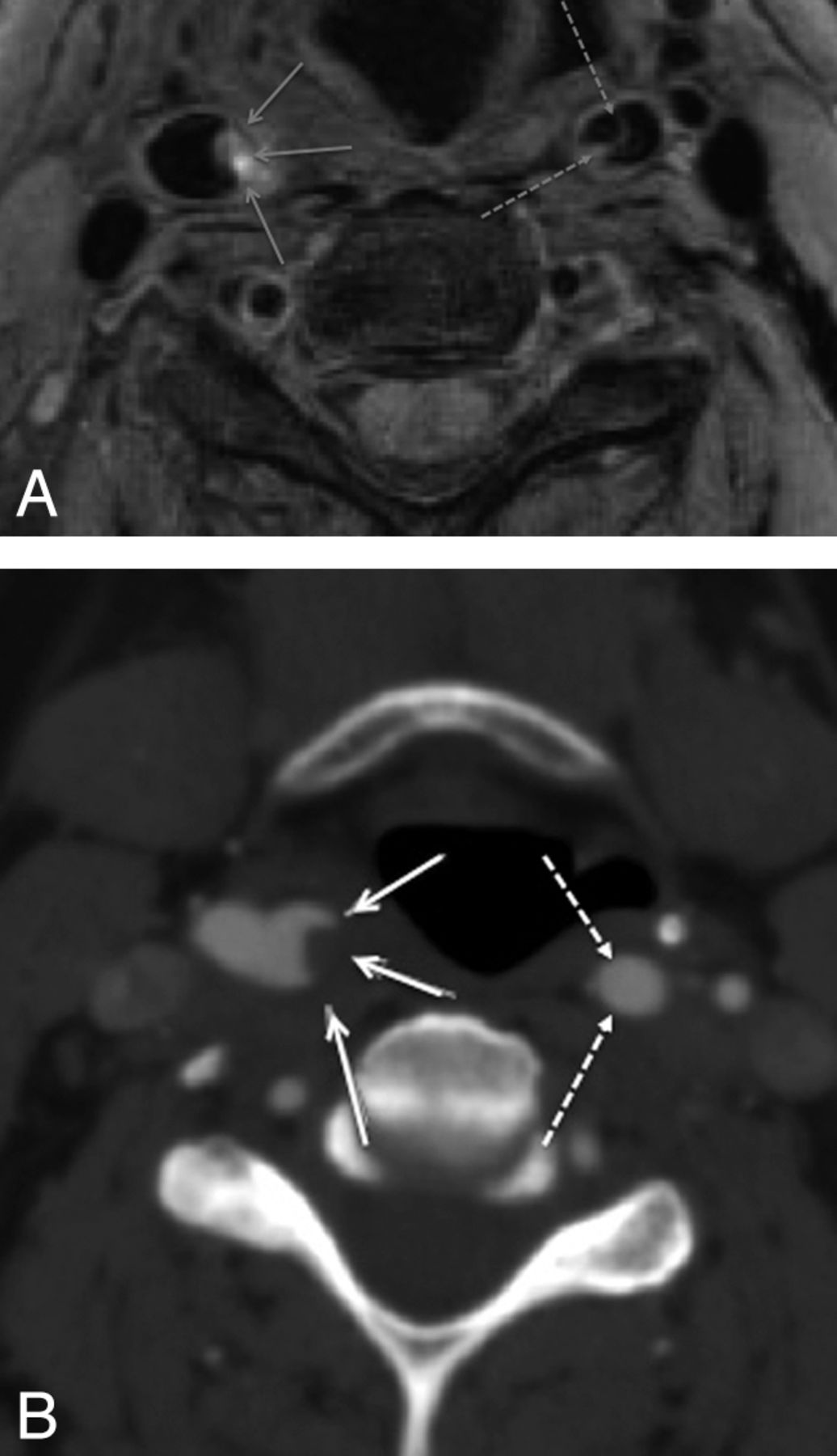
BBI (A) performed 1 day subsequent to CTA (B) demonstrates thrombus within the lumen of the right internal carotid artery (continuous arrows) but also a dissection flap in the left internal carotid artery not appreciated on CTA.

Fig 2.
  • Download figure
  • Open in new tab
  • Download powerpoint
Fig 2.

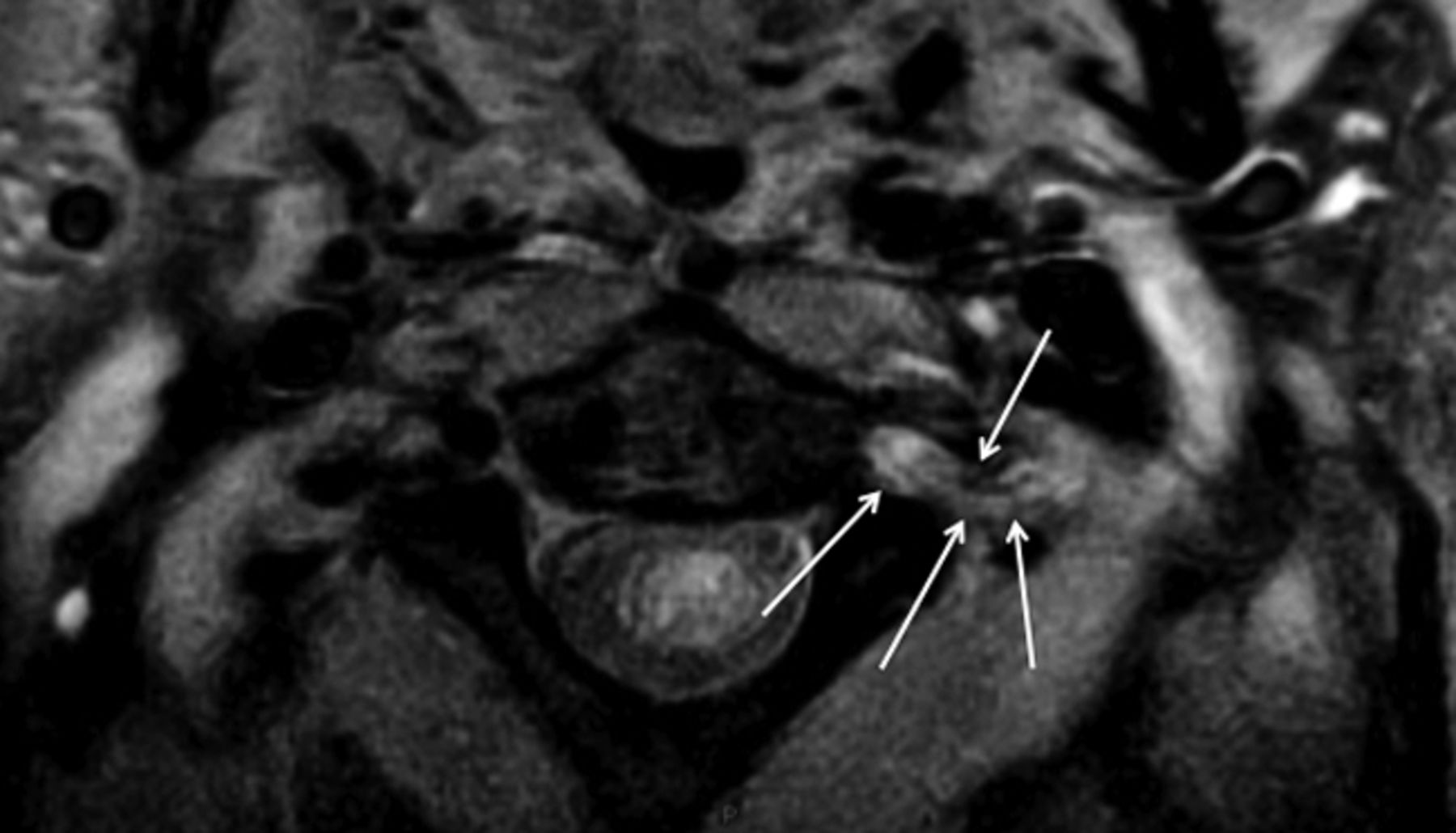
Double inversion recovery BBI demonstrates crescentic intramural high T1 methemoglobin signal intensity (arrows) in the dissected left VA.

Fig 3.
  • Download figure
  • Open in new tab
  • Download powerpoint
Fig 3.

T2* sequence demonstrates crescentic intramural deoxyhemoglobin signal intensity (arrows) in the dissected right VA.

Fig 4.
  • Download figure
  • Open in new tab
  • Download powerpoint
Fig 4.

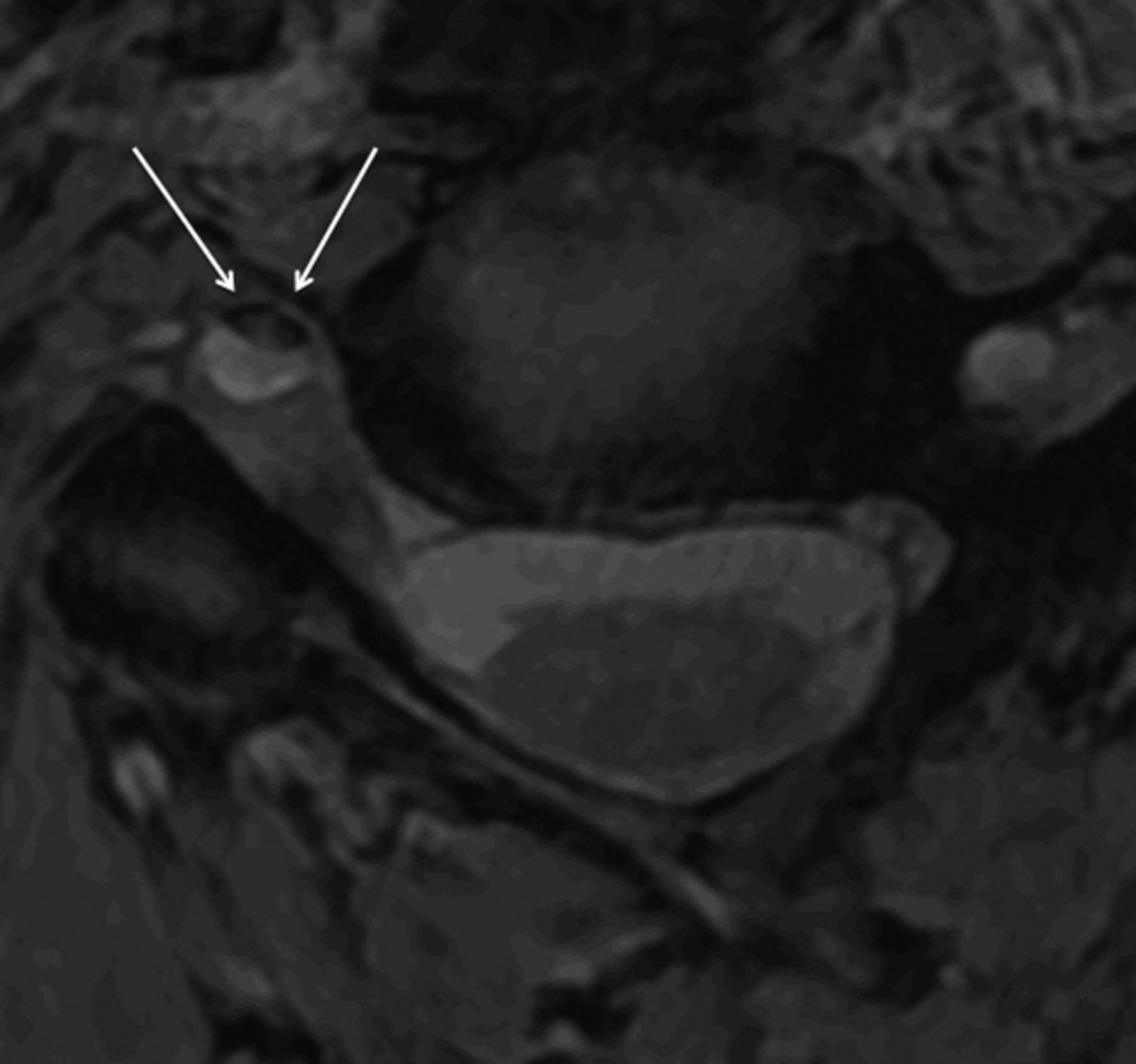
“Target sign” appearance on BBI caused by hematoma in subintimal dissection. The media (continuous arrow) returns low signal intensity; the hematoma (discontinuous arrow), high signal intensity.

Fig 5.
  • Download figure
  • Open in new tab
  • Download powerpoint
Fig 5.

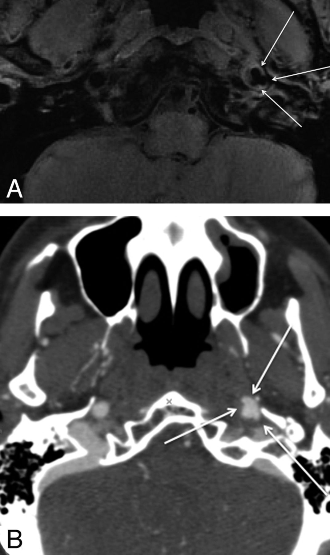
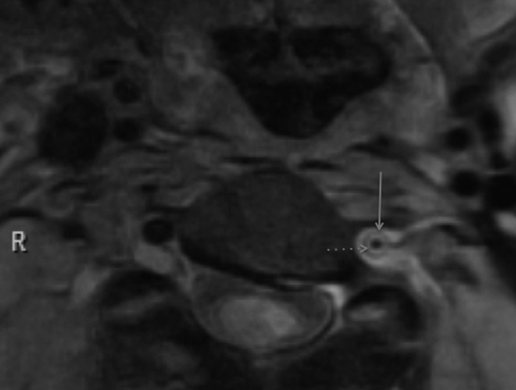
Both BBI (A) and CTA (B) demonstrate pseudoaneurysm formation in the skull base of the dissected left internal carotid artery.

There was no difference in the ability of CTA and MR imaging to detect occlusion (CTA, n = 8; MR imaging, n = 7) or stenosis (CTA, n = 13; MR imaging, n = 15) (McNemar test, no significant difference). A pseudoaneurysm was seen in only 1 dissected CA, on both modalities (Fig 2 A, -B).

Observational note was made of T2 hyperintense periadventitial edematous signal-intensity change, which tracked into the adjacent parenchyma (dissected vessels, n = 20; normal vessels, n = 0; χ2 test, P < .001) (Fig 6 ).

Fig 6.
  • Download figure
  • Open in new tab
  • Download powerpoint
Fig 6.

Periadventitial signal-intensity change (arrows), presumed due to inflammatory response, surrounding the dissected left VA.

Discussion

This study confirms that BBI is a useful technique for detection of the pathologic findings of dissection, allowing a confident diagnosis to be made.

We chose to perform this technique on subjects with known dissection, as confirmed by CTA, providing a high concentration of pathologic vessels on which to evaluate this sequence. While our study was not designed to compare the sensitivity and specificity of CTA and MR imaging in diagnosing dissection,8 BBI did offer improved detection of intimal flaps and wall thickening, suggesting it might offer some improved sensitivity. This technique allows clear visualization of the vessel lumen: Flowing blood is saturated by inversion pulses, giving a true black-blood appearance, improved over spin-echo sequences and offering a high sensitivity to the detection of intraluminal pathology. It also offers excellent spatial and contrast resolution of the vessel wall, allowing better detection of wall thickening and hemorrhage. High T1 contrast resolution allows detection of subacute hemorrhage. We did notice some variation in the strength of T1 hyperintensity, as yet unexplained and not in our data, related to symptom duration. We included a T2*-weighted sequence to identify the low signal intensity of deoxyhemoglobin in acute dissection, though the numbers were too small to analyze.

The high-resolution DIR-BBI technique may allow us to examine the pathogenesis of dissection further. The traditional belief is that a tear occurs within the intima, allowing entry of arterial blood, creating a false plane of cleavage. This explains why patients with inherent structural collagen weakness9,10 are more prone to the condition, but in total, this accounts for only 1%–5% of those affected; in our series only 1 patient (5%) had such connective tissue disease. Contrary to this theory is that of spontaneous rupture of the vasa vasorum. Following this initial event, dissection can extend either between the intima and media, resulting in stenosis or occlusion, or alternatively and less frequently between media and thinner adventitia, which may lead to pseudoaneurysm formation and occasionally rupture and subarachnoid hemorrhage if intracranial.11 Spontaneous rupture of the vasa vasorum, as an underlying cause of dissection might explain why a flap is uncommonly seen at autopsy. However, we identified a flap in nearly one-third of subjects, suggesting that its presence is more common than previously thought and adding support for an intimal tear as a significant primary pathogenic process of arterial dissection.

With the high-resolution DIR-BBI technique, we found it possible to clearly differentiate intramural hematoma from intraluminal thrombus; furthermore, we could sometimes differentiate intramedial from subintimal hemorrhage.

An observational finding commonly noted was T2 hyperintense periadventitial signal-intensity change. We postulate that during acute dissection, the artery is surrounded by a variable degree of inflammatory edema. A similar response has been demonstrated in spontaneous coronary artery dissection,12,13 and our study confirms similar observation in cervical dissection, as recently reported.14

Conclusions

3T DIR-BBI is useful in the evaluation of acute cervical artery dissection, offering excellent visualization of the lumen, wall, and periadventitia. This technique could be used either for primary diagnosis or as an adjunct in difficult or inconclusive cases. Furthermore, its high spatial and contrast resolution offers a mode of further exploration into the pathogenesis of dissection.

Footnotes

  • Paper previously presented at: Annual Meeting of the American Society of Neuroradiology, May 31–June 5, 2008; New Orleans, Louisiana.

References

  1. 1.↵
    1. DeMarco JK,
    2. Ota H,
    3. Underhill HR,
    4. et al
    . MR carotid plaque imaging and contrast-enhanced MR angiography identifies lesions associated with recent ipsilateral thromboembolic symptoms: an in vivo study at 3T. AJNR Am J Neuroradiol 31: 1395–402
  2. 2.↵
    1. Schievink WI
    . Spontaneous dissection of the carotid and vertebral arteries. N Engl J Med 2001; 344: 898– 906
    CrossRefPubMed
  3. 3.↵
    1. Schievink WI,
    2. Mokri B,
    3. O'Fallon WM
    . Recurrent spontaneous cervical artery dissection. N Engl J Med 1994; 330: 393– 97
    CrossRefPubMed
  4. 4.↵
    1. Schievink WI,
    2. Mokri B,
    3. Whisnant JP
    . Internal carotid artery dissection in a community: Rochester, Minnesota, 1987–1992. Stroke 1993; 24: 1678– 80
    Abstract/FREE Full Text
  5. 5.↵
    1. Provenzale JM
    . Dissection of the internal carotid and vertebral arteries: imaging features. AJR Am J Roentgenol 1995: 165; 1099– 1104
    CrossRefPubMed
  6. 6.↵
    1. Chen CJ,
    2. Tseng YC,
    3. Lee TH,
    4. et al
    . Multisection CT angiography compared with catheter angiography in diagnosing vertebral artery dissection. AJNR Am J Neuroradiol 2004; 25: 769– 74
    Abstract/FREE Full Text
  7. 7.↵
    1. Ozdoba C,
    2. Sturzenegger M,
    3. Schroth G
    . Internal carotid artery dissection: MR imaging features and clinical-radiologic correlation. Radiology 1996; 199: 191– 98
    PubMed
  8. 8.↵
    1. Provenzale JM,
    2. Sarikaya B
    . Comparison of test performance characteristics of MRI, MR angiography, and CT angiography in the diagnosis of carotid and vertebral dissection: a review of the medical literature. AJR Am J Roentgenol 2009; 193: 1167– 74
    CrossRefPubMed
  9. 9.↵
    1. Schievink WI,
    2. Michels VV,
    3. Piepgras DG
    . Neurovascular manifestations of heritable connective tissue disorders: a review. Stroke 1994; 25: 889– 903
    Abstract/FREE Full Text
  10. 10.↵
    1. Brandt T,
    2. Orberk E,
    3. Weber R,
    4. et al
    . Pathogenesis of cervical artery dissections: association with connective tissue abnormalities. Neurology 2001; 57: 24–30
    Abstract/FREE Full Text
  11. 11.↵
    1. Dziewas R,
    2. Konrad C,
    3. Drager B,
    4. et al
    . Cervical artery dissection: clinical features, risk factors, therapy and outcome in 126 patients. J Neurol 2003; 250: 1179– 84
    CrossRefPubMed
  12. 12.↵
    1. Bateman AC,
    2. Gallagher PJ,
    3. Vincenti AC
    . Sudden death from coronary artery dissection. J Clin Pathol 1995; 48: 781– 84
    Abstract/FREE Full Text
  13. 13.↵
    1. Robinowitz M,
    2. Virmani R,
    3. McAllister HA
    . Spontaneous coronary artery dissection and eosinophilic inflammation: a cause and effect relationship? Am J Med 1982; 72: 923– 28
    CrossRefPubMed
  14. 14.↵
    1. Bachmann R,
    2. Nassenstein I,
    3. et al
    . Spontaneous acute dissection of the internal carotid artery: high-resolution magnetic resonance imaging at 3.0 Tesla with a dedicated surface coil. Invest Radiol 2006; 41: 105– 11
    CrossRefPubMed
  • Received November 21, 2010.
  • Accepted after revision February 4, 2011.
  • © 2012 by American Journal of Neuroradiology
View Abstract
PreviousNext
Back to top

In this issue

American Journal of Neuroradiology: 33 (11)
American Journal of Neuroradiology
Vol. 33, Issue 11
1 Dec 2012
  • Table of Contents
  • Index by author
Advertisement
Print
Download PDF
Email Article

Thank you for your interest in spreading the word on American Journal of Neuroradiology.

NOTE: We only request your email address so that the person you are recommending the page to knows that you wanted them to see it, and that it is not junk mail. We do not capture any email address.

Enter multiple addresses on separate lines or separate them with commas.
High-Resolution Double Inversion Recovery Black-Blood Imaging of Cervical Artery Dissection Using 3T MR Imaging
(Your Name) has sent you a message from American Journal of Neuroradiology
(Your Name) thought you would like to see the American Journal of Neuroradiology web site.
CAPTCHA
This question is for testing whether or not you are a human visitor and to prevent automated spam submissions.
Citation Tools
High-Resolution Double Inversion Recovery Black-Blood Imaging of Cervical Artery Dissection Using 3T MR Imaging
M.A. Hunter, C. Santosh, E. Teasdale, K.P. Forbes
American Journal of Neuroradiology Dec 2012, 33 (11) E133-E137; DOI: 10.3174/ajnr.A2599

Citation Manager Formats

  • BibTeX
  • Bookends
  • EasyBib
  • EndNote (tagged)
  • EndNote 8 (xml)
  • Medlars
  • Mendeley
  • Papers
  • RefWorks Tagged
  • Ref Manager
  • RIS
  • Zotero
Share
High-Resolution Double Inversion Recovery Black-Blood Imaging of Cervical Artery Dissection Using 3T MR Imaging
M.A. Hunter, C. Santosh, E. Teasdale, K.P. Forbes
American Journal of Neuroradiology Dec 2012, 33 (11) E133-E137; DOI: 10.3174/ajnr.A2599
Reddit logo Twitter logo Facebook logo Mendeley logo
  • Tweet Widget
  • Facebook Like
  • Google Plus One
Purchase

Jump to section

  • Article
    • Abstract
    • ABBREVIATIONS:
    • Methods and Technique
    • Discussion
    • Conclusions
    • Footnotes
    • References
  • Figures & Data
  • Info & Metrics
  • References
  • PDF

Related Articles

  • No related articles found.
  • PubMed
  • Google Scholar

Cited By...

  • Standard Diffusion-Weighted Imaging in the Brain Can Detect Cervical Internal Carotid Artery Dissections
  • Blunt Cerebrovascular Injuries: Advances in Screening, Imaging, and Management Trends
  • Crossref
  • Google Scholar

This article has not yet been cited by articles in journals that are participating in Crossref Cited-by Linking.

More in this TOC Section

  • 3D Fast Spin-Echo T1 Black-Blood Imaging for the Diagnosis of Cervical Artery Dissection
  • The Kety-Schmidt Technique for Quantitative Perfusion and Oxygen Metabolism Measurements in the MR Imaging Environment
  • A Swine Model to Analyze Arterial Structural Changes Induced by Mechanical Thrombectomy
Show more Technical Note

Similar Articles

Advertisement

News and Updates

  • Lucien Levy Best Research Article Award
  • Thanks to our 2022 Distinguished Reviewers
  • Press Releases

Resources

  • Evidence-Based Medicine Level Guide
  • How to Participate in a Tweet Chat
  • AJNR Podcast Archive
  • Ideas for Publicizing Your Research
  • Librarian Resources
  • Terms and Conditions

Opportunities

  • Share Your Art in Perspectives
  • Get Peer Review Credit from Publons
  • Moderate a Tweet Chat

American Society of Neuroradiology

  • Neurographics
  • ASNR Annual Meeting
  • Fellowship Portal
  • Position Statements

© 2023 by the American Society of Neuroradiology | Print ISSN: 0195-6108 Online ISSN: 1936-959X

Powered by HighWire