Advertisement

Main menu

  • Home
  • Content
    • Current Issue
    • Publication Preview--Ahead of Print
    • Past Issue Archive
    • Case of the Week Archive
    • Classic Case Archive
    • Case of the Month Archive
    • COVID-19 Content and Resources
  • For Authors
  • About Us
    • About AJNR
    • Editors
    • American Society of Neuroradiology
  • Submit a Manuscript
  • Podcasts
    • Subscribe on iTunes
    • Subscribe on Stitcher
  • More
    • Subscribers
    • Permissions
    • Advertisers
    • Alerts
    • Feedback
  • Other Publications
    • ajnr

User menu

  • Subscribe
  • Alerts
  • Log in

Search

  • Advanced search
American Journal of Neuroradiology

American Journal of Neuroradiology

  • Subscribe
  • Alerts
  • Log in

Advanced Search

  • Home
  • Content
    • Current Issue
    • Publication Preview--Ahead of Print
    • Past Issue Archive
    • Case of the Week Archive
    • Classic Case Archive
    • Case of the Month Archive
    • COVID-19 Content and Resources
  • For Authors
  • About Us
    • About AJNR
    • Editors
    • American Society of Neuroradiology
  • Submit a Manuscript
  • Podcasts
    • Subscribe on iTunes
    • Subscribe on Stitcher
  • More
    • Subscribers
    • Permissions
    • Advertisers
    • Alerts
    • Feedback
  • Follow AJNR on Twitter
  • Visit AJNR on Facebook
  • Follow AJNR on Instagram
  • Join AJNR on LinkedIn
  • RSS Feeds
Research ArticleExtracranial Vascular

Carotid Artery Wall Imaging: Perspective and Guidelines from the ASNR Vessel Wall Imaging Study Group and Expert Consensus Recommendations of the American Society of Neuroradiology

L. Saba, C. Yuan, T.S. Hatsukami, N. Balu, Y. Qiao, J.K. DeMarco, T. Saam, A.R. Moody, D. Li, C.C. Matouk, M.H. Johnson, H.R. Jäger, M. Mossa-Basha, M.E. Kooi, Z. Fan, D. Saloner, M. Wintermark, D.J. Mikulis and B.A. Wasserman on behalf of the Vessel Wall Imaging Study Group of the American Society of Neuroradiology
American Journal of Neuroradiology February 2018, 39 (2) E9-E31; DOI: https://doi.org/10.3174/ajnr.A5488
L. Saba
aFrom the Department of Medical Imaging (L.S.), University of Cagliari, Cagliari, Italy
  • Find this author on Google Scholar
  • Find this author on PubMed
  • Search for this author on this site
  • ORCID record for L. Saba
C. Yuan
bDepartments of Radiology (C.Y., N.B., M.M.-B.)
  • Find this author on Google Scholar
  • Find this author on PubMed
  • Search for this author on this site
  • ORCID record for C. Yuan
T.S. Hatsukami
cSurgery (T.S.H.), University of Washington, Seattle, Washington
  • Find this author on Google Scholar
  • Find this author on PubMed
  • Search for this author on this site
  • ORCID record for T.S. Hatsukami
N. Balu
bDepartments of Radiology (C.Y., N.B., M.M.-B.)
  • Find this author on Google Scholar
  • Find this author on PubMed
  • Search for this author on this site
  • ORCID record for N. Balu
Y. Qiao
dThe Russell H. Morgan Department of Radiology and Radiological Sciences (Y.Q., B.A.W.), Johns Hopkins Hospital, Baltimore, Maryland
  • Find this author on Google Scholar
  • Find this author on PubMed
  • Search for this author on this site
  • ORCID record for Y. Qiao
J.K. DeMarco
eDepartment of Radiology (J.K.D.), Walter Reed National Military Medical Center, Bethesda, Maryland
  • Find this author on Google Scholar
  • Find this author on PubMed
  • Search for this author on this site
  • ORCID record for J.K. DeMarco
T. Saam
fDepartment of Radiology (T.S.), Ludwig-Maximilian University Hospital, Munich, Germany
  • Find this author on Google Scholar
  • Find this author on PubMed
  • Search for this author on this site
  • ORCID record for T. Saam
A.R. Moody
hDepartment of Medical Imaging (A.R.M.), Sunnybrook Health Sciences Centre, University of Toronto, Toronto, Ontario, Canada
  • Find this author on Google Scholar
  • Find this author on PubMed
  • Search for this author on this site
  • ORCID record for A.R. Moody
D. Li
iBiomedical Imaging Research Institute (D.L., Z.F.), Cedars-Sinai Medical Center, Los Angeles, California
  • Find this author on Google Scholar
  • Find this author on PubMed
  • Search for this author on this site
  • ORCID record for D. Li
C.C. Matouk
jDepartments of Neurosurgery, Neurovascular and Stroke Programs (C.C.M., M.H.J.)
kRadiology and Biomedical Imaging (C.C.M., M.H.J.)
  • Find this author on Google Scholar
  • Find this author on PubMed
  • Search for this author on this site
  • ORCID record for C.C. Matouk
M.H. Johnson
jDepartments of Neurosurgery, Neurovascular and Stroke Programs (C.C.M., M.H.J.)
kRadiology and Biomedical Imaging (C.C.M., M.H.J.)
lSurgery (M.H.J.), Yale University School of Medicine, New Haven, Connecticut
  • Find this author on Google Scholar
  • Find this author on PubMed
  • Search for this author on this site
  • ORCID record for M.H. Johnson
H.R. Jäger
mNeuroradiological Academic Unit (H.R.J.), Department of Brain Repair and Rehabilitation, University College London Institute of Neurology, London, UK
  • Find this author on Google Scholar
  • Find this author on PubMed
  • Search for this author on this site
  • ORCID record for H.R. Jäger
M. Mossa-Basha
bDepartments of Radiology (C.Y., N.B., M.M.-B.)
  • Find this author on Google Scholar
  • Find this author on PubMed
  • Search for this author on this site
  • ORCID record for M. Mossa-Basha
M.E. Kooi
nDepartment of Radiology (M.E.K.), CARIM School for Cardiovascular Diseases, Maastricht University Medical Center, Maastricht, the Netherlands
  • Find this author on Google Scholar
  • Find this author on PubMed
  • Search for this author on this site
  • ORCID record for M.E. Kooi
Z. Fan
iBiomedical Imaging Research Institute (D.L., Z.F.), Cedars-Sinai Medical Center, Los Angeles, California
  • Find this author on Google Scholar
  • Find this author on PubMed
  • Search for this author on this site
  • ORCID record for Z. Fan
D. Saloner
oDepartment of Radiology and Biomedical Imaging (D.S.), University of California, San Francisco, California
  • Find this author on Google Scholar
  • Find this author on PubMed
  • Search for this author on this site
  • ORCID record for D. Saloner
M. Wintermark
pDepartment of Radiology (M.W.), Neuroradiology Division, Stanford University, Stanford, California.
  • Find this author on Google Scholar
  • Find this author on PubMed
  • Search for this author on this site
  • ORCID record for M. Wintermark
D.J. Mikulis
gDivision of Neuroradiology (D.J.M.), Department of Medical Imaging, University Health Network
  • Find this author on Google Scholar
  • Find this author on PubMed
  • Search for this author on this site
  • ORCID record for D.J. Mikulis
B.A. Wasserman
dThe Russell H. Morgan Department of Radiology and Radiological Sciences (Y.Q., B.A.W.), Johns Hopkins Hospital, Baltimore, Maryland
  • Find this author on Google Scholar
  • Find this author on PubMed
  • Search for this author on this site
  • ORCID record for B.A. Wasserman
  • Article
  • Figures & Data
  • Info & Metrics
  • References
  • PDF
Loading

Abstract

SUMMARY: Identification of carotid artery atherosclerosis is conventionally based on measurements of luminal stenosis and surface irregularities using in vivo imaging techniques including sonography, CT and MR angiography, and digital subtraction angiography. However, histopathologic studies demonstrate considerable differences between plaques with identical degrees of stenosis and indicate that certain plaque features are associated with increased risk for ischemic events. The ability to look beyond the lumen using highly developed vessel wall imaging methods to identify plaque vulnerable to disruption has prompted an active debate as to whether a paradigm shift is needed to move away from relying on measurements of luminal stenosis for gauging the risk of ischemic injury. Further evaluation in randomized clinical trials will help to better define the exact role of plaque imaging in clinical decision-making. However, current carotid vessel wall imaging techniques can be informative. The goal of this article is to present the perspective of the ASNR Vessel Wall Imaging Study Group as it relates to the current status of arterial wall imaging in carotid artery disease.

ABBREVIATIONS:

CE
contrast-enhanced
DIR
double inversion recovery
FC
fibrous cap
IPH
intraplaque hemorrhage
IVUS
intravascular ultrasound
LRNC
lipid-rich necrotic core
MATCH
multicontrast atherosclerosis characterization
SNAP
simultaneous noncontrast angiography and intraplaque hemorrhage
SPACE
sampling perfection with application-optimized contrasts using different flip angle evolution
US
ultrasound

The word “atherosclerosis” is derived from the 2 Greek words “athera” (mush) and “sclerosis” (hardening), indicating hardening of the vascular wall. This disease is highly prevalent in developed countries, with carotid artery narrowing reported in up to 75% of men and 62% of women 65 years of age and older.1 Stroke is the second most common cause of death worldwide,2 and approximately 18%–25% of all strokes are due to carotid atherosclerotic disease.3

Conventionally, identification of atherosclerosis affecting the carotid artery is based on measurements of the degree of luminal stenosis and surface irregularities4,5 by sonography, catheter-based angiography, and, nowadays, CTA or MRA.6,7 However, histopathologic studies initially performed on coronary arteries and subsequently on carotid arteries demonstrate considerable differences between plaques with identical degrees of stenosis. These observations led to research indicating that certain plaque features are associated with increased risk for ischemic events.8⇓–10 The more recent introduction of fast multidetector row CT technology, high-field MR imaging, and advanced ultrasound (US) systems has enabled accurate characterization of plaque features that relate to risk of ischemic injury.11⇓⇓–14 The ability to look beyond the lumen using advanced wall imaging methods to identify “vulnerable plaque”15,16 is spurring a paradigm shift away from simple measurement of percent luminal stenosis for gauging the risk of ischemic injury. Currently, characterization of the vessel wall and atherosclerotic plaque is the focus of several ongoing research studies that are investigating the optimal approach to vulnerable plaque imaging.17⇓–19

Further evaluation in randomized clinical trials is needed to establish the exact role of plaque imaging in clinical decision-making. However, carotid vessel wall imaging techniques may be beneficial at present. For example, improved visualization of the location and extent of atherosclerotic plaque would assist in surgical planning before endarterectomy or carotid artery stent placement. Vessel wall imaging may also be helpful in borderline clinical cases. Identification of carotid plaque harboring a large lipid-rich necrotic core (LRNC) with ulceration and intraplaque hemorrhage (IPH) despite guideline-based medical therapy in a patient with repeat strokes and 50% carotid stenosis may lead to consideration for carotid endarterectomy. In asymptomatic patients, vessel wall imaging with a large LRNC may represent the phenotype of atherosclerotic disease amenable to more intensive lipid-lowering therapy.20 Similarly, progressive vulnerable plaque features with increasing IPH in asymptomatic carotid stenosis may benefit from more intensive lipid-lowering therapy.21,22

The goal of this article is therefore to present the perspective of the ASNR Vessel Wall Imaging Study Group on the current status of arterial wall imaging in carotid artery disease.

Clinical Background and Physiology

For several years, digital subtraction angiography remained the primary imaging method for studying carotid arteries for detecting stenosis secondary to atherosclerotic plaque.23,24 The method provides optimal spatial resolution for defining the opacified lumen, the associated degree of luminal stenosis, and plaque-related luminal changes that include lumen irregularity and plaque ulcerations.25,26

Carotid endarterectomy trials were undertaken during the 1980s to mid-1990s that quantified the benefit of carotid endarterectomy according to the degree of luminal stenosis.4,27⇓⇓–30 These studies became the basis for considering degree of stenosis as the primary metric for stratifying subsequent stroke risk and selecting the optimal therapeutic approach (surgery versus best medical management).27 In particular, 3 multicenter randomized studies, the European Carotid Surgery Trial (ECST), NASCET, and Asymptomatic Carotid Atherosclerosis Study (ACAS) evaluated cutoffs for the degree of carotid stenosis as they relate to stroke risk reduction by carotid endarterectomy.28⇓–30

NASCET, ECST, and ACAS used DSA to assess the percent reduction in the luminal diameter of the artery. The methodology for carotid stenosis quantification is debated because NASCET and ECST used indirect ratio-percent methods.31 Stenosis measurements with NASCET and ECST differ substantially: With the ECST method, twice as many stenoses were classified as severe, and less than one-third of the number of stenoses, as mild compared with the NASCET method.32 Techniques that enable identification of both outer and inner walls of the artery might lead to more accurate assessment of risk. Bartlett et al33 suggested the use of this direct diameter–based measurement to overcome the limitations of the percent-based methods, and the results they found suggest that this technique could be efficient.

The degree of luminal stenosis as a marker of atherosclerotic disease severity has been criticized because of observations that plaques producing only mild-to-moderate stenosis may still lead to acute cerebral infarction.34⇓⇓⇓⇓⇓⇓–41 Histopathologic evaluation of these plaques showed that plaque erosion and disruption were common morphologic features found in symptomatic lesions, indicating that luminal narrowing was not the sole predictor of cerebrovascular events.36⇓–38

These studies introduced the following new concepts: 1) The degree of carotid stenosis is a weak indicator of the volume and extension of carotid plaque42⇓–44; 2) a set of plaque features identifiable by imaging are closely linked to the development of ischemic symptoms; and 3) these features can significantly increase the risk of stroke regardless of the degree of stenosis.45⇓⇓–48 Thanks to advancements in the imaging techniques to specifically target the vessel wall as opposed to the vessel lumen, considerable research effort is underway to identify those plaque-related parameters that, together with the degree of stenosis, can more accurately predict the presence of vulnerable plaque and the associated risk of ischemic events.

More than 30 years ago, Imparato et al8 found that there were certain plaque features, such as IPH, that were associated with an increased risk of plaque rupture and distal embolization. Since that time, roughly 1000 articles have been published on IPH as well as ones characterizing additional features associated with plaque vulnerability, including the thickness of the fibrous cap, rupture of the cap, the presence and size of the LRNC, and the presence of active plaque inflammation. Vulnerable plaques also tend to be characterized by an eccentric distribution, an irregular surface of the intimal layer, or superficial ulcerations with intimal exposure.

Imaging Features of Plaques at Risk for Stroke

US can assess plaque composition based on echogenicity with classification systems proposed by Geroulakos et al49 and Bluth.50 The presence of echogenic/hypoechogenic elements is associated with the LRNC,51,52 whereas hyperechogenic regions or the presence of acoustic shadowing is indicative of calcification. US is sensitive in identifying calcification, but when present, the ensuing acoustic shadowing limits visualization of tissues deep to the calcification.53

CT has been used to type plaques based on Hounsfield attenuation. de Weert et al54,55 categorized plaques as fatty for attenuation values of <60 HU, mixed for attenuation values between 60 and 130 HU, and calcified for attenuation values of >130 HU. By applying these thresholds, it is possible to identify those plaques with an LRNC from others with a prevalent expression of myofibroblasts, hemorrhage, or calcification.56 Based on this analysis, calcified plaques were found to be 21 times less likely to be symptomatic than noncalcified plaques,57 whereas fatty plaques were clearly associated with an increased risk of rupture.48,58

MR imaging has the ability to distinguish plaque components such as the LRNC, fibrous tissue, and IPH with high accuracy.59⇓⇓⇓⇓–64 The identification of calcified components can be more challenging than CT, but MR imaging typically offers good results.65,66

Luminal Morphology and Ulcerations

The morphology of the luminal surface of carotid plaques can be classified as smooth, irregular, or ulcerated.67 A smooth surface is identified as plain luminal morphology without any sign of ulceration or irregularity. An irregular surface indicates the presence of small alterations of the luminal surface on the luminal profile of the plaque; this condition is considered a risk factor for embolism and is associated with an increased risk of TIA/stroke.61 The third type of morphology is ulceration. Plaque ulceration has been defined as “an intimal defect larger than 1 mm in width, exposing the necrotic core of the atheromatous plaque”68; however, other authors suggested other (smaller) sizes.69⇓–71 The NASCET study demonstrated a significantly increased risk of cerebrovascular events in plaques with ulcerations.4

Intraplaque Hemorrhage

IPH is a common feature of atherosclerotic plaques and is considered one of the identifying features of vulnerable plaque.40 A number of studies have found a statistically significant association between the presence of IPH and cerebrovascular events,72,73 and IPH has been implicated in plaque progression.74 It is thought that the rupture of neovessels or plaque rupture itself causes IPH; and some conditions such as inflammation, metabolic disease, or diabetes may precipitate this event.75 Recent literature also indicates a potential role of blood pressure.76,77

Fissured Fibrous Cap and Lipid Rich Necrotic Core

The fibrous cap (FC), which separates LRNC from the vessel lumen, is considered one of the most important features of the carotid artery vulnerable plaque model. The FC is a layer of fibrous connective tissue and contains macrophages and smooth-muscle cells within a collagen-proteoglycan matrix associated with T-lymphocytes.78 Vulnerable plaques are characterized by the presence of a thin FC covering a large LRNC containing macrophages and inflammatory cells. In both cross-sectional and longitudinal studies,21,47 the LRNC size was found to be a strong predictor of fibrous cap disruption. The fissuring or rupture of the FC exposes the LRNC to luminal blood, activating the thromboembolic cascade. Therefore, LRNC and FC status are expected to be associated with a risk of cerebrovascular events, as shown in a single-center experience.73,79,80 The intact thick FC is associated with a low risk of plaque rupture, a thin FC is associated with a mild risk, while a fissured FC is associated with a high risk of plaque rupture.81 Additionally, percent LRNC area exceeding 40% of vessel wall area indicates a high risk for plaque rupture, while percent luminal stenosis did not correlate with plaque rupture.21

Neovascularization and Inflammation

Intraplaque neovascularization arises from newly formed microvessels that grow into the intima through breaks in the medial wall and are characterized by leaky capillaries with an endothelial lining that is immature and imperfect due to the harsh atherosclerotic environment. Histopathologic studies have demonstrated that neovessels can be found within carotid artery plaques, and the degree of neovascularity is associated with the “activity” of the plaque in terms of inflammation and increased risk of neovessel rupture and hemorrhage (IPH).82,83 Inflammation of unstable “vulnerable” atherosclerotic plaques was first identified in coronary artery lesions and subsequently demonstrated in carotid artery plaques.84,85

The recruitment of inflammatory cells in atherosclerotic lesions is a constitutive phenomenon seen throughout the process of lesion initiation and plaque growth. In addition, inflammation appears to play a role in the process of plaque disruption.86 Inflammatory cells are typically found in the plaque shoulder, cap, or both.85 In many instances and particularly in advanced plaques with a complex architecture, inflammatory cells tend to accumulate focally within plaques.85,86

Several types of inflammatory cells are detected in the carotid artery vulnerable plaque, and some studies have found that the presence of macrophages is significantly associated with the risk of plaque rupture87⇓–89; therefore, the identification of macrophages has become the target of imaging studies devoted to the detection of plaque inflammation.90⇓⇓⇓–94

Plaque Remodeling (Positive versus Negative)

The concept of plaque remodeling was initially described for atherosclerotic lesions in coronary arteries but is largely accepted for other vascular beds, including the carotid arteries.95,96 Carotid plaques can show either positive or negative remodeling or both. Positive remodeling is dilation of the vessel wall in response to an increase in plaque volume with little or no compromise of the vessel lumen as the vessel initially attempts to maintain normal lumen diameter.97 Negative remodeling is present when the vessel lumen diameter is decreased (stenosis).

Plaque Volume

Recent studies have demonstrated that the volume of the carotid artery plaque could play a role in determining plaque “vulnerability” and risk of cardiovascular events.98,99 Increasing plaque volume predicted cardiovascular events.43,100 Some authors have hypothesized that the plaque volume may be a better indicator of the severity of atherosclerotic disease than the degree of stenosis.21 Noninvasive in vivo assessment of atherosclerotic plaque volume and the relative contribution of different plaque components clearly have important clinical implications as they relate to risk assessment for ischemic events. In addition, it has been shown that higher LRNC volumes appear to be associated with the presence of plaque ulcerations, representing a significant risk factor for the development of cerebrovascular events.101 Furthermore, plaque composition is known to change with increasing plaque volume. More specifically, there is an increase in the proportion of lipid and calcification with increasing plaque volume.101 Plaque length, which relates more directly with plaque volume than the degree of stenosis, has been shown to be an independent risk factor for periprocedural complications and an excess restenosis rate in a secondary data analysis of the Carotid and Vertebral Artery Transluminal Angioplasty Study (CAVATAS).102

Summary Concepts

It is clear that there are several plaque features of increased clinical risk supported by associations with endarterectomy specimen analyses. It is of utmost importance to test management strategies based on these MR imaging–defined features of risk before treatment guidelines can be established. Currently, there are several prospective trials intended to examine the value of prospective plaque imaging (Atherosclerosis Risk in Communities [ARIC], Plaque At RISK [PARISK], Carotid Plaque Imaging in Acute Stroke [CAPIAS], Chinese Atherosclerosis Risk Evaluation-Phase II [CARE-II], and Canadian Atherosclerosis Imaging Network [CAIN]).103⇓⇓⇓⇓–108 In the meantime, it is possible that carotid plaque characterization may be of immediate clinical value today. Given that the presence of IPH, large LRNC, and/or a thin-ruptured FC is associated with a higher risk of future cardiovascular events, the presence of these plaque features may warrant closer clinical follow-up and consideration for more intensive medical therapy. Despite attempts to encourage all physicians to manage atherosclerosis medically with current evidence-based guidelines, many patients are not receiving high-intensity lipid-lowering therapy, even when indicated. Providing additional information based on carotid plaque MR imaging identification of IPH, large LRNC, and/or a thin-ruptured FC may improve patient/physician compliance with current therapeutic guidelines. If patients receiving standard-of-care medical therapy have repeat strokes ipsilateral to carotid plaque harboring “vulnerable” plaque features, they may warrant surgical intervention even if they do not meet the stenosis thresholds by the NASCET criteria.

Current Imaging State of the Art

In this section of the paper, we will summarize the imaging techniques that can be used in the imaging of the carotid artery wall.

US

US is generally accepted as the standard imaging technique for first-line diagnosis of atherosclerosis of the carotid artery.67 US has shown very good results in the identification and characterization of high-risk plaques in patients with atherosclerosis.109 In particular, the use of microbubble contrast material facilitates assessment of vulnerable plaque features such as the presence/absence of neovascularization.110,111 The recently introduced volumetric US technology seems to add further value to this technique by improving the interobserver concordance and increasing the spatial coverage.112 Another US technique that could be used for carotid plaque characterization is intravascular US (IVUS). The advantage of IVUS is excellent spatial resolution, which is possible given the short distance between the probe and the carotid plaque, which permits the use of high-frequency (up to 50 MHz) insonation without excessive attenuation.

US, however, suffers from some key limitations. In patients with short muscular necks, it may be very difficult to identify the carotid bifurcation.113 In obese patients or in patients that have had radiation therapy, US assessment of the carotid arteries can be challenging. Another limitation of US is the evaluation of highly calcified plaques that create acoustic shadowing that can reduce visualization of the lesion.114 Furthermore, US is less capable of detecting additional, more distally located (“tandem”) stenoses than CTA or MRA. It is important to underline that IVUS is invasive and is only performed in selected cases that are largely treated with carotid artery stent placement; thus, no pathologic correlate is available. IVUS identification of the fibrous cap or visualization of friable plaque may correlate with increased risk of emboli. Moreover, the small cohorts of assessed IVUS patients as well as the potential risk related to the procedure need further analysis before IVUS can be included in the routine clinical work-up.115

Luminal Morphology and Ulcerations.

It has recently been shown that 3D-US could be effective in the detection of ulceration in carotid artery plaques.116,117

Intraplaque Hemorrhage.

A few articles have assessed US performance in the detection of IPH, and the results demonstrated low sensitivity and specificity.118,119

Fibrous Cap Status.

Some authors have explored the potential of conventional US to characterize the FC, but the results obtained were suboptimal.120⇓–122 Recent articles123,124 have suggested that intravascular ultrasonography can assess in detail plaque structure and the FC but with associated procedural risk.

Neovascularization and Inflammation.

Several recently published110,111,125,126 contrast-enhanced sonography studies found that sonographic enhancement correlates with intraplaque neovascularization in carotid endarterectomy specimens. However, the reproducibility and utility of this technique for clinical care are not well-established.127

It is important to underline that US can also be used to assess those initial subtle wall alterations in the very early phases of atherosclerosis progression, for example, the intima-media thickness that is considered a significant predictor of coronary and cerebrovascular events.128,129

Recent studies assessed the reliability of US for assessing certain plaque features. Bar et al130 assessed plaques in 30 patients. Interrater agreement values for the following plaque features were as follows: homogeneity, 96% (κ = 0.84; P < .001); surface characteristics, 90% (κ = 0.77; P < 001); and echogenicity, 86% (κ = 0.60; P < .001). The correlation coefficient for plaque content and volume measurement agreement was 0.81, and measurements did not differ significantly (P = not significant). In an article published in 1999 by Hartmann et al,131 the κ values and 95% confidence intervals for interrater reproducibility were 0.05 (−0.07 to 0.16) for plaque surface structure, 0.15 (0.02 to 0.28) for plaque heterogeneity, 0.18 (0.09 to 0.29) for plaque echogenicity, and 0.29 (0.19 to 0.39) for plaque calcification. The upper bounds of all the confidence intervals were below the 0.40 level, indicating very low reliability.

CT

Modern CT scanners can provide exquisite, rapid high-resolution imaging of the carotid artery lumen and the arterial wall. The introduction of multienergy technology provided a tremendous boost to the development of CT techniques; and constant advances in detector technology, in spatial and temporal resolution, and release of advanced software for image reconstruction have helped to consolidate this technique as a reliable tool for the evaluation of arterial pathology, with particular success in the detection and characterization of carotid atherosclerosis.

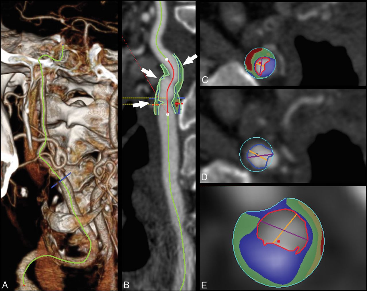
Because of its spatial resolution, CT imaging seems to be a promising tool for the quantification of the volume of the carotid plaque as well as for the volume quantification of plaque subcomponents (fatty, mixed, calcified) (Fig 1). Moreover, the introduction of multienergy technology is opening new options in tissue characterization because the different tissue components show different attenuation levels with varying kiloelectron volt values.132,133

Fig 1.
  • Download figure
  • Open in new tab
  • Download powerpoint
Fig 1.

Plaque volume analysis in a 75-year-old man with a TIA. In the volume-rendered image, the carotid is traced (A), and in the curved-planar-reconstructed postprocessed image (CTA module, Aquarius iNtuition Edition, Version 44121382907; TeraRecon, San Mateo, California) (B), the plaque is identified based on the green contours (white arrows). The volume analysis with automated boundary detection and tissue segmentation is shown in panels C, D, and E (corresponding to the 3 arrows, proximal-to-distal) with contours delineating the lumen (red contour), outer wall (blue contour), and shading of calcium (blue), mixed tissue (green), and lipid component (red).

Luminal Morphology and Ulcerations.

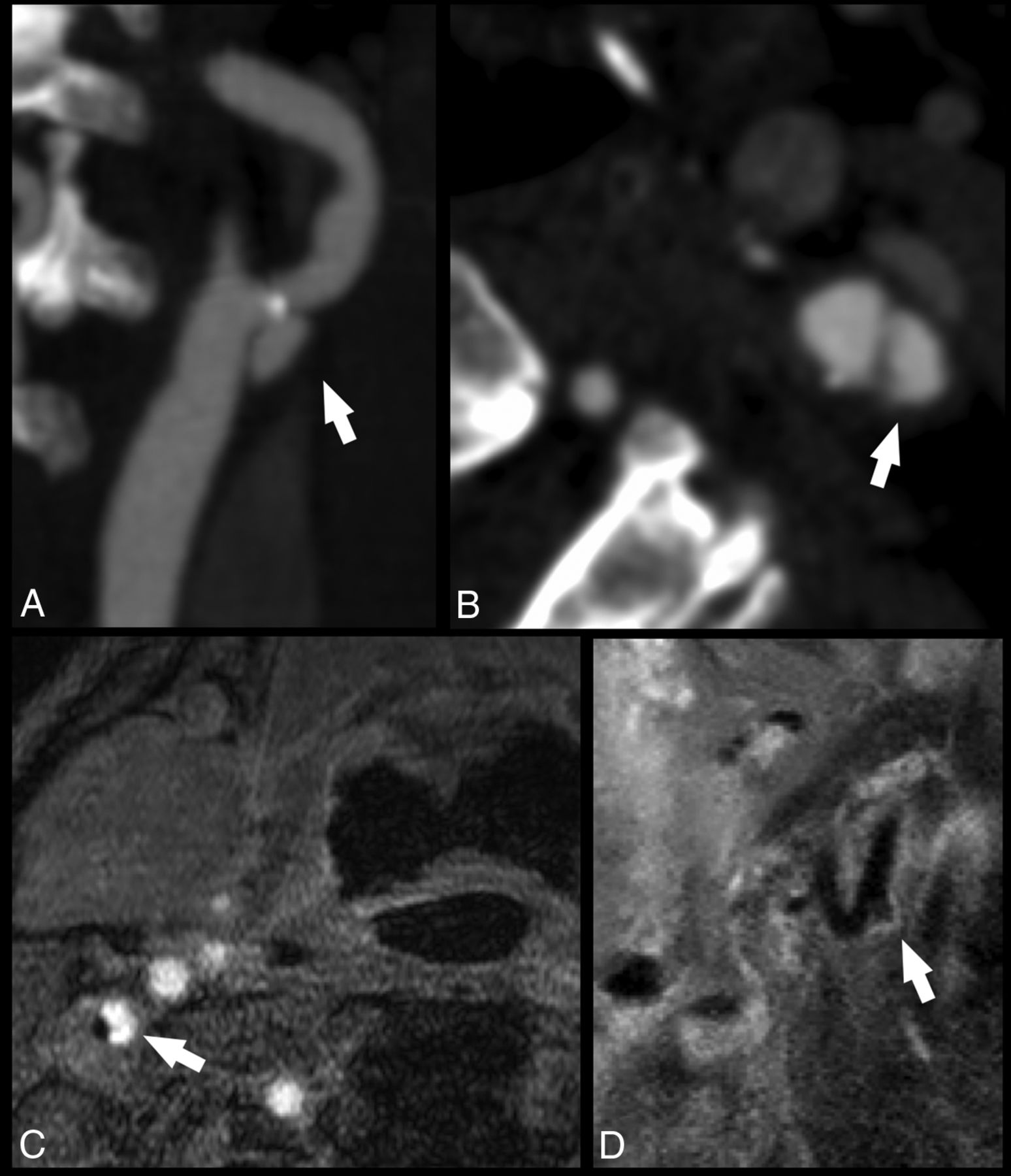
CT offers very good results in detecting ulcerations when compared with histopathology (Fig 2), with performance significantly better than US,134⇓–136 but the presence of a halo or edge blur may hinder detection of smaller ulcerations.

Fig 2.
  • Download figure
  • Open in new tab
  • Download powerpoint
Fig 2.

Ulcerated carotid artery plaques detected with CT and MR imaging. In the first case, the CTA of a 74-year-old man with a TIA demonstrates an ulcerated carotid artery plaque (white arrows) in the left internal carotid artery (white arrow) in the MIP (A) and axial source (B) images. In the second case, an MR imaging analysis of a 63-year-old man with a TIA shows a tiny ulceration (white arrows) in the right internal carotid artery visible in the axial (C) and paracoronal (D) planes.

Intraplaque Hemorrhage.

Detection of IPH using CT is challenging, and conflicting results have been reported. While some authors have found that CT density is slightly higher in fatty plaques with IPH identified by MR imaging compared with plaques without IPH,137 others found no significant differences in Hounsfield units of fatty plaques with and without IPH identified by MR imaging.104 Other authors found a correlation between the presence of IPH and low Hounsfield unit values (<30 HU),56,138,139 which might be explained by the associated presence of LRNC. Recently, some authors suggested that the rim sign on CTA (soft-tissue plaque with adventitial calcifications) as well as maximum soft-plaque thickness could be predictive of carotid IPH.140

Fibrous Cap Status.

The assessment of the status of the FC using CT is complex because of the artifacts related to the edge blur and halo effects, but authors suggest that CT can be used to assess the FC status, in particular to identify rupture.141,142 Notably, it seems that the rupture of the FC correlates with the presence of postcontrast plaque enhancement in CTA analysis.45

Neovascularization and Inflammation.

The degree of postcontrast plaque enhancement has been shown to be associated with the extent of neovascularization on CT.143 The adventitial neovascularization has been assessed with both MR imaging144 and CT.145,146 Romero et al145 showed adventitial neovascularity on CTA to be significantly more common in symptomatic than in asymptomatic patients with ≥70% stenosis and for stenosis between 50% and 70%,146 and similar findings have been reported by MR imaging.144

Plaque Remodeling.

CT studies have found that positive carotid remodeling was significantly greater in patients with cerebral ischemic symptoms than in asymptomatic patients and that the extent of expansive remodeling may indicate underlying atherosclerotic plaque vulnerability.147,148

Plaque Volume.

CT can calculate the volume of the carotid artery plaque and determine the volume of the subcomponents, according to the Hounsfield unit threshold.149 Further, it has been shown in a CT/MR imaging comparison study that the best discriminating factor for predicting a complicated American Heart Association type VI plaque is the thickness of the fatty plaque component with a receiver operating characteristic area under the curve of 0.89.104

CT Limitations.

CT imaging has 3 main limitations: 1) the radiation dose delivered to patients, 2) the risk related to the administration of contrast material, and 3) the limited fatty tissue contrast. Diagnostic radiation exposure and the consequent potential radiation hazards represent a significant issue,150,151 particularly when longitudinal monitoring is required. The second limiting factor of CT is the potential anaphylactic reaction to contrast material152,153 and contrast-induced nephropathy, which is a common form of hospital-acquired acute renal failure.154,155 A further limitation of CT is the limited published information concerning the reliability of this technique and the prospective value of CT-based plaque features on stroke risk and/or stroke recurrence.

MR

The use of a high field strength (1.5T–3T) and dedicated surface radiofrequency coils improves the signal-to-noise ratio, which allows for the evaluation of plaque components and investigation beyond the simple assessment of stenosis measurements. Multicontrast carotid MR imaging (including T1WI, T2WI, and proton density and TOF) can characterize plaque components (eg, fibrous cap, LRNC, calcification, and intraplaque hemorrhage) without administration of contrast agents. Contrast-enhanced MR imaging improves tissue characterization60,61 and offers information on the presence of neovascularization (Figs 3 and 4).

Fig 3.
  • Download figure
  • Open in new tab
  • Download powerpoint
Fig 3.

Carotid atherosclerotic plaque MR imaging and a specimen from a 73-year-old man with stenosis of the carotid bulb measuring 69% by the NASCET criteria demonstrated on a contrast-enhanced MRA (A). The precontrast (mask) image from the contrast-enhanced MRA demonstrates bright signal indicative of intraplaque hemorrhage, specifically subacute blood, or methemoglobin (B, arrow). Subacute blood is also identified as bright signal on the precontrast T1-weighted black-blood image (C, arrow). A rim of hemosiderin is identified as hypointense signal on the postcontrast black-blood image (D) and a hemosiderin-sensitive sequence (E) and is confirmed on the endarterectomy specimen (F and G). The fibrous cap is also delineated (green arrow, D and F). Black-blood imaging was achieved by using 2D cardiac-gated double inversion recovery turbo spin-echo. ECA indicates external carotid artery; CCA, common carotid artery; TFE, turbo field echo.

Fig 4.
  • Download figure
  • Open in new tab
  • Download powerpoint
Fig 4.

Carotid atherosclerotic plaque MR imaging and a specimen from a 76-year-old woman with transient ischemic attacks ipsilateral to carotid bulb stenosis, measuring 47% by the NASCET criteria demonstrated on a contrast-enhanced MRA. A, Narrowing is caused by the plaque characterized by 2D cardiac-gated double inversion recovery black-blood MR imaging (B). Regional enhancement (green arrow) within the lipid core (yellow arrow) suggests focal inflammation with neovascularity as confirmed on the endarterectomy specimen (C, green circle). Contrast enhancement is also useful for delineating the fibrous cap (B and C, orange arrowheads). Calcification is identified as areas of hypointensity (B, red arrows, and C, red circle).

Luminal Morphology and Ulcerations.

MR imaging detects plaque ulcerations with a sensitivity similar to CT (Fig 2).156 Etesami et al156 demonstrated that the use of contrast-enhanced MRA techniques improved the sensitivity for ulcerations by 37.5% compared with an unenhanced time-of-flight sequence.

Intraplaque Hemorrhage.

MR imaging is considered the best imaging technique for the detection of IPH (Figs 3 and 5). Several studies have shown that the appearance of IPH depends on the oxidative state of hemoglobin.157⇓–159 Because of the sensitivity of MR imaging in detecting IPH and the risk attributed to this feature, some authors suggest that MR imaging is the best technique for imaging carotid artery vulnerable plaque.12,40,63,160,161 During the subacute and chronic phases, IPH appears bright on T1-weighted imaging due to the relatively short T1 of methemoglobin. This phenomenon has been exploited using widely available sequences such as MPRAGE, though other heavily T1-weighted techniques have been developed to satisfy this purpose such as multicontrast atherosclerosis characterization (MATCH) and simultaneous noncontrast angiography and intraplaque hemorrhage (SNAP) (Figs 6 and 7). In MATCH, hyper-T1 contrast weighting is achieved using inversion preparation and data acquisition at the background nulling point, and thus IPH can be exclusively visualized with a near-dark background; on the other hand, background tissues can readily be visualized on other contrast weightings, thanks to the inherently coregistered multicontrast acquisition (Fig 7).162,163 IPH detection using 3D SNAP enables greater conspicuity of the lumen boundary compared with MPRAGE (Figs 5⇓–7).164 SNAP provides the advantage of 3D isotropic resolution as well as simultaneous bright-blood angiography to detect stenosis or ulceration that may be colocalized with IPH.165 It is worth noting that it is not necessary to restrict carotid wall imaging to dedicated, small-FOV surface coils for IPH detection since this can be achieved at a lower spatial resolution using large-FOV neck coils (Fig 3).63,79,166 In fact, IPH detection can be achieved on the mask sequence acquired as part of a routine contrast-enhanced MRA (Fig 3).63

Fig 5.
  • Download figure
  • Open in new tab
  • Download powerpoint
Fig 5.

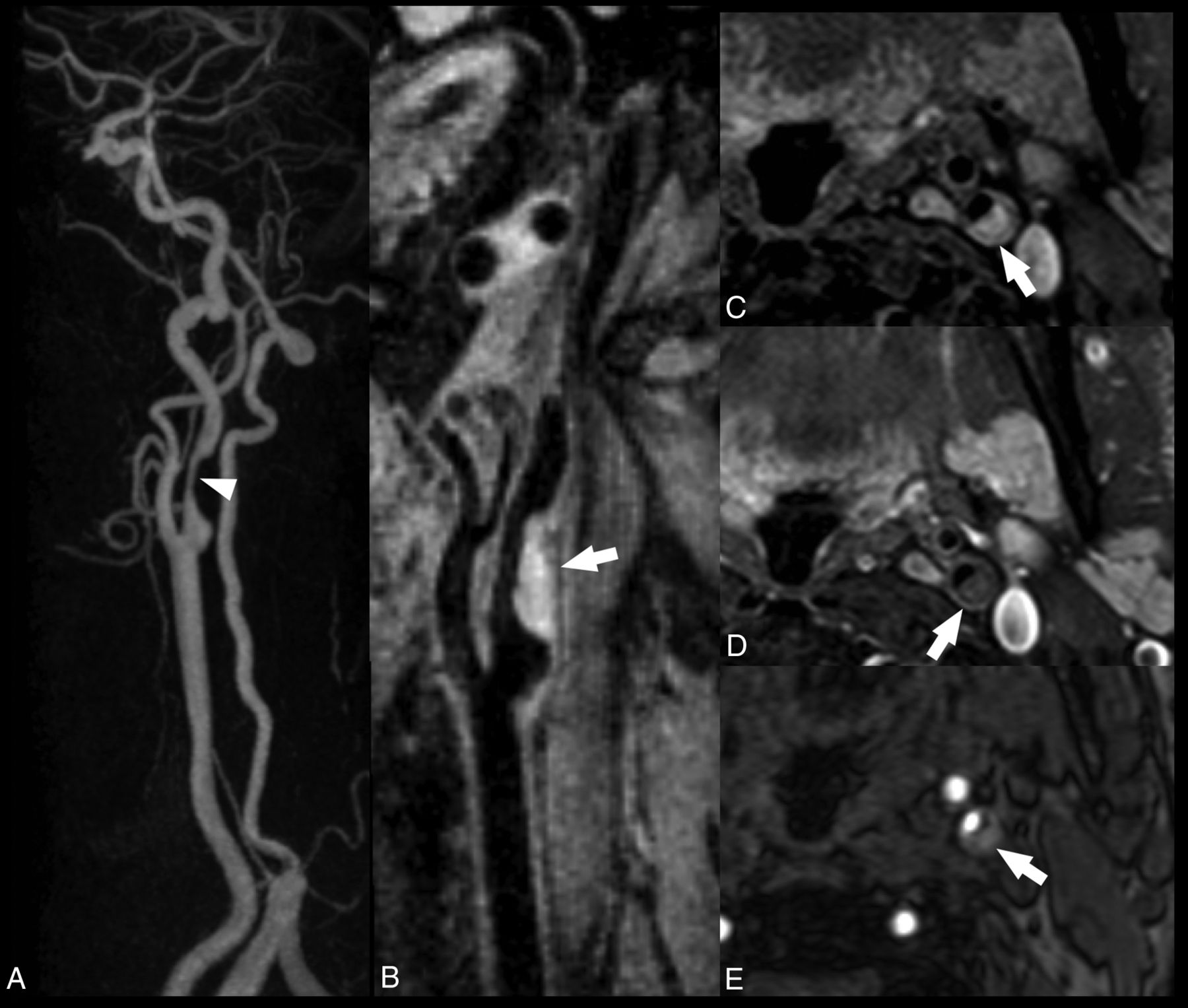
Smooth left internal carotid artery stenosis with intraplaque hemorrhage. All images were acquired with a 16-channel neurovascular coil at 3T. The CE-MRA demonstrates a smooth, nonulcerated stenosis in the bulbous and postbulbous parts of the left internal carotid artery (white arrowhead, A). Oblique reformat of a coronally acquired MPRAGE image shows extensive intraplaque hemorrhage, which appears hyperintense (white arrow, B). The IPH is hyperintense on the nonenhanced T1 fat-saturated spin-echo image (C) and isointense on the gadolinium-enhanced T1 fat-saturated spin-echo image (D). On the TOF MRA source data, the IPH also appears hyperintense but to a lesser degree than the intraluminal flow signal (E).

Fig 6.
  • Download figure
  • Open in new tab
  • Download powerpoint
Fig 6.

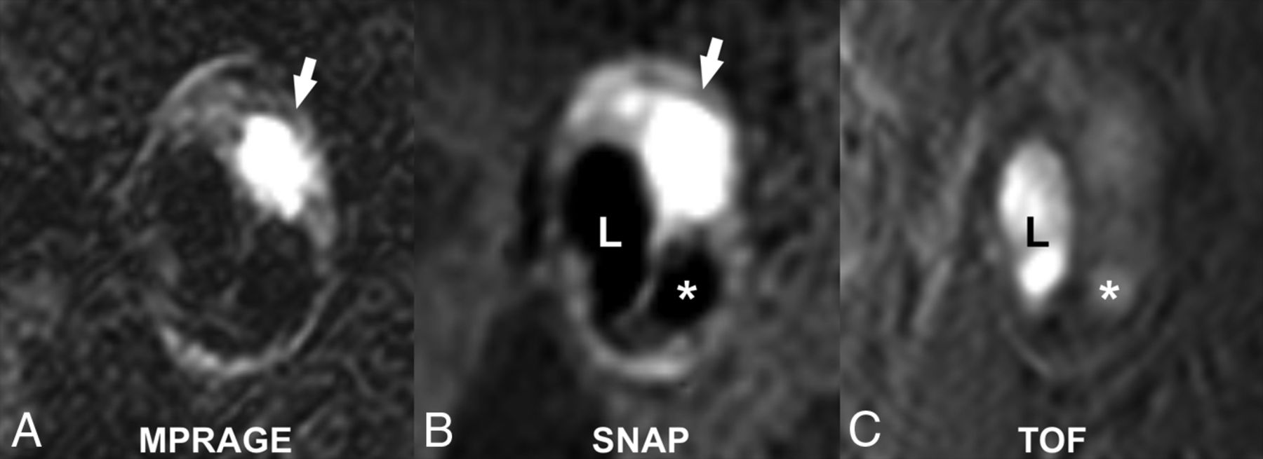
Matched cross-sectional images of a carotid plaque with high signal intensity (white arrows), consistent with the presence of intraplaque hemorrhage on MPRAGE (A) and SNAP MR imaging (B). Note the greater conspicuity of the carotid lumen (L) on SNAP compared with the MPRAGE image. There is a penetrating ulcer (asterisk) that is more easily detected on SNAP compared with the TOF MRA image (C).

Fig 7.
  • Download figure
  • Open in new tab
  • Download powerpoint
Fig 7.

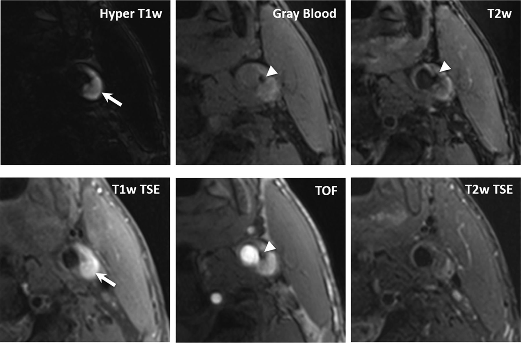
In a 68-year-old male patient, coexistent plaque components, fresh intraplaque hemorrhage (arrows), and superficial calcifications (arrowheads) are detected by MATCH (first row) and the conventional multicontrast protocol (second row). Compared with T1-weighted TSE and TOF, MATCH provides more conspicuous depiction of intraplque hemorrhage on the hyper-T1-weighted image and calcification on the gray blood image. Notice that the calcification is also visible on the MATCH T2-weighted image but not on the T2-weighted TSE image.

Lipid-Rich Necrotic Core.

Initial research demonstrated that LRNC could be detected as a focal hypointense region on T2WI (Fig 8).167,168 Multiple studies have confirmed the improved detection of LRNC seen as a focal nonenhancing region on contrast-enhanced T1WI within the carotid vessel wall.60,61 Larger LRNC size correlates with future ipsilateral carotid symptoms. All validation of LRNC detection/quantification and predictive features has been based on multicontrast carotid plaque MR imaging protocol using dedicated carotid coils. Recent work has suggested the ability to detect a large LRNC using commercially available 3D T1WI sequences and large-FOV neck coils. The Canadian Atherosclerosis Imaging Network has recently completed a prospective, multi-institution study using large-FOV neck coils and commercial sequences from a variety of MR imaging vendors to detect LRNC and IPH. When fully analyzed, CAIN may give us additional information about the ability of IPH and LRNC size detected with large-FOV neck coils and commercial sequences to predict future ipsilateral events.

Fig 8.
  • Download figure
  • Open in new tab
  • Download powerpoint
Fig 8.

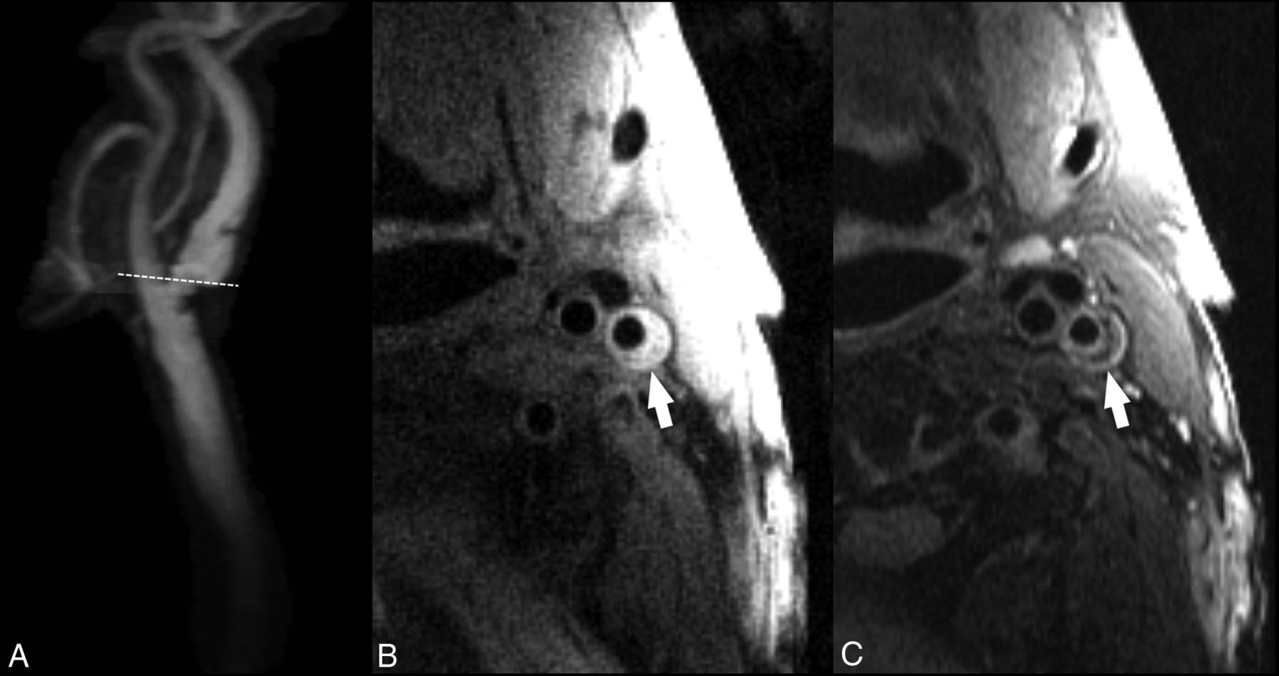
A, Contrast-enhanced MRA of the extracranial carotid bifurcation indicating the level of 2D-FSE images obtained with 1.5T. B, T1-weighted double inversion recovery black-blood FSE image shows an eccentric plaque (arrow) in the internal carotid artery. C, T2-weighted double inversion recovery black-blood FSE image at the same level shows a crescentic, hypointense signal from the necrotic core, which is separated by a higher intensity fibrous cap from the flow lumen.

Fibrous Cap Status.

MR imaging can assess fibrous cap status,61,169 as opposed to the other noninvasive imaging modalities such as CT and US.141 A regular (thick) FC is characterized by the presence of a juxtaluminal band of low signal on time-of-flight MR images and/or a hyperintense juxtaluminal region on contrast-enhanced T1WI, whereas a thin FC is present when this band of low signal on TOF or the hyperintense region on contrast-enhanced (CE)-T1WI is not visible or when the juxtaluminal hyperintense region on CE-T1-weighted MR imaging is interrupted. The fissured fibrous cap is characterized by 2 distinct features: 1) the absence of the juxtaluminal band of low signal, and 2) the presence of a bright gray region adjacent to the lumen, corresponding to plaque hemorrhage and/or mural thrombus.60,170 As shown by Wasserman et al61 and Cai et al,60 contrast-enhanced imaging could be useful for improving delineation of the cap compared with noncontrast (T2), and CE-T1WI can be used to quantify the fibrous cap and the LRNC. Although resolving a thin fibrous cap defined by pathologic criteria would necessitate a higher field strength to overcome signal constraints, distinguishing thin/ruptured from thick fibrous cap thicknesses can be achieved at 1.5T (Fig 8).171,172

Consistent visualization of the FC requires dedicated carotid surface coils. Yuan et al170 and others have shown that the fissured FC has a statistically significant association with the presence of cerebrovascular symptoms81 and is associated with a higher risk of ischemic symptoms in prospective studies.79,80

Neovascularization and Inflammation.

There are new contrast agents using iron particles (ultrasmall superparamagnetic iron oxide or P947)173,174 that can evaluate plaque inflammation via uptake by phagocytic cells within the inflamed vessel wall. Small particle–based MR imaging contrast agents (iron oxide) can be used to evaluate the presence of plaque inflammation. These iron oxide particles enter atherosclerotic plaques, with the agents accumulating in macrophages transformed from blood monocytes attracted by inflammatory mediators.90 High-risk inflamed plaques contain a focal area of signal loss on MR images, due to iron oxide accumulation.91 Iron nanoparticles (10- to 300-nm-sized) are also bound to antibodies, drugs, peptides, and polysaccharides; and avidin-biotin cross-linked with polymers is used to assess endothelial function in animal models. Polymer hydroxyl acidic core (polylactic acid) and dendrimers (polyamidoamine, diaminobutane) have been described as suitable to functionalize the surface of superparamagnetic iron oxide175 (15- to 60-nm superparamagnetic iron oxide) particles, allowing for ligand binding. Ligand-bound superparamagnetic iron oxide (anti-VCAM-1 and anti-E-selectin antibody conjugated superparamagnetic iron oxide) can cause dephasing and loss of T2* signal intensity due to susceptibility effects and is suitable for passive targeted imaging of inflammation in cardiovascular tissue.175 In addition to iron oxide nanoparticles, various other nanoparticles are being used for molecular imaging of atherosclerosis in animal models, eg, liposome vesicles (50–70 nm) for US176/MR imaging177; perfluorocarbon core emulsions (200–300 nm) for MR imaging, US, fluorescence, and nuclear imaging; chemo-thermo-immuno178; and high-density lipoprotein and low-density lipoprotein micelles for MR imaging.179 Other types of particles such as gold, carbon nanotube fullerenes (4 nm), quantum dots cadmium selenide spheres (2–10 nm), and metal-based agents are in the process of standardization and may be useful in fluorescent imaging.180 Moreover, other investigators have reported the possibility of viral capsid protein cages with gadolinium as potential nanospheres for drug encapsulation and imaging.181

A recent MR imaging study182 showed that enhancement of carotid plaque after administration of gadolinium is associated with neovascularization (P < .001) (Fig 4). The correlation between the degree of plaque enhancement and the degree of neovascularization, which is itself linked to the degree of plaque inflammation, was also confirmed at histology in a recent study by Millon et al.182 Investigations have shown that inflammatory cells are also present at the interface with the underlying necrotic core and in the plaque shoulder region.183,184 From the imaging point of view, it is possible to distinguish 2 different types of neovascularization: 1) adventitial neovascularization, and 2) intraplaque neovascularization. The adventitial neovascularization has been assessed with MR imaging.144 Ectopic neovascularization in the intima and media is a hallmark of advanced atherosclerotic lesions, but the adventitial layer is a fundamental target because it serves as the main source of in-growth of new vessels. The degree of neovascularity measured using gadolinium perfusion methods correlated with adventitial perfusion as measured by its transfer constant (Fig 9).185 Wasserman171 categorized the circumferential enhancement (0, absent; 1, <50%; 2, ≥50%) on postcontrast MR imaging by finding an association between the grade of adventitial enhancement and cerebrovascular events. Plaque perfusion imaging using dynamic contrast-enhanced MR imaging has been shown to give reproducible physiologic measurements of the vasa vasorum.186,187 However, protocol compliance may be more important for functional imaging such as dynamic contrast-enhanced MR imaging as compared with anatomic imaging.

Fig 9.
  • Download figure
  • Open in new tab
  • Download powerpoint
Fig 9.

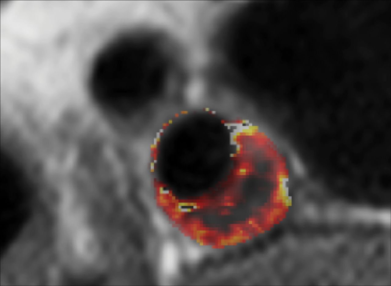
Volume transfer constant (Ktrans) map of a patient with carotid plaque. Maps were generated using pharmacokinetic modeling of dynamic contrast-enhanced MR images. The parametric map is overlaid on the anatomic MR image, and voxel Ktrans values (Patlak model) are color-coded. The necrotic core exhibits low Ktrans values at the center of the plaque, while the highly vascularized adventitia at the outer rim exhibits high Ktrans values. There is another region of higher Ktrans values near the inner rim of the plaque.

Plaque Volume.

Recently published studies show the utility of MR imaging for this type of quantification.103,188 In general, the reliability of MR imaging for plaque assessment has been very good. A study published by Wasserman in 2010103 found that the scan reliability for common carotid artery lumen area was 0.94, whereas for the ICA lumen area, it was 0.89. In the assessment of the total wall volume, the value was 0.79, but in the assessment of LRNC volume, the value was very low (0.3). The authors found that overall reliability is primarily related to reader variability rather than scan acquisition. The coefficient of variation values for the plaque area or plaque volume are between 3% and 6%, as demonstrated by Saam et al.189,190

Imaging studies have documented changes in atherosclerotic plaque volume and composition and progression of subclinical lesions into rupture-prone plaques.191⇓⇓⇓⇓–196 The ability to monitor these changes might contribute to our ability to estimate risk and assess pharmaceutical treatment efficacy.197 For example, changes in plaque structure that correspond with a clinical event help to identify that plaque as a culprit lesion, which puts it at a higher risk for future stroke.34,122,198 Several studies have reported using MR imaging for longitudinal analysis of carotid plaque variations,191⇓–193 with fewer reports using CTA.194,195

MR Imaging Limitations.

An important limitation to contrast-enhanced MR imaging evaluation of plaque that has recently emerged is the potential for gadolinium toxicity, particularly when longitudinal monitoring is required. Recent studies have reported the accumulation of gadolinium in various tissues of patients without renal impairment, including in bone, brain, and kidneys,199⇓–201 and in July 2015, the US Food and Drug Administration published a safety announcement that it is investigating the risk of brain deposits associated with the repeat use of gadolinium contrast agents in MR imaging,202 stating: “To reduce the potential for gadolinium accumulation, health care professionals should consider limiting GBCA [gadolinium-based contrast agents] use to clinical circumstances in which the additional information provided by the contrast is necessary. Health care professionals are also urged to reassess the necessity of repetitive GBCA MRIs in established treatment protocols.” This risk must now be weighed against the potential radiation hazard described earlier that limits longitudinal plaque monitoring by CT.

Advanced Algorithms to Carotid Artery Plaque Characterization

With the development of deep learning technology and plaque characterization algorithms applied to medical imaging, it is now possible to identify, classify, and quantify target features from imaging datasets such as total carotid artery plaque volume and plaque subcomponent detection (calcium, IPH, lipid core).203,204 Deep-learning technology has experienced rapid progress in health care over recent years, with early reports of implementation in carotid imaging204 raising the prospect of routine use in the clinical setting once validated.

Functional–Molecular Imaging

“Molecular” imaging techniques have been gaining popularity. The objective of molecular imaging is to provide biologic insight into the identification and classification of carotid artery plaques, especially those at high risk. In atherosclerotic plaques, multiple and complex reactions take place at the molecular and cellular level, with various atherosclerosis-related biomarkers present at different stages of disease progression.205 Conventional imaging with US, MR, or CT cannot identify these components because of limited imaging contrast; therefore, several methods have been proposed that use external contrast agents targeting these specific biomarkers.

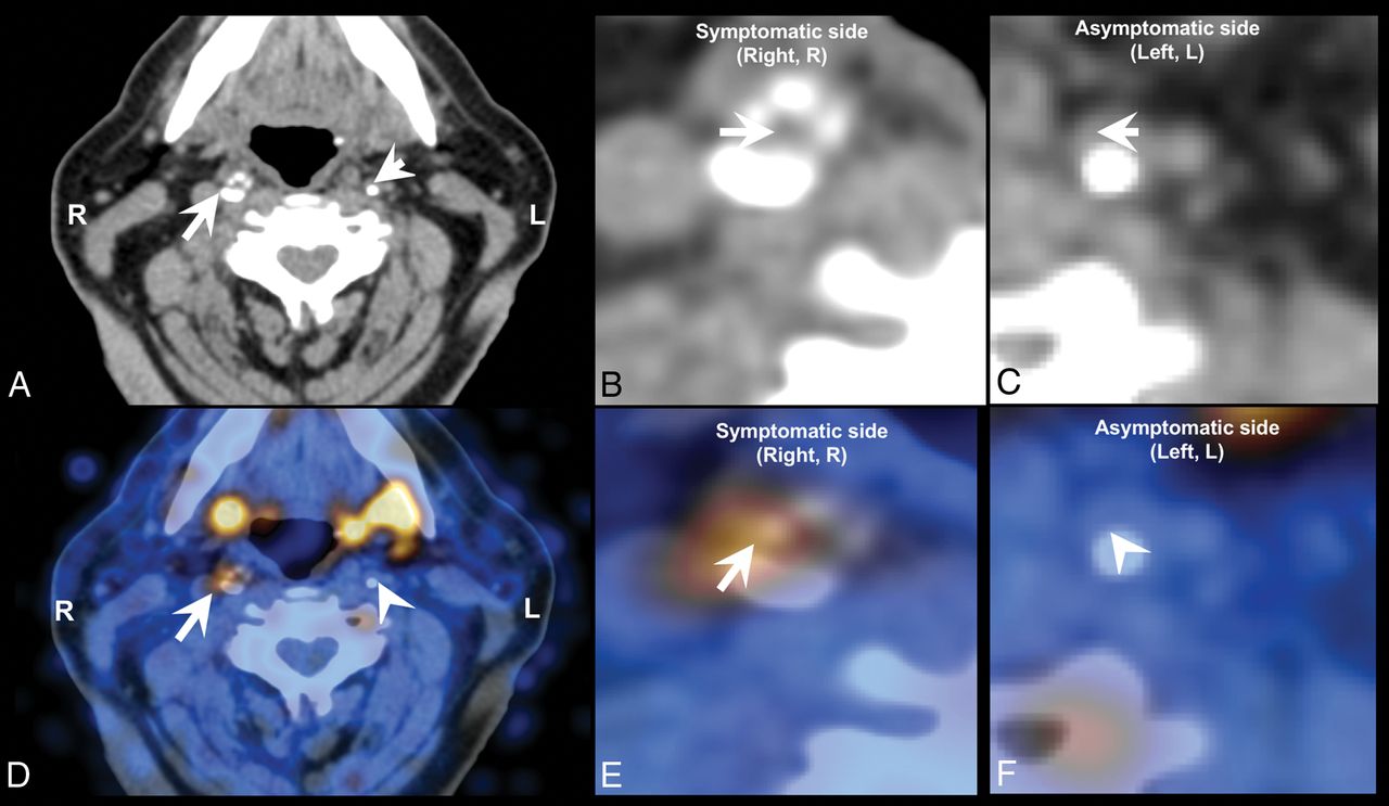
A wide variety of studies has assessed the diagnostic potential of nuclear medicine techniques for imaging and quantifying plaque inflammation, such as by PET using the widely available [18F] FDG or newer radiotracers such as [18F] fluorocholine (Fig 10).206⇓–208 Nuclear medicine tracer techniques have also shown efficacy in the identification of neovascularization.209,210

Fig 10.
  • Download figure
  • Open in new tab
  • Download powerpoint
Fig 10.

[18F] fluorocholine positron-emission tomography CT (18F-FCH PET CT) image of a symptomatic (arrow) and contralateral asymptomatic (arrowhead) carotid plaque of a patient who experienced right-sided stroke. A, Diagnostic contrast-enhanced CT shows a significant stenosis in the right internal carotid artery because of a calcified plaque, whereas a noncalcified atherosclerotic plaque can be seen on the contralateral internal carotid artery. B, CT, inset on the symptomatic plaque. C, CT, inset on the asymptomatic plaque. D, The fused PET CT image denotes a focal area of high [18F] FCH uptake in the right symptomatic carotid plaque, whereas there is no visible [18F] FCH uptake in the left asymptomatic carotid plaque. E, Fused PET CT, inset on the symptomatic plaque. F, Fused PET-CT, inset on the asymptomatic plaque.

Because vulnerable plaques are infiltrated by lymphocytes and macrophages, with the latter cell population capable of taking up [18F] FDG from the interstitial spaces, [18F] FDG PET can be used to directly detect plaque inflammation in various anatomic locations.211 In recent years, a number of studies have assessed the diagnostic potential of [18F] FDG PET to image and quantify plaque inflammation206,207 as well as monitoring the reduction of plaque inflammation resulting from statin therapy.208

Recommendations

Carotid MRI

Background.

Results from recently published meta-analyses support the hypothesis that MR imaging detection of carotid IPH is associated with increased risk for future primary and recurrent ischemic neurologic events.79,212,213 Furthermore, the absence of IPH portends a benign clinical course, even among patients with symptomatic 50%–99% carotid stenosis.213 Other plaque features associated with increased risk include identification of a large LRNC and a thin or ruptured fibrous cap.79

Goals.

1) To provide general guidelines for carotid MR vessel wall imaging with recommended imaging sequences, spatial resolution, and coverage. Guideline considerations are that the protocol can be applied broadly across a spectrum of clinical scanners and not require specialized software or research keys for implementation, and 2) to recommend future areas for technical development and clinical expansion needs.

Essential Features for Identification with Carotid Plaque Imaging.

Any MR imaging protocol for plaque imaging should be able to identify the following atherosclerotic plaque characteristics:

  1. Stenosis and luminal surface condition (fibrous cap and ulceration).

  2. Presence of intraplaque hemorrhage.

  3. Presence of lipid rich necrotic core.

  4. Plaque burden and distribution.

Minimum MR Imaging Protocol Requirements for Identification of Essential Plaque Features (1.5T and 3T).

Recommended minimum sequence requirements are the following:

  • Resolution: In-plane 0.6 mm, through-plane 2 mm

  • Longitudinal coverage: 3–4 cm centered on the carotid bifurcation

  • Effective blood suppression for a plaque burden visualization sequence.

The protocol may include any combination of sequences that meets the minimum requirements set forth above. The sequences used can be either 2D or 3D or a combination, provided that they together meet the minimum sequence requirements above. Overall scan time can be adjusted based on field strength and the availability of specialized hardware such as carotid phased array coils. 3T scanners are recommended for improved SNR.

Example Protocols.

Four protocols are presented based on considerations for 2D and 3D imaging and the use of gadolinium contrast agents (Tables 1⇓⇓–4). If patients are able to undergo gadolinium contrast injection, its use is recommended for the detection and quantification of LRNC and the delineation of the fibrous cap.60,214 Use of large-coverage 3D sequences can detect plaques extending beyond the 4-cm coverage centered on the bifurcation and is preferable. Carotid coils are recommended for use with all protocols, though large-FOV neck coils can detect IPH. It is possible to add a 4-minute 3D MPRAGE sequence to routine clinical carotid MRA protocol. The protocol is similar to Table 1, but with 0.8-mm isotropic resolution using a large-FOV neck coil instead of 0.6 mm using dedicated carotid surface coils. Focal regions of T1 hyperintensity within the carotid plaque that are 15× greater than the adjacent sternocleidomastoid muscle can be used to identify IPH.

View this table:
  • View inline
  • View popup
Table 1:

3D Noncontrast protocol

View this table:
  • View inline
  • View popup
Table 2:

3D contrast protocol

View this table:
  • View inline
  • View popup
Table 3:

2D noncontrast protocol

View this table:
  • View inline
  • View popup
Table 4:

2D contrast protocol

Motion-sensitized driven-equilibrium215/flow-sensitive dephasing216 flow suppression is required for 3D sampling perfection with application-optimized contrasts using different flip angle evolution (SPACE; Siemens, Erlangen, Germany)/Cube (GE Healthcare, Milwaukee, Wisconsin)/volume isotropic turbo spin-echo acquisition (VISTA; Philips Healthcare, Best, the Netherlands) to ensure effective blood suppression to accurately identify plaque lumen boundaries. Good blood suppression postcontrast requires the use of motion-sensitized driven-equilibrium or double inversion recovery (DIR)/quadruple inversion recovery217 flow suppression. For DIR, the inversion time can be calculated based on estimated T1 values of blood at 5-minute intervals following contrast administration (0.1 mmol/kg) for 1.5T or 3T scanners.218 We recommend a TI of 250 ms for 3T scanners for a TR triggered at a 1 RR interval, which generally produces adequate flow suppression beginning 5 minutes after injection despite variations in postinjection scan time and heart rate, which will affect the T1 blood values.

Discussion.

Lumen. Quantifying luminal narrowing is a prerequisite, as stenosis severity is the cornerstone for treatment decisions in current clinical guidelines. Furthermore, detection of ulceration provides prognostic value. Use of TOF MRA for lumen assessment avoids the need for IV contrast and may provide confirmatory evidence of intraplaque hemorrhage and sometimes calcification. Addition or substitution with CE-MRA should be considered for those without contraindication for contrast administration. This would also provide an opportunity to perform post-contrast-enhanced imaging of the vessel wall for direct identification of the LRNC and identification/confirmation of fibrous cap status and ulcerations.

IPH. MR imaging techniques are available for IPH detection across scanner platforms, and the predictive value of IPH for ischemic events has been extensively evaluated, both with and without custom carotid coils. In a review performed by Gupta et al,79 studies were stratified by those utilizing multisequence, carotid coil–dependent protocols and those using a single sequence with standard large-FOV neck coils for IPH detection. Using either technique, IPH was associated with significantly increased risk for TIA or stroke (hazard ratio, 440; 95% CI, 210–923; and hazard ratio, 504; 95% CI, 215-1185, respectively). While IPH can be identified on T1WI sequences such as T1WI fast spin-echo, T1WI SPACE, TOF, and so forth, a highly T1-weighted sequence such as MPRAGE can provide higher sensitivity and specificity for IPH detection.219

Lipid-Rich Necrotic Core. T2-weighted imaging can be used to detect the presence of LRNC.167,168 Direct assessment of the LRNC can also be done in patients undergoing contrast administration using a postcontrast T1WI scan. CE-MRA followed by post-CE vessel wall imaging in patients without contraindication will improve detection and quantification of the LRNC and delineation of the fibrous cap.214

Plaque Burden and Distribution. Knowledge of the location and distribution of plaque assists in preprocedural planning. Time-efficient 3D large-coverage black-blood MR imaging may be better suited for this purpose.

Future Improvements and Needs for MR Imaging.

Technical developments are urgently needed in the following areas:

  1. Improved spatial resolution both in-plane and through-plane to better characterize finer structures such as fibrous caps.

  2. More effective blood flow suppression for large-spatial-coverage imaging acquisition and pre- and postcontrast administration.

  3. Dedicated carotid coils that are integrated with head and neck coils for extensive coverage.

  4. Improved techniques for identifying the lipid rich necrotic core, especially without the need for contrast application.

  5. More effective methods to deal with motion.

  6. A streamlined imaging protocol that is able to identify multiple imaging targets in 1 or 2 imaging sequences.

  7. Effective image-processing tools for efficient quantitative identification of imaging targets.

  8. Development of training programs for MR imaging specialists on image acquisition and for radiologists on vessel wall image interpretation.

  9. Ultimately, a guideline that clearly calls for the need for carotid plaque imaging and 1 simple protocol that can meet all the needs.

Currently, there are many new techniques being developed for carotid plaque imaging. 3D-SNAP provides non-contrast-enhanced MRA and simultaneous IPH detection.164 3D spoiled gradient-recalled echo pulse sequence for hemorrhage assessment using inversion recovery and multiple echoes (3D-SHINE) provides information about the state of IPH in addition to IPH detection.220 IPH can also be identified on a precontrast mask of CE-MRA if available.63 MATCH provides comprehensive information regarding plaque composition in a single sequence.163 3D Multiple Echo Recombined Gradient Echo (3D-MERGE)221 and 3D delay alternating with nutation for tailored excitation with fast low-angle shot (3D-DASH)222 provide large-coverage blood suppression for plaque burden measurements. Diffusion-weighted imaging can detect LRNC without the use of contrast media.223 Self-gating has been used to reject data acquired during swallowing motion.224 T1-insensitive blood-suppression techniques such as quadruple inversion recovery217 provide good blood suppression for postcontrast imaging. However, these supplementary techniques require specialized equipment (3T, custom carotid coils, custom sequences) and more intensive interpreter training.

Carotid CT

Background.

Currently no meta-analyses or prospective trials have suggested that some specific CT features are associated with an increased risk for future primary and recurrent ischemic neurologic events, even if there are several prospective trials on their way or that have been published that examine the value of plaque imaging prospectively (PARISK, CAPIAS, CARE-II).104⇓–106 However, cross-sectional studies have found that some CT characteristics (Hounsfield unit attenuation, the presence of neovascularization) are associated with increased risk of cerebrovascular events.48,225

Goals.

1) To provide general guidelines for carotid CT vessel wall imaging with recommended desirable imaging techniques, tissue contrast, spatial resolution, and coverage. Guideline considerations are that the protocol can be applied broadly across a spectrum of clinical CT scanners and not require specialized software or research keys for implementation. 2) To recommend future areas for technical development and clinical expansion needs.

Essential Features for Identification with Carotid Plaque Imaging.

Any CT protocol for plaque imaging should be able to identify the following atherosclerotic plaque characteristics:

  1. Stenosis and luminal surface condition (plaque morphology and ulceration).

  2. Type of plaque (fatty versus mixed versus calcified).

  3. Presence of plaque enhancement.

  4. Plaque burden and distribution.

Minimum CT Protocol Requirements for Identification of Essential Plaque Features.

Recommended minimum parameter requirements are the following:

  • Resolution: isotropic voxel with 1-mm resolution

  • Longitudinal coverage: from the aortic arch to intracranial vessels

  • CT generation: third with at least 16-detector-row.

Example Protocols.

Four protocols are presented (Tables 5⇓⇓–8). No CT study of carotid arteries must be performed without the administration of contrast material. The use of a biphasic approach (unenhanced scan followed by a contrast scan) allows the assessment of the carotid plaque neovascularization. This is becoming more important but is not considered currently necessary. To reduce the radiation dose delivered to the patients, the z-length of the basal scan should cover only the carotid artery plaque bifurcation (4-cm coverage centered on the bifurcation). The dual-energy CT technique143 allows a virtual unenhanced image to assess plaque enhancement without the need for a biphasic approach.

View this table:
  • View inline
  • View popup
Table 5:

Aquilion Visiona

View this table:
  • View inline
  • View popup
Table 6:

Somatom Sensation 64a

View this table:
  • View inline
  • View popup
Table 7:

ICT

View this table:
  • View inline
  • View popup
Table 8:

LightSpeed VCTa

Discussion.

Lumen. Quantifying luminal narrowing is a prerequisite. To correctly assess the degree of stenosis, by avoiding the halo or edge blur, the correct window settings should be used.226 At the current level of technology, the status of the FC cannot be adequately explored by CT.

Type of Plaque. According to Hounsfield unit attenuation, the carotid plaque can be categorized as fatty (<60 HU), mixed (between 60 and 130 HU), and calcified (>130 HU). By applying these thresholds, it is possible to identify those plaques with a LRNC from others. Applying the Hounsfield unit classification, however, creates 2 problems that have recently come to light: 1) The Hounsfield unit value of the plaque is dependent on the level of energy applied, as demonstrated by Saba et al132 using multienergy systems, and 2) the carotid artery plaques may show contrast enhancement (by comparing the attenuation values of the basal and postcontrast scans), suggesting that the attenuation value of the plaque obtained after administration of contrast material represents 2 different parameters: the type of the plaque and the degree of neovascularization of the tissue.143,227 This is not a problem if pre- and postcontrast scans or dual-energy is applied (capable of distinguishing plaque from contrast enhancement), but this is not usually done clinically secondary to an increase in x-ray dose.

Carotid Plaque Enhancement. Assessment of plaque enhancement is limited in the case of single-phase CTA, and multiphase CTA is rarely performed outside of research studies due to radiation concerns. An unenhanced axial CT scan obtained over 4 cm centered on the carotid bifurcation, followed by CTA, would theoretically be ideal in assessing plaque enhancement but carries a greater radiation penalty. Alternatively, some authors have employed dual-energy techniques with the use of the virtual nonenhanced image to assess plaque enhancement with less radiation dose.143

Plaque Burden and Distribution. Knowledge of the location and distribution of plaque assists in preprocedural planning. Moreover, CT can calculate the volume of the carotid artery plaque and determine the volume of the subcomponents, according to the Hounsfield unit threshold.149

Future Improvements and Needs for CT.

Technical developments are urgently needed in the following areas:

  1. Improved contrast resolution for greater discrimination of tissue types in plaque.

  2. Improved techniques such as multienergy applications for identifying the lipid-rich necrotic core, especially without the need for contrast application.

  3. Evidence-based guidelines that invoke the need for carotid plaque imaging, preferably using 1 simple universal protocol that can meet all needs.

Currently, most of the research on carotid artery CT is focusing on methods that 1) reduce the radiation dose delivered to the patients, and 2) improve carotid artery plaque characterization using multienergy tools that promise more accurate detection of plaque components.

Conclusions

In the last 20 years, there has been a paradigm shift in the imaging of the atherosclerotic carotid artery, from the assessment of the degree of luminal stenosis to the characterization of plaque. Several features have been identified that are potentially associated with plaque rupture, and imaging has been used to identify these features in vivo.

Researchers and clinicians now have several imaging modalities that allow in-depth exploration of carotid artery plaque and its components. Sonography should be considered as a first-line examination, at least for screening, whereas CT and MR imaging improve identification of several plaque features associated with vulnerability.

Also promising are nuclear medicine and molecular imaging techniques that can further explore assessment of plaque vulnerability, especially inflammation, but these approaches are still investigational and not part of the main diagnostic algorithm of carotid atherosclerosis. In the future, larger prospective longitudinal studies investigating these technologic advances may fully exploit the clinical potential of vessel wall imaging.

Acknowledgments

The authors would like to thank Drs Stefan Vöö and Wei Zu for their help in image selection for this article.

Footnotes

  • The views expressed in this article are those of the authors and do not reflect the official policy of the Department of Army/Navy/Air Force, Department of Defense, or US Government. The identification of specific products or scientific instrumentation does not constitute endorsement or implied endorsement on the part of the authors, Department of Defense, or any component agency.

References

  1. 1.↵
    1. Yanez ND,
    2. Burke GL,
    3. Manolio T, et al
    ; CHS Collaborative Research Group. Sibling history of myocardial infarction or stroke and risk of cardiovascular disease in the elderly: the Cardiovascular Health Study. Ann Epidemiol 2009;19:858–66 doi:10.1016/j.annepidem.2009.07.095 pmid:19944349
    CrossRefPubMed
  2. 2.↵
    1. Truelsen T,
    2. Begg S,
    3. Mathers C
    . The global burden of cerebrovascular disease. Geneva: World Health Organization; 2000. http://www.who.int/healthinfo/statistics/bod_cerebrovasculardiseasestroke.pdf. Accessed December 6, 2017.
  3. 3.↵
    1. Ooi YC,
    2. Gonzalez NR
    . Management of extracranial carotid artery disease. Cardiol Clin 2015;33:1–35 doi:10.1016/j.ccl.2014.09.001 pmid:25439328
    CrossRefPubMed
  4. 4.↵
    1. Barnett HJ,
    2. Taylor DW,
    3. Eliasziw M, et al
    . Benefit of carotid endarterectomy in patients with symptomatic moderate or severe stenosis: North American Symptomatic Carotid Endarterectomy Trial Collaborators. N Engl J Med 1998;339:1415–25 doi:10.1056/NEJM199811123392002 pmid:9811916
    CrossRefPubMed
  5. 5.↵
    1. Howarth JC,
    2. Klotz JG
    . The diagnostic value of carotid arteriography: a preliminary report. Cleve Clin Q 1951;18:179–83 doi:10.3949/ccjm.18.3.179 pmid:14848967
    CrossRefPubMed
  6. 6.↵
    1. von Arbin M,
    2. Britton M,
    3. de Faire U, et al
    . Non invasive assessment of the internal carotid artery in stroke patients. Scand J Clin Lab Invest 1983;43:275–83 doi:10.3109/00365518309168257 pmid:6635531
    CrossRefPubMed
  7. 7.↵
    1. Weinberger J
    . Clinical applications of noninvasive carotid artery testing. J Am Coll Cardiol 1985;5:137–48 doi:10.1016/S0735-1097(85)80097-8 pmid:3880566
    FREE Full Text
  8. 8.↵
    1. Imparato AM,
    2. Riles TS,
    3. Gorstein F
    . The carotid bifurcation plaque: pathologic findings associated with cerebral ischemia. Stroke 1979;10:238–45 doi:10.1161/01.STR.10.3.238 pmid:462508
    Abstract/FREE Full Text
  9. 9.↵
    1. Redgrave JN,
    2. Lovett JK,
    3. Gallagher PJ, et al
    . Histological assessment of 526 symptomatic carotid plaques in relation to the nature and timing of ischemic symptoms: the Oxford plaque study. Circulation 2006;113:2320–28 doi:10.1161/CIRCULATIONAHA.105.589044 pmid:16651471
    Abstract/FREE Full Text
  10. 10.↵
    1. Jeziorska M,
    2. Woolley DE
    . Local neovascularization and cellular composition within vulnerable regions of atherosclerotic plaques of human carotid arteries. J Pathol 1999;188:189–96 pmid:10398163
    CrossRefPubMed
  11. 11.↵
    1. Choi CJ,
    2. Kramer CM
    . MR imaging of atherosclerotic plaque. Radiol Clin North Am 2002;40:887–98 doi:10.1016/S0033-8389(02)00019-2 pmid:12171190
    CrossRefPubMed
  12. 12.↵
    1. Moody AR
    . Magnetic resonance direct thrombus imaging. J Thromb Haemost 2003;1:1403–09 doi:10.1046/j.1538-7836.2003.00333.x pmid:12871274
    CrossRefPubMed
  13. 13.↵
    1. Oliver TB,
    2. Lammie GA,
    3. Wright AR, et al
    . Atherosclerotic plaque at the carotid bifurcation: CT angiographic appearance with histopathologic correlation. AJNR Am J Neuroradiol 1999;20:897–901 pmid:10369363
    Abstract/FREE Full Text
  14. 14.↵
    1. Bluth EI
    . Evaluation and characterization of carotid plaque. Semin Ultrasound CT MR 1997;18:57–65 doi:10.1016/S0887-2171(97)90038-X pmid:9143066
    CrossRefPubMed
  15. 15.↵
    1. Naghavi M,
    2. Libby P,
    3. Falk E, et al
    . From vulnerable plaque to vulnerable patient: a call for new definitions and risk assessment strategies, part II. Circulation 2003;108:1772–88 doi:10.1161/01.CIR.0000087481.55887.C9 pmid:14557340
    Abstract/FREE Full Text
  16. 16.↵
    1. Naghavi M,
    2. Libby P,
    3. Falk E, et al
    . From vulnerable plaque to vulnerable patient: a call for new definitions and risk assessment strategies, part I. Circulation 2003;108:1664–72 doi:10.1161/01.CIR.0000087480.94275.97 pmid:14530185
    Abstract/FREE Full Text
  17. 17.↵
    1. Zavodni AE,
    2. Wasserman BA,
    3. McClelland RL, et al
    . Carotid artery plaque morphology and composition in relation to incident cardiovascular events: the Multi-Ethnic Study of Atherosclerosis (MESA). Radiology 2014;271:381–89 doi:10.1148/radiol.14131020 pmid:24592924
    CrossRefPubMed
  18. 18.↵
    1. van Dijk AC,
    2. Truijman MT,
    3. Hussain B, et al
    . Intraplaque hemorrhage and the plaque surface in carotid atherosclerosis: the Plaque At RISK Study (PARISK). AJNR Am J Neuroradiol 2015;36:2127–33 doi:10.3174/ajnr.A4414 pmid:26251429
    Abstract/FREE Full Text
  19. 19.↵
    1. Zhao XQ,
    2. Hatsukami TS,
    3. Hippe DS, et al
    ; AIM-HIGH Carotid MRI Sub-study Investigators. Clinical factors associated with high-risk carotid plaque features as assessed by magnetic resonance imaging in patients with established vascular disease (from the AIM-HIGH Study). Am J Cardiol 2014;114:1412–19 doi:10.1016/j.amjcard.2014.08.001 pmid:25245415
    CrossRefPubMed
  20. 20.↵
    1. Underhill HR,
    2. Yuan C
    . Carotid MRI: a tool for monitoring individual response to cardiovascular therapy? Expert Rev Cardiovasc Ther 2011;9:63–80 doi:10.1586/erc.10.172 pmid:21166529
    CrossRefPubMed
  21. 21.↵
    1. Xu D,
    2. Hippe DS,
    3. Underhill HR, et al
    . Prediction of high-risk plaque development and plaque progression with the carotid atherosclerosis score. JACC Cardiovasc Imaging 2014;7:366–73 doi:10.1016/j.jcmg.2013.09.022 pmid:24631510
    Abstract/FREE Full Text
  22. 22.↵
    1. DeMarco JK,
    2. Spence JD
    . Plaque assessment in the management of patients with asymptomatic carotid stenosis. Neuroimaging Clin N Am 2016;26:111–27 doi:10.1016/j.nic.2015.09.009 pmid:26610664
    CrossRefPubMed
  23. 23.↵
    1. Croft RJ,
    2. Ellam LD,
    3. Harrison MJ
    . Accuracy of carotid angiography in the assessment of atheroma of the internal carotid artery. Lancet 1980;1:997–1000 pmid:6103388
    PubMed
  24. 24.↵
    1. Wardlaw JM,
    2. Chappell FM,
    3. Best JJ, et al
    ; NHS Research and Development Health Technology Assessment Carotid Stenosis Imaging Group. Non-invasive imaging compared with intra-arterial angiography in the diagnosis of symptomatic carotid stenosis: a meta-analysis. Lancet 2006;367:1503–12 pmid:16679163
    CrossRefPubMed
  25. 25.↵
    1. Liapis CD,
    2. Paraskevas KI
    . Do carotid surface irregularities correlate with the development of cerebrovascular symptoms? An analysis of the supporting studies, the opposing studies, and the possible pathomechanism. Vascular 2006;14:88–92 doi:10.2310/6670.2006.00016 pmid:16956477
    CrossRefPubMed
  26. 26.↵
    1. Comerota AJ,
    2. Katz ML,
    3. White JV, et al
    . The preoperative diagnosis of the ulcerated carotid atheroma. J Vasc Surg 1990;11:505–10 pmid:2182913
    CrossRefPubMed
  27. 27.↵
    1. Rothwell PM,
    2. Eliasziw M,
    3. Gutnikov SA, et al
    ; Carotid Endarterectomy Trialists' Collaboration. Analysis of pooled data from the randomised controlled trials of endarterectomy for symptomatic carotid stenosis. Lancet 2003;361:107–16 doi:10.1016/S0140-6736(03)12228-3 pmid:12531577
    CrossRefPubMed
  28. 28.↵
    European Carotid Surgery Trialists' Collaborative Group. Randomised trial of endarterectomy for recently symptomatic carotid stenosis: final results of the MRC European Carotid Surgery Trial (ECST). Lancet 1998;351:1379–87 doi:10.1016/S0140-6736(97)09292-1 pmid:9593407
    CrossRefPubMed
  29. 29.↵
    1. Barnett HJ,
    2. Taylor DW,
    3. Haynes RB, et al
    ; North American Symptomatic Carotid Endarterectomy Trial Collaborators. Beneficial effect of carotid endarterectomy in symptomatic patients with high-grade carotid stenosis. N Engl J Med 1991;325:445–53 pmid:1852179
    CrossRefPubMed
  30. 30.↵
    Executive Committee for the Asymptomatic Carotid Atherosclerosis Study. Endarterectomy for asymptomatic carotid artery stenosis. JAMA 1995;273:1421–28 doi:10.1001/jama.1995.03520420037035 pmid:7723155
    CrossRefPubMed
  31. 31.↵
    1. Bladin CF,
    2. Alexandrov AV,
    3. Murphy J, et al
    . Carotid Stenosis Index: a new method of measuring internal carotid artery stenosis. Stroke 1995;26:230–34 doi:10.1161/01.STR.26.2.230 pmid:7831693
    Abstract/FREE Full Text
  32. 32.↵
    1. Rothwell PM,
    2. Gibson RJ,
    3. Slattery J, et al
    . Equivalence of measurements of carotid stenosis: a comparison of three methods on 1001 angiograms—European Carotid Surgery Trialists' Collaborative Group. Stroke 1994;5:2435–39 doi:10.1161/01.STR.25.12.2435 pmid:7974586
    CrossRefPubMed
  33. 33.↵
    1. Bartlett ES,
    2. Walters TD,
    3. Symons SP, et al
    . Carotid stenosis index revisited with direct CT angiography measurement of carotid arteries to quantify carotid stenosis. Stroke 2007;38:286–91 doi:10.1161/01.STR.0000254596.81137.51 pmid:17170356
    Abstract/FREE Full Text
  34. 34.↵
    1. Wasserman BA,
    2. Wityk RJ,
    3. Trout HH 3rd., et al
    . Low-grade carotid stenosis: looking beyond the lumen with MRI. Stroke 2005;36:2504–13 doi:10.1161/01.STR.0000185726.83152.00 pmid:16239630
    Abstract/FREE Full Text
  35. 35.↵
    1. Astor BC,
    2. Sharrett AR,
    3. Coresh J, et al
    . Remodeling of carotid arteries detected with MR imaging: atherosclerosis risk in communities carotid MRI study. Radiology 2010;256:879–86 doi:10.1148/radiol.10091162 pmid:20651061
    CrossRefPubMed
  36. 36.↵
    1. Lovett JK,
    2. Gallagher PJ,
    3. Hands LJ, et al
    . Histological correlates of carotid plaque surface morphology on lumen contrast imaging. Circulation 2004;110:2190–97 doi:10.1161/01.CIR.0000144307.82502.32 pmid:15466633
    Abstract/FREE Full Text
  37. 37.↵
    1. Spagnoli LG,
    2. Mauriello A,
    3. Sangiorgi G, et al
    . Extracranial thrombotically active carotid plaque as a risk factor for ischemic stroke. JAMA 2004;292:1845–52 pmid:15494582
    CrossRefPubMed
  38. 38.↵
    1. Lovett JK,
    2. Gallagher PJ,
    3. Rothwell PM
    . Reproducibility of histological assessment of carotid plaque: implications for studies of carotid imaging. Cerebrovasc Dis 2004;18:117–23 doi:10.1159/000079259 pmid:15218276
    CrossRefPubMed
  39. 39.↵
    1. Redgrave JN,
    2. Lovett JK,
    3. Rothwell PM
    . Histological features of symptomatic carotid plaques in relation to age and smoking: the oxford plaque study. Stroke 2010;41:2288–94 doi:10.1161/STROKEAHA.110.587006 pmid:20814007
    Abstract/FREE Full Text
  40. 40.↵
    1. Altaf N,
    2. Daniels L,
    3. Morgan PS, et al
    . Detection of intraplaque hemorrhage by magnetic resonance imaging in symptomatic patients with mild to moderate carotid stenosis predicts recurrent neurological events. J Vasc Surg 2008;47:337–42 doi:10.1016/j.jvs.2007.09.064 pmid:18164171
    CrossRefPubMed
  41. 41.↵
    1. Freilinger TM,
    2. Schindler A,
    3. Schmidt C, et al
    . Prevalence of nonstenosing, complicated atherosclerotic plaques in cryptogenic stroke. JACC Cardiovasc Imaging 2012;5:397–405 doi:10.1016/j.jcmg.2012.01.012 pmid:22498329
    Abstract/FREE Full Text
  42. 42.↵
    1. Mono ML,
    2. Karameshev A,
    3. Slotboom J, et al
    . Plaque characteristics of asymptomatic carotid stenosis and risk of stroke. Cerebrovasc Dis 2012;34:343–50 doi:10.1159/000343227 pmid:23154753
    CrossRefPubMed
  43. 43.↵
    1. Rozie S,
    2. de Weert TT,
    3. de Monyé C, et al
    . Atherosclerotic plaque volume and composition in symptomatic carotid arteries assessed with multidetector CT angiography; relationship with severity of stenosis and cardiovascular risk factors. Eur Radiol 2009;19:2294–301 doi:10.1007/s00330-009-1394-6 pmid:19384548
    CrossRefPubMed
  44. 44.↵
    1. Saam T,
    2. Yuan C,
    3. Chu B, et al
    . Predictors of carotid atherosclerotic plaque progression as measured by noninvasive magnetic resonance imaging. Atherosclerosis 2007;194:e34–42 doi:10.1016/j.atherosclerosis.2006.08.016 pmid:16978632
    CrossRefPubMed
  45. 45.↵
    1. Saba L,
    2. Tamponi E,
    3. Raz E, et al
    . Correlation between fissured fibrous cap and contrast enhancement: preliminary results with the use of CTA and histologic validation. AJNR Am J Neuroradiol 2014;35:754–59 doi:10.3174/ajnr.A3759 pmid:24157737
    Abstract/FREE Full Text
  46. 46.↵
    1. Albuquerque LC,
    2. Narvaes LB,
    3. Maciel AA, et al
    . Intraplaque hemorrhage assessed by high-resolution magnetic resonance imaging and C-reactive protein in carotid atherosclerosis. J Vasc Surg 2007;46:1130–37 doi:10.1016/j.jvs.2007.07.041 pmid:17920229
    CrossRefPubMed
  47. 47.↵
    1. Ota H,
    2. Yu W,
    3. Underhill HR, et al
    . Hemorrhage and large lipid-rich necrotic cores are independently associated with thin or ruptured fibrous caps: an in vivo 3T MRI study. Arterioscler Thromb Vasc Biol 2009;29:1696–701 doi:10.1161/ATVBAHA.109.192179 pmid:19608971
    Abstract/FREE Full Text
  48. 48.↵
    1. Saba L,
    2. Montisci R,
    3. Sanfilippo R, et al
    . Multidetector row CT of the brain and carotid artery: a correlative analysis. Clin Radiol 2009;64:767–78 doi:10.1016/j.crad.2009.03.009 pmid:19589415
    CrossRefPubMed
  49. 49.↵
    1. Geroulakos G,
    2. Ramaswami G,
    3. Nicolaides A, et al
    . Characterization of symptomatic and asymptomatic carotid plaques using high-resolution real-time ultrasonography. Br J Surg 1993;80:1274–77 doi:10.1002/bjs.1800801016 pmid:8242296
    CrossRefPubMed
  50. 50.↵
    1. Bluth EI,
    2. Kay D,
    3. Merritt CR, et al
    . Sonographic characterization of carotid plaque: detection of hemorrhage. AJR Am J Roentgenol 1986;146:1061–65 doi:10.2214/ajr.146.5.1061 pmid:3515878
    CrossRefPubMed
  51. 51.↵
    1. Grønholdt ML,
    2. Wiebe BM,
    3. Laursen H, et al
    . Lipid-rich carotid artery plaques appear echolucent on ultrasound B-mode images and may be associated with intraplaque haemorrhage. Eur J Vasc Endovasc Surg 1997;14:439–45 doi:10.1016/S1078-5884(97)80121-9 pmid:9467517
    CrossRefPubMed
  52. 52.↵
    1. Noritomi T,
    2. Sigel B,
    3. Swami V, et al
    . Carotid plaque typing by multiple-parameter ultrasonic tissue characterization. Ultrasound Med Biol 1997;23:643–50 doi:10.1016/S0301-5629(97)00013-6 pmid:9253812
    CrossRefPubMed
  53. 53.↵
    1. Schminke U,
    2. Motsch L,
    3. Hilker L, et al
    . Three-dimensional ultrasound observation of carotid artery plaque ulceration. Stroke 2000;31:1651–55 doi:10.1161/01.STR.31.7.1651 pmid:10884468
    Abstract/FREE Full Text
  54. 54.↵
    1. de Weert TT,
    2. Ouhlous M,
    3. Zondervan PE, et al
    . In vitro characterization of atherosclerotic carotid plaque with multidetector computed tomography and histopathological correlation. Eur Radiol 2005;15:1906–14 doi:10.1007/s00330-005-2712-2 pmid:15806368
    CrossRefPubMed
  55. 55.↵
    1. de Weert TT,
    2. Ouhlous M,
    3. Meijering E, et al
    . In vivo characterization and quantification of atherosclerotic carotid plaque components with multidetector computed tomography and histopathological correlation. Arterioscler Thromb Vasc Biol 2006;26:2366–72 doi:10.1161/01.ATV.0000240518.90124.57 pmid:16902158
    Abstract/FREE Full Text
  56. 56.↵
    1. Ajduk M,
    2. Pavić L,
    3. Bulimbasić S, et al
    . Multidetector-row computed tomography in evaluation of atherosclerotic carotid plaques complicated with intraplaque hemorrhage. Ann Vasc Surg 2009;23:186–93 doi:10.1016/j.avsg.2008.05.008 pmid:18657388
    CrossRefPubMed
  57. 57.↵
    1. Nandalur KR,
    2. Hardie AD,
    3. Raghavan P, et al
    . Composition of the stable carotid plaque: insights from a multidetector computed tomography study of plaque volume. Stroke 2007;38:935–40 pmid:17272781
    Abstract/FREE Full Text
  58. 58.↵
    1. Nandalur KR,
    2. Baskurt E,
    3. Hagspiel KD, et al
    . Calcified carotid atherosclerotic plaque is associated less with ischemic symptoms than is noncalcified plaque on MDCT. AJR Am J Roentgenol 2005;184:295–98 doi:10.2214/ajr.184.1.01840295 pmid:15615991
    CrossRefPubMed
  59. 59.↵
    1. Saam T,
    2. Ferguson MS,
    3. Yarnykh VL, et al
    . Quantitative evaluation of carotid plaque composition by in vivo MRI. Arterioscler Thromb Vasc Biol 2005;25:234–39 pmid:15528475
    Abstract/FREE Full Text
  60. 60.↵
    1. Cai J,
    2. Hatsukami TS,
    3. Ferguson MS, et al
    . In vivo quantitative measurement of intact fibrous cap and lipid-rich necrotic core size in atherosclerotic carotid plaque: comparison of high-resolution, contrast-enhanced magnetic resonance imaging and histology. Circulation 2005;112:3437–44 doi:10.1161/CIRCULATIONAHA.104.528174 pmid:16301346
    Abstract/FREE Full Text
  61. 61.↵
    1. Wasserman BA,
    2. Smith WI,
    3. Trout HH 3rd., et al
    . Carotid artery atherosclerosis: in vivo morphologic characterization with gadolinium-enhanced double-oblique MR imaging initial results. Radiology 2002;223:566–73 doi:10.1148/radiol.2232010659 pmid:11997569
    CrossRefPubMed
  62. 62.↵
    1. Wasserman BA,
    2. Haacke EM,
    3. Li D
    . Carotid plaque formation and its evaluation with angiography, ultrasound, and MR angiography. J Magn Reson Imaging 1994;4:515–27 doi:10.1002/jmri.1880040403 pmid:7949676
    CrossRefPubMed
  63. 63.↵
    1. Qiao Y,
    2. Etesami M,
    3. Malhotra S, et al
    . Identification of intraplaque hemorrhage on MR angiography images: a comparison of contrast-enhanced mask and time-of-flight techniques. AJNR Am J Neuroradiol 2011;32:454–59 doi:10.3174/ajnr.A2320 pmid:21233234
    Abstract/FREE Full Text
  64. 64.↵
    1. Moody AR,
    2. Murphy RE,
    3. Morgan PS, et al
    . Characterization of complicated carotid plaque with magnetic resonance direct thrombus imaging in patients with cerebral ischemia. Circulation 2003;107:3047–52 doi:10.1161/01.CIR.0000074222.61572.44 pmid:12796133
    Abstract/FREE Full Text
  65. 65.↵
    1. Cai JM,
    2. Hatsukami TS,
    3. Ferguson MS, et al
    . Classification of human carotid atherosclerotic lesions with in vivo multicontrast magnetic resonance imaging. Circulation 2002;106:1368–73 doi:10.1161/01.CIR.0000028591.44554.F9 pmid:12221054
    Abstract/FREE Full Text
  66. 66.↵
    1. Hofman JM,
    2. Branderhorst WJ,
    3. ten Eikelder HM, et al
    . Quantification of atherosclerotic plaque components using in vivo MRI and supervised classifiers. Magn Reson Med 2006;55:790–99 pmid:16526020
    CrossRefPubMed
  67. 67.↵
    1. Saba L,
    2. Anzidei M,
    3. Marincola BC, et al
    . Imaging of the carotid artery vulnerable plaque. Cardiovasc Intervent Radiol 2014;37:572–85 doi:10.1007/s00270-013-0711-2 pmid:23912494
    CrossRefPubMed
  68. 68.↵
    1. Sitzer M,
    2. Müller W,
    3. Siebler M, et al
    . Plaque ulceration and lumen thrombus are the main sources of cerebral microemboli in high-grade internal carotid artery stenosis. Stroke 1995;26:1231–33 doi:10.1161/01.STR.26.7.1231 pmid:7604420
    Abstract/FREE Full Text
  69. 69.↵
    1. Troyer A,
    2. Saloner D,
    3. Pan XM, et al
    ; Assessment of Carotid Stenosis by Comparison with Endarterectomy Plaque Trial Investigators. Major carotid plaque surface irregularities correlate with neurologic symptoms. J Vasc Surg 2002;35:741–47 doi:10.1067/mva.2002.121210 pmid:11932673
    CrossRefPubMed
  70. 70.↵
    1. Polak JF,
    2. O'Leary DH,
    3. Kronmal RA, et al
    . Sonographic evaluation of carotid artery atherosclerosis in the elderly: relationship of disease severity to stroke and transient ischemic attack. Radiology 1993;188:363–70 doi:10.1148/radiology.188.2.8327679 pmid:8327679
    CrossRefPubMed
  71. 71.↵
    1. Anderson GB,
    2. Ashforth R,
    3. Steinke DE, et al
    . CT angiography for the detection and characterization of carotid artery bifurcation disease. Stroke 2000;31:2168–74 doi:10.1161/01.STR.31.9.2168 pmid:10978047
    Abstract/FREE Full Text
  72. 72.↵
    1. Altaf N,
    2. MacSweeney ST,
    3. Gladman J, et al
    . Carotid intraplaque hemorrhage predicts recurrent symptoms in patients with high-grade carotid stenosis. Stroke 2007;38:1633–35 doi:10.1161/STROKEAHA.106.473066 pmid:17379827
    Abstract/FREE Full Text
  73. 73.↵
    1. Takaya N,
    2. Yuan C,
    3. Chu B, et al
    . Association between carotid plaque characteristics and subsequent ischemic cerebrovascular events: a prospective assessment with MRI–initial results. Stroke 2006;37:818–23 doi:10.1161/01.STR.0000204638.91099.91 pmid:16469957
    Abstract/FREE Full Text
  74. 74.↵
    1. Takaya N,
    2. Yuan C,
    3. Chu B, et al
    . Presence of intraplaque hemorrhage stimulates progression of carotid atherosclerotic plaques: a high-resolution magnetic resonance imaging study. Circulation 2005;111:2768–75 doi:10.1161/CIRCULATIONAHA.104.504167 pmid:15911695
    Abstract/FREE Full Text
  75. 75.↵
    1. Pasterkamp G,
    2. van der Steen AF
    . Intraplaque hemorrhage: an imaging marker for atherosclerotic plaque destabilization? Arterioscler Thromb Vasc Biol 2012;32:167–68 doi:10.1161/ATVBAHA.111.241414 pmid:22258898
    FREE Full Text
  76. 76.↵
    1. Selwaness M,
    2. van den Bouwhuijsen QJ,
    3. Verwoert GC, et al
    . Blood pressure parameters and carotid intraplaque hemorrhage as measured by magnetic resonance imaging: the Rotterdam Study. Hypertension 2013;61:76–81 doi:10.1161/HYPERTENSIONAHA.112.198267 pmid:23213192
    CrossRefPubMed
  77. 77.↵
    1. Sun J,
    2. Canton G,
    3. Balu N, et al
    . Blood pressure is a major modifiable risk factor implicated in pathogenesis of intraplaque hemorrhage: an in vivo magnetic resonance imaging study. Arterioscler Thromb Vasc Biol 2016;36:743–49 doi:10.1161/ATVBAHA.115.307043 pmid:26848155
    Abstract/FREE Full Text
  78. 78.↵
    1. Cury RC,
    2. Houser SL,
    3. Furie KL, et al
    . Vulnerable plaque detection by 3.0 Tesla magnetic resonance imaging. Invest Radiol 2006;41:112–15 doi:10.1097/01.rli.0000186419.55504.30 pmid:16428981
    CrossRefPubMed
  79. 79.↵
    1. Gupta A,
    2. Baradaran H,
    3. Schweitzer AD, et al
    . Carotid plaque MRI and stroke risk: a systematic review and meta-analysis. Stroke 2013;44:3071–77 doi:10.1161/STROKEAHA.113.002551 pmid:23988640
    Abstract/FREE Full Text
  80. 80.↵
    1. Kwee RM,
    2. van Oostenbrugge RJ,
    3. Mess WH, et al
    . MRI of carotid atherosclerosis to identify TIA and stroke patients who are at risk of a recurrence. J Magn Reson Imaging 2013;37:1189–94 doi:10.1002/jmri.23918 pmid:23166040
    CrossRefPubMed
  81. 81.↵
    1. Demarco JK,
    2. Ota H,
    3. Underhill HR, et al
    . MR carotid plaque imaging and contrast-enhanced MR angiography identifies lesions associated with recent ipsilateral thromboembolic symptoms: an in vivo study at 3T. AJNR Am J Neuroradiol 2010;31:1395–402 doi:10.3174/ajnr.A2213 pmid:20651015
    Abstract/FREE Full Text
  82. 82.↵
    1. Virmani R,
    2. Kolodgie FD,
    3. Burke AP, et al
    . Atherosclerotic plaque progression and vulnerability to rupture: angiogenesis as a source of intraplaque hemorrhage. Arterioscler Thromb Vasc Biol 2005;25:2054–61 doi:10.1161/01.ATV.0000178991.71605.18 pmid:16037567
    Abstract/FREE Full Text
  83. 83.↵
    1. McCarthy MJ,
    2. Loftus IM,
    3. Thompson MM, et al
    . Angiogenesis and the atherosclerotic carotid plaque: an association between symptomatology and plaque morphology. J Vasc Surg 1999;30:261–68 doi:10.1016/S0741-5214(99)70136-9 pmid:10436445
    CrossRefPubMed
  84. 84.↵
    1. Schroeder AP,
    2. Falk E
    . Vulnerable and dangerous coronary plaques. Atherosclerosis 1995;118(suppl):S141–49 doi:10.1016/0021-9150(95)90081-0 pmid:8821473
    CrossRefPubMed
  85. 85.↵
    1. de Boer OJ,
    2. van der Wal AC,
    3. Teeling P, et al
    . Leucocyte recruitment in rupture prone regions of lipid-rich plaques: a prominent role for neovascularization? Cardiovasc Res 1999;41:443–49 doi:10.1016/S0008-6363(98)00255-7 pmid:10341843
    CrossRefPubMed
  86. 86.↵
    1. van der Wal AC,
    2. Becker AE,
    3. van der Loos CM, et al
    . Site of intimal rupture or erosion of thrombosed coronary atherosclerotic plaques is characterized by an inflammatory process irrespective of the dominant plaque morphology. Circulation 1994;89:36–44 doi:10.1161/01.CIR.89.1.36 pmid:8281670
    Abstract/FREE Full Text
  87. 87.↵
    1. Fleiner M,
    2. Kummer M,
    3. Mirlacher M, et al
    . Arterial neovascularization and inflammation in vulnerable patients: early and late signs of symptomatic atherosclerosis. Circulation 2004;110:2843–50 doi:10.1161/01.CIR.0000146787.16297.E8 pmid:15505090
    Abstract/FREE Full Text
  88. 88.↵
    1. Moulton KS,
    2. Vakili K,
    3. Zurakowski D, et al
    . Inhibition of plaque neovascularization reduces macrophage accumulation and progression of advanced atherosclerosis. Proc Natl Acad Sci U S A 2003;100:4736–41 doi:10.1073/pnas.0730843100 pmid:12682294
    Abstract/FREE Full Text
  89. 89.↵
    1. Lennartz MR,
    2. Aggarwal A,
    3. Michaud TM, et al
    . Ligation of macrophage Fcγ receptors recapitulates the gene expression pattern of vulnerable human carotid plaques. PLoS One 2011;6:e21803 doi:10.1371/journal.pone.0021803 pmid:21814555
    CrossRefPubMed
  90. 90.↵
    1. Ruehm SG,
    2. Corot C,
    3. Vogt P, et al
    . Ultrasmall superparamagnetic iron oxide-enhanced MR imaging of atherosclerotic plaque in hyperlipidemic rabbits. Acad Radiol 2002;9(suppl 1):S143–44 doi:10.1016/S1076-6332(03)80422-1 pmid:12019852
    CrossRefPubMed
  91. 91.↵
    1. Trivedi RA,
    2. Mallawarachi C,
    3. U-King-Im JM, et al
    . Identifying inflamed carotid plaques using in vivo USPIO-enhanced MR imaging to label plaque macrophages. Arterioscler Thromb Vasc Biol 2006;26:1601–06 doi:10.1161/01.ATV.0000222920.59760.df pmid:16627809
    Abstract/FREE Full Text
  92. 92.↵
    1. Annovazzi A,
    2. Bonanno E,
    3. Arca M, et al
    . 99mTc-interleukin-2 scintigraphy for the in vivo imaging of vulnerable atherosclerotic plaques. Eur J Nucl Med Mol Imaging 2006;33:117–26 doi:10.1007/s00259-005-1899-4 pmid:16220305
    CrossRefPubMed
  93. 93.↵
    1. Tang TY,
    2. Howarth SP,
    3. Miller SR, et al
    . Correlation of carotid atheromatous plaque inflammation using USPIO-enhanced MR imaging with degree of luminal stenosis. Stroke 2008;39:2144–47 doi:10.1161/STROKEAHA.107.504753 pmid:18451355
    Abstract/FREE Full Text
  94. 94.↵
    1. Howarth SP,
    2. Tang TY,
    3. Trivedi R, et al
    . Utility of USPIO-enhanced MR imaging to identify inflammation and the fibrous cap: a comparison of symptomatic and asymptomatic individuals. Eur J Radiol 2009;70:555–60 doi:10.1016/j.ejrad.2008.01.047 pmid:18356000
    CrossRefPubMed
  95. 95.↵
    1. Fukuda K,
    2. Iihara K,
    3. Maruyama D, et al
    . Relationship between carotid artery remodeling and plaque vulnerability with T1-weighted magnetic resonance imaging. J Stroke Cerebrovasc Dis 2014;23:1462–70 doi:10.1016/j.jstrokecerebrovasdis.2013.12.010 pmid:24582790
    CrossRefPubMed
  96. 96.↵
    1. Achenbach S,
    2. Ropers D,
    3. Hoffmann U, et al
    . Assessment of coronary remodeling in stenotic and nonstenotic coronary atherosclerotic lesions by multidetector spiral computed tomography. J Am Coll Cardiol 2004;43:842–47 doi:10.1016/j.jacc.2003.09.053 pmid:14998627
    FREE Full Text
  97. 97.↵
    1. Virmani R,
    2. Burke AP,
    3. Kolodgie FD, et al
    . Pathology of the thin-cap fibroatheroma: a type of vulnerable plaque. J Interv Cardiol 2003;16:267–72 doi:10.1034/j.1600-0854.2003.8042.x pmid:12800406
    CrossRefPubMed
  98. 98.↵
    1. Miura T,
    2. Matsukawa N,
    3. Sakurai K, et al
    . Plaque vulnerability in internal carotid arteries with positive remodeling. Cerebrovasc Dis Extra 2011;1:54–65 doi:10.1159/000328645 pmid:22566983
    CrossRefPubMed
  99. 99.↵
    1. Vukadinovic D,
    2. Rozie S,
    3. van Gils M, et al
    . Automated versus manual segmentation of atherosclerotic carotid plaque volume and components in CTA: associations with cardiovascular risk factors. Int J Cardiovasc Imaging 2012;28:877–87 doi:10.1007/s10554-011-9890-6 pmid:21614484
    CrossRefPubMed
  100. 100.↵
    1. Wannarong T,
    2. Parraga G,
    3. Buchanan D, et al
    . Progression of carotid plaque volume predicts cardiovascular events. Stroke 2013;44:1859–65 doi:10.1161/STROKEAHA.113.001461 pmid:23735956
    Abstract/FREE Full Text
  101. 101.↵
    1. Saba L,
    2. Sanfilippo R,
    3. Sannia S, et al
    . Association between carotid artery plaque volume, composition, and ulceration: a retrospective assessment with MDCT. AJR Am J Roentgenol 2012;199:151–56 doi:10.2214/AJR.11.6955 pmid:22733906
    CrossRefPubMed
  102. 102.↵
    1. Bonati LH,
    2. Ederle J,
    3. Dobson J, et al
    ; CAVATAS Investigators. Length of carotid stenosis predicts peri-procedural stroke or death and restenosis in patients randomized to endovascular treatment or endarterectomy. Int J Stroke 2014;9:297–305 doi:10.1111/ijs.12084 pmid:23895672
    CrossRefPubMed
  103. 103.↵
    1. Wasserman BA,
    2. Astor BC,
    3. Sharrett AR, et al
    . MRI measurements of carotid plaque in the atherosclerosis risk in communities (ARIC) study: methods, reliability and descriptive statistics. J Magn Reson Imaging 2010;31:406–15 doi:10.1002/jmri.22043 pmid:20099354
    CrossRefPubMed
  104. 104.↵
    1. Trelles M,
    2. Eberhardt KM,
    3. Buchholz M, et al
    . CTA for screening of complicated atherosclerotic carotid plaque–American Heart Association type VI lesions as defined by MRI. AJNR Am J Neuroradiol 2013;34:2331–37 doi:10.3174/ajnr.A3607 pmid:23868157
    Abstract/FREE Full Text
  105. 105.↵
    1. Bayer-Karpinska A,
    2. Schwarz F,
    3. Wollenweber FA, et al
    . The carotid plaque imaging in acute stroke (CAPIAS) study: protocol and initial baseline data. BMC Neurol 2013;13:201 doi:10.1186/1471-2377-13-201 pmid:24330333
    CrossRefPubMed
  106. 106.↵
    1. Truijman MT,
    2. Kooi ME,
    3. van Dijk AC, et al
    . Plaque At RISK (PARISK): prospective multicenter study to improve diagnosis of high-risk carotid plaques. Int J Stroke 2014;9:747–54 doi:10.1111/ijs.12167 pmid:24138596
    CrossRefPubMed
  107. 107.↵
    Chinese Atherosclerosis Risk Evaluation-Phase II (CARE-II). https://clinicaltrials.gov/ct2/show/NCT02017756. Accessed December 6, 2017.
  108. 108.↵
    1. Tardif JC,
    2. Spence JD,
    3. Heinonen TM, et al
    . Atherosclerosis imaging and the Canadian Atherosclerosis Imaging Network. Can J Cardiol 2013;29:297–303 doi:10.1016/j.cjca.2012.09.017 pmid:23245557
    CrossRefPubMed
  109. 109.↵
    1. Bluth EI
    . Value of US in selecting patients for carotid angioplasty and stent placement. Radiology 2005;237:374–75; author reply 375 doi:10.1148/radiol.2371050432 pmid:16183944
    CrossRefPubMed
  110. 110.↵
    1. Akkus Z,
    2. Hoogi A,
    3. Renaud G, et al
    . New quantification methods for carotid intra-plaque neovascularization using contrast-enhanced ultrasound. Ultrasound Med Biol 2014;40:25–36 doi:10.1016/j.ultrasmedbio.2013.09.010 pmid:24161799
    CrossRefPubMed
  111. 111.↵
    1. van den Oord SC,
    2. van der Burg J,
    3. Akkus Z, et al
    . Impact of gender on the density of intraplaque neovascularization: a quantitative contrast-enhanced ultrasound study. Atherosclerosis 2014;233:461–66 doi:10.1016/j.atherosclerosis.2013.12.054 pmid:24530780
    CrossRefPubMed
  112. 112.↵
    1. Chiu B,
    2. Shamdasani V,
    3. Entrekin R, et al
    . Characterization of carotid plaques on 3-dimensional ultrasound imaging by registration with multicontrast magnetic resonance imaging. J Ultrasound Med 2012;31:1567–80 doi:10.7863/jum.2012.31.10.1567 pmid:23011620
    Abstract/FREE Full Text
  113. 113.↵
    1. Gaitini D,
    2. Soudack M
    . Diagnosing carotid stenosis by Doppler sonography: state of the art. J Ultrasound Med 2005;24:1127–36 doi:10.7863/jum.2005.24.8.1127 pmid:16040828
    Abstract/FREE Full Text
  114. 114.↵
    1. Grant EG,
    2. Benson CB,
    3. Moneta GL, et al
    ; Society of Radiologists in Ultrasound. Carotid artery stenosis: grayscale and Doppler ultrasound diagnosis—Society of Radiologists in Ultrasound consensus conference. Ultrasound Q 2003;19:190–98 doi:10.1097/00013644-200312000-00005 pmid:14730262
    CrossRefPubMed
  115. 115.↵
    1. Dembo T,
    2. Tanahashi N
    . Recurring extracranial internal carotid artery vasospasm detected by intravascular ultrasound. Intern Med 2012;51:1249–53 doi:10.2169/internalmedicine.51.7066 pmid:22687799
    CrossRefPubMed
  116. 116.↵
    1. Heliopoulos J,
    2. Vadikolias K,
    3. Piperidou C, et al
    . Detection of carotid artery plaque ulceration using 3-dimensional ultrasound. J Neuroimaging 2011;21:126–31 doi:10.1111/j.1552-6569.2009.00450.x pmid:19888925
    CrossRefPubMed
  117. 117.↵
    1. Madani A,
    2. Beletsky V,
    3. Tamayo A, et al
    . High-risk asymptomatic carotid stenosis: ulceration on 3D ultrasound vs TCD microemboli. Neurology 2011;77:744–50 doi:10.1212/WNL.0b013e31822b0090 pmid:21849642
    Abstract/FREE Full Text
  118. 118.↵
    1. Ajduk M,
    2. Bulimbasić S,
    3. Pavić L, et al
    . Comparison of multidetector-row computed tomography and duplex Doppler ultrasonography in detecting atherosclerotic carotid plaques complicated with intraplaque hemorrhage. Coll Antropol 2013;37:213–19 pmid:23697276
    PubMed
  119. 119.↵
    1. Kume S,
    2. Hama S,
    3. Yamane K, et al
    . Vulnerable carotid arterial plaque causing repeated ischemic stroke can be detected with B-mode ultrasonography as a mobile component: jellyfish sign. Neurosurg Rev 2010;33:419–30 doi:10.1007/s10143-010-0270-9 pmid:20563621
    CrossRefPubMed
  120. 120.↵
    1. Huibers A,
    2. de Borst GJ,
    3. Wan S, et al
    . Non-invasive carotid artery imaging to identify the vulnerable plaque: current status and future goals. Eur J Vasc Endovasc Surg 2015;50:563–72 doi:10.1016/j.ejvs.2015.06.113 pmid:26298222
    CrossRefPubMed
  121. 121.↵
    1. Varetto G,
    2. Gibello L,
    3. Castagno C, et al
    . Use of contrast-enhanced ultrasound in carotid atherosclerotic disease: limits and perspectives. Biomed Res Int 2015;2015:293163 doi:10.1155/2015/293163 pmid:26180793
    CrossRefPubMed
  122. 122.↵
    1. Funaki T,
    2. Iihara K,
    3. Miyamoto S, et al
    . Histologic characterization of mobile and nonmobile carotid plaques detected with ultrasound imaging. J Vasc Surg 2011;53:977–83 doi:10.1016/j.jvs.2010.10.105 pmid:21215581
    CrossRefPubMed
  123. 123.↵
    1. Sangiorgi G,
    2. Bedogni F,
    3. Sganzerla P, et al
    . The Virtual histology In CaroTids Observational RegistrY (VICTORY) study: a European prospective registry to assess the feasibility and safety of intravascular ultrasound and virtual histology during carotid interventions. Int J Cardiol 2013;168:2089–93 doi:10.1016/j.ijcard.2013.01.159 pmid:24120235
    CrossRefPubMed
  124. 124.↵
    1. Hitchner E,
    2. Zayed MA,
    3. Lee G, et al
    . Intravascular ultrasound as a clinical adjunct for carotid plaque characterization. J Vasc Surg 2014;59:774–80 doi:10.1016/j.jvs.2013.09.028 pmid:24571941
    CrossRefPubMed
  125. 125.↵
    1. Hoogi A,
    2. Zurakhov G,
    3. Adam D
    . Evaluation of a 3D technique for quantifying neovascularization within plaques imaged by contrast enhanced ultrasound. Conf Proc IEEE Eng Med Biol Soc 2013;2013:1124–27 doi:10.1109/EMBC.2013.6609703 pmid:24109890
    CrossRefPubMed
  126. 126.↵
    1. van den Oord SC,
    2. Akkus Z,
    3. Roeters van Lennep JE, et al
    . Assessment of subclinical atherosclerosis and intraplaque neovascularization using quantitative contrast-enhanced ultrasound in patients with familial hypercholesterolemia. Atherosclerosis 2013;231:107–13 doi:10.1016/j.atherosclerosis.2013.08.040 pmid:24125419
    CrossRefPubMed
  127. 127.↵
    1. Jaipersad AS,
    2. Shantsila A,
    3. Silverman S, et al
    . Evaluation of carotid plaque neovascularization using contrast ultrasound. Angiology 2013;64:447–50 doi:10.1177/0003319712457013 pmid:22923703
    CrossRefPubMed
  128. 128.↵
    1. O'Leary DH,
    2. Polak JF,
    3. Kronmal RA, et al
    ; Cardiovascular Health Study Collaborative Research Group. Carotid-artery intima and media thickness as a risk factor for myocardial infarction and stroke in older adults. N Engl J Med 1999;340:14–22 doi:10.1056/NEJM199901073400103 pmid:9878640
    CrossRefPubMed
  129. 129.↵
    1. Polak JF,
    2. Pencina MJ,
    3. Pencina KM, et al
    . Carotid-wall intima-media thickness and cardiovascular events. N Engl J Med 2011;365:213–21 doi:10.1056/NEJMoa1012592 pmid:21774709
    CrossRefPubMed
  130. 130.↵
    1. Bar M,
    2. Roubec M,
    3. Farana R, et al
    . Inter-rater reliability of carotid atherosclerotic plaque quantification by 3-dimensional sonography. J Ultrasound Med 2014;33:1273–78 doi:10.7863/ultra.33.7.1273 pmid:24958414
    Abstract/FREE Full Text
  131. 131.↵
    1. Hartmann A,
    2. Mohr JP,
    3. Thompson JL, et al
    . Interrater reliability of plaque morphology classification in patients with severe carotid artery stenosis. Acta Neurol Scand 1999;99:61–64 pmid:9925240
    PubMed
  132. 132.↵
    1. Saba L,
    2. Argiolas GM,
    3. Siotto P, et al
    . Carotid artery plaque characterization using CT multienergy imaging. AJNR Am J Neuroradiol 2013;34:855–59 doi:10.3174/ajnr.A3285 pmid:23042919
    Abstract/FREE Full Text
  133. 133.↵
    1. Ertl-Wagner BB,
    2. Hoffmann RT,
    3. Bruning R, et al
    . Multi-detector row CT angiography of the brain at various kilovoltage settings. Radiology 2004;231:528–35 doi:10.1148/radiol.2312030543 pmid:15044744
    CrossRefPubMed
  134. 134.↵
    1. Saba L,
    2. Caddeo G,
    3. Sanfilippo R, et al
    . CT and ultrasound in the study of ulcerated carotid plaque compared with surgical results: potentialities and advantages of multidetector row CT angiography. AJNR Am J Neuroradiol 2007;28:1061–66 doi:10.3174/ajnr.A0486 pmid:17569958
    Abstract/FREE Full Text
  135. 135.↵
    1. Saba L,
    2. Caddeo G,
    3. Sanfilippo R, et al
    . Efficacy and sensitivity of axial scans and different reconstruction methods in the study of the ulcerated carotid plaque using multidetector-row CT angiography: comparison with surgical results. AJNR Am J Neuroradiol 2007;28:716–23 pmid:17416828
    Abstract/FREE Full Text
  136. 136.↵
    1. Kuk M,
    2. Wannarong T,
    3. Beletsky V, et al
    . Volume of carotid artery ulceration as a predictor of cardiovascular events. Stroke 2014;45:1437–41 doi:10.1161/STROKEAHA.114.005163 pmid:24713529
    Abstract/FREE Full Text
  137. 137.↵
    1. U-King-Im JM,
    2. Fox AJ,
    3. Aviv RI, et al
    . Characterization of carotid plaque hemorrhage: a CT angiography and MR intraplaque hemorrhage study. Stroke 2010;41:1623–29 doi:10.1161/STROKEAHA.110.579474 pmid:20576955
    Abstract/FREE Full Text
  138. 138.↵
    1. Wintermark M,
    2. Jawadi SS,
    3. Rapp JH, et al
    . High-resolution CT imaging of carotid artery atherosclerotic plaques. AJNR Am J Neuroradiol 2008;29:875–82 doi:10.3174/ajnr.A0950 pmid:18272562
    Abstract/FREE Full Text
  139. 139.↵
    1. Saba L,
    2. Francone M,
    3. Bassareo PP, et al
    . CT attenuation analysis of carotid intraplaque hemorrhage. AJNR Am J Neuroradiol 2017 Nov 30. [Epub ahead of print] doi:10.3174/ajnr.A5461 pmid:29191874
    Abstract/FREE Full Text
  140. 140.↵
    1. Eisenmenger LB,
    2. Aldred BW,
    3. Kim SE, et al
    . Prediction of carotid intraplaque hemorrhage using adventitial calcification and plaque thickness on CTA. AJNR Am J Neuroradiol 2016;37:1496–503 doi:10.3174/ajnr.A4765 pmid:27102316
    Abstract/FREE Full Text
  141. 141.↵
    1. Saba L,
    2. Potters F,
    3. van der Lugt A, et al
    . Imaging of the fibrous cap in atherosclerotic carotid plaque. Cardiovasc Intervent Radiol 2010;33:681–89 doi:10.1007/s00270-010-9828-8 pmid:20237780
    CrossRefPubMed
  142. 142.↵
    1. Saba L,
    2. Mallarini G
    . Fissured fibrous cap of vulnerable carotid plaques and symptomaticity: are they correlated? Preliminary results by using multi-detector-row CT angiography. Cerebrovasc Dis 2009;27:322–27 doi:10.1159/000202008 pmid:19218797
    CrossRefPubMed
  143. 143.↵
    1. Saba L,
    2. Lai ML,
    3. Montisci R, et al
    . Association between carotid plaque enhancement shown by multidetector CT angiography and histologically validated microvessel density. Eur Radiol 2012;22:2237–45 doi:10.1007/s00330-012-2467-5 pmid:22572988
    CrossRefPubMed
  144. 144.↵
    1. Qiao Y,
    2. Etesami M,
    3. Astor BC, et al
    . Carotid plaque neovascularization and hemorrhage detected by MR imaging are associated with recent cerebrovascular ischemic events. AJNR Am J Neuroradiol 2012;33:755–60 doi:10.3174/ajnr.A2863 pmid:22194363
    Abstract/FREE Full Text
  145. 145.↵
    1. Romero JM,
    2. Babiarz LS,
    3. Forero NP, et al
    . Arterial wall enhancement overlying carotid plaque on CT angiography correlates with symptoms in patients with high grade stenosis. Stroke 2009;40:1894–96 doi:10.1161/STROKEAHA.108.529008 pmid:19182087
    Abstract/FREE Full Text
  146. 146.↵
    1. Romero JM,
    2. Pizzolato R,
    3. Atkinson W, et al
    . Vasa vasorum enhancement on computerized tomographic angiography correlates with symptomatic patients with 50% to 70% carotid artery stenosis. Stroke 2013;44:3344–49 doi:10.1161/STROKEAHA.113.002400 pmid:24172578
    Abstract/FREE Full Text
  147. 147.↵
    1. Hardie AD,
    2. Kramer CM,
    3. Raghavan P, et al
    . The impact of expansive arterial remodeling on clinical presentation in carotid artery disease: a multidetector CT angiography study. AJNR Am J Neuroradiol 2007;28:1067–70 doi:10.3174/ajnr.A0508 pmid:17569959
    Abstract/FREE Full Text
  148. 148.↵
    1. Ohara T,
    2. Toyoda K,
    3. Otsubo R, et al
    . Eccentric stenosis of the carotid artery associated with ipsilateral cerebrovascular events. AJNR Am J Neuroradiol 2008;29:1200–03 doi:10.3174/ajnr.A0997 pmid:18339721
    Abstract/FREE Full Text
  149. 149.↵
    1. Adraktas DD,
    2. Tong E,
    3. Furtado AD, et al
    . Evolution of CT imaging features of carotid atherosclerotic plaques in a 1-year prospective cohort study. J Neuroimaging 2014;24:1–6 doi:10.1111/j.1552-6569.2012.00705.x pmid:22985127
    CrossRefPubMed
  150. 150.↵
    1. Brenner DJ,
    2. Hall EJ
    . Computed tomography–an increasing source of radiation exposure. N Engl J Med 2007;357:2277–84 doi:10.1056/NEJMra072149 pmid:18046031
    CrossRefPubMed
  151. 151.↵
    1. Nickoloff EL,
    2. Alderson PO
    . Radiation exposures to patients from CT: reality, public perception, and policy. AJR Am J Roentgenol 2001;177:285–87 doi:10.2214/ajr.177.2.1770285 pmid:11461846
    CrossRefPubMed
  152. 152.↵
    1. Cashman JD,
    2. McCredie J,
    3. Henry DA
    . Intravenous contrast media: use and associated mortality. Med J Aust 1991;155:618–23 pmid:1943961
    PubMed
  153. 153.↵
    1. Morcos SK
    . Acute serious and fatal reactions to contrast media: our current understanding. Br J Radiol 2005;78:686–93 doi:10.1259/bjr/26301414 pmid:16046418
    Abstract/FREE Full Text
  154. 154.↵
    1. Rudnick MR,
    2. Kesselheim A,
    3. Goldfarb S
    . Contrast-induced nephropathy: how it develops, how to prevent it. Cleve Clin J Med 2006;73:75–80, 83–87 pmid:16444919
    Abstract/FREE Full Text
  155. 155.↵
    1. Chappell FM,
    2. Wardlaw JM,
    3. Young GR, et al
    . Carotid artery stenosis: accuracy of noninvasive tests–individual patient data meta-analysis. Radiology 2009;251:493–502 doi:10.1148/radiol.2512080284 pmid:19276319
    CrossRefPubMed
  156. 156.↵
    1. Etesami M,
    2. Hoi Y,
    3. Steinman DA, et al
    . Comparison of carotid plaque ulcer detection using contrast-enhanced and time-of-flight MRA techniques. AJNR Am J Neuroradiol 2013;34:177–84 doi:10.3174/ajnr.A3132 pmid:22627797
    Abstract/FREE Full Text
  157. 157.↵
    1. Moreno PR,
    2. Purushothaman KR,
    3. Sirol M, et al
    . Neovascularization in human atherosclerosis. Circulation 2006;113:2245–52 doi:10.1161/CIRCULATIONAHA.105.578955 pmid:16684874
    FREE Full Text
  158. 158.↵
    1. Cappendijk VC,
    2. Cleutjens KB,
    3. Kessels AG, et al
    . Assessment of human atherosclerotic carotid plaque components with multisequence MR imaging: initial experience. Radiology 2005;234:487–92 doi:10.1148/radiol.2342032101 pmid:15671004
    CrossRefPubMed
  159. 159.↵
    1. Chu B,
    2. Kampschulte A,
    3. Ferguson MS, et al
    . Hemorrhage in the atherosclerotic carotid plaque: a high-resolution MRI study. Stroke 2004;35:1079–84 doi:10.1161/01.STR.0000125856.25309.86 pmid:15060318
    Abstract/FREE Full Text
  160. 160.↵
    1. Bitar R,
    2. Moody AR,
    3. Leung G, et al
    . In vivo 3D high-spatial-resolution MR imaging of intraplaque hemorrhage. Radiology 2008;249:259–67 doi:10.1148/radiol.2491071517 pmid:18796681
    CrossRefPubMed
  161. 161.↵
    1. Moody AR,
    2. Allder S,
    3. Lennox G, et al
    . Direct magnetic resonance imaging of carotid artery thrombus in acute stroke. Lancet 1999;353:122–23 pmid:10023906
    CrossRefPubMed
  162. 162.↵
    1. Dai Y,
    2. Lv P,
    3. Lin J, et al
    . Comparison study between multicontrast atherosclerosis characterization (MATCH) and conventional multicontrast MRI of carotid plaque with histology validation. J Magn Reson Imaging 2017;45:764–70 doi:10.1002/jmri.25444 pmid:27556726
    CrossRefPubMed
  163. 163.↵
    1. Fan Z,
    2. Yu W,
    3. Xie Y, et al
    . Multi-contrast atherosclerosis characterization (MATCH) of carotid plaque with a single 5-min scan: technical development and clinical feasibility. J Cardiovasc Magn Reson 2014;16:53 doi:10.1186/s12968-014-0053-5 pmid:25184808
    CrossRefPubMed
  164. 164.↵
    1. Wang J,
    2. Börnert P,
    3. Zhao H, et al
    . Simultaneous noncontrast angiography and intraplaque hemorrhage (SNAP) imaging for carotid atherosclerotic disease evaluation. Magn Reson Med 2013;69:337–45 doi:10.1002/mrm.24254 pmid:22442116
    CrossRefPubMed
  165. 165.↵
    1. Chen S,
    2. Zhao H,
    3. Li J, et al
    . Evaluation of carotid atherosclerotic plaque surface characteristics utilizing simultaneous noncontrast angiography and intraplaque hemorrhage (SNAP) technique. J Magn Reson Imaging 2017 Aug 2. [Epub ahead of print] doi:10.1002/jmri.25815 pmid:28766810
    CrossRefPubMed
  166. 166.↵
    1. Wintermark M,
    2. Rapp JH,
    3. Tan J, et al
    . Unmasking complicated atherosclerotic plaques on carotid magnetic resonance angiography: a report of three cases. J Vasc Surg 2006;44:884–87 doi:10.1016/j.jvs.2006.06.011 pmid:17012014
    CrossRefPubMed
  167. 167.↵
    1. Trivedi RA,
    2. U-King-Im JM,
    3. Graves MJ, et al
    . MRI-derived measurements of fibrous-cap and lipid-core thickness: the potential for identifying vulnerable carotid plaques in vivo. Neuroradiology 2004;46:738–43 doi:10.1007/s00234-004-1247-6 pmid:15309350
    CrossRefPubMed
  168. 168.↵
    1. Toussaint JF,
    2. LaMuraglia GM,
    3. Southern JF, et al
    . Magnetic resonance images lipid, fibrous, calcified, hemorrhagic, and thrombotic components of human atherosclerosis in vivo. Circulation 1996;94:932–38 doi:10.1161/01.CIR.94.5.932 pmid:8790028
    Abstract/FREE Full Text
  169. 169.↵
    1. Hatsukami TS,
    2. Ross R,
    3. Polissar NL, et al
    . Visualization of fibrous cap thickness and rupture in human atherosclerotic carotid plaque in vivo with high-resolution magnetic resonance imaging. Circulation 2000;102:959–64 doi:10.1161/01.CIR.102.9.959 pmid:10961958
    Abstract/FREE Full Text
  170. 170.↵
    1. Yuan C,
    2. Zhang SX,
    3. Polissar NL, et al
    . Identification of fibrous cap rupture with magnetic resonance imaging is highly associated with recent transient ischemic attack or stroke. Circulation 2002;105:181–85 doi:10.1161/hc0202.102121 pmid:11790698
    Abstract/FREE Full Text
  171. 171.↵
    1. Wasserman BA
    . Advanced contrast-enhanced MRI for looking beyond the lumen to predict stroke: building a risk profile for carotid plaque. Stroke 2010;41(10 suppl):S12–16 doi:10.1161/STROKEAHA.110.596288 pmid:20876485
    Abstract/FREE Full Text
  172. 172.↵
    1. Kwee RM,
    2. van Engelshoven JM,
    3. Mess WH, et al
    . Reproducibility of fibrous cap status assessment of carotid artery plaques by contrast-enhanced MRI. Stroke 2009;40:3017–21 doi:10.1161/STROKEAHA.109.555052 pmid:19556528
    Abstract/FREE Full Text
  173. 173.↵
    1. Ouimet T,
    2. Lancelot E,
    3. Hyafil F, et al
    . Molecular and cellular targets of the MRI contrast agent P947 for atherosclerosis imaging. Mol Pharm 2012;9:850–61 doi:10.1021/mp2003863 pmid:22352457
    CrossRefPubMed
  174. 174.↵
    1. Chan JM,
    2. Monaco C,
    3. Wylezinska-Arridge M, et al
    . Imaging of the vulnerable carotid plaque: biological targeting of inflammation in atherosclerosis using iron oxide particles and MRI. Eur J Vasc Endovasc Surg 2014;47:462–69 doi:10.1016/j.ejvs.2014.01.017 pmid:24594295
    CrossRefPubMed
  175. 175.↵
    1. Hayek SS,
    2. Sharma R,
    3. Kwon S, et al
    . Temperature and magnetic resonance characteristics of zinc, manganese, gadolinium, gold, iron magnetic nanoparticles and cytokine synergy in hyperthermia. J Biomed Sci Eng 2008;1:182–89 doi:10.4236/jbise.2008.13031
    CrossRef
  176. 176.↵
    1. Demos SM,
    2. Alkan-Onyuksel H,
    3. Kane BJ, et al
    . In vivo targeting of acoustically reflective liposomes for intravascular and transvascular ultrasonic enhancement. J Am Coll Cardiol 1999;33:867–75 doi:10.1016/S0735-1097(98)00607-X pmid:10080492
    FREE Full Text
  177. 177.↵
    1. Flacke S,
    2. Fischer S,
    3. Scott MJ, et al
    . Novel MRI contrast agent for molecular imaging of fibrin: implications for detecting vulnerable plaques. Circulation 2001;104:1280–85 doi:10.1161/hc3601.094303 pmid:11551880
    Abstract/FREE Full Text
  178. 178.↵
    1. Neubauer AM,
    2. Sim H,
    3. Winter PM, et al
    . Nanoparticle pharmacokinetic profiling in vivo using magnetic resonance imaging. Magn Reson Med 2008;60:1353–61 pmid:19025903
    CrossRefPubMed
  179. 179.↵
    1. Li H,
    2. Gray BD,
    3. Corbin I, et al
    . MR and fluorescent imaging of low-density lipoprotein receptors. Acad Radiol 2004;11:1251–59 doi:10.1016/j.acra.2004.08.007 pmid:15561572
    CrossRefPubMed
  180. 180.↵
    1. Michalet X,
    2. Pinaud FF,
    3. Bentolila LA, et al
    . Quantum dots for live cells, in vivo imaging, and diagnostics. Science 2005;307:538–44 doi:10.1126/science.1104274 pmid:15681376
    Abstract/FREE Full Text
  181. 181.↵
    1. Winter PM,
    2. Cai K,
    3. Chen J, et al
    . Targeted PARACEST nanoparticle contrast agent for the detection of fibrin. Magn Reson Med 2006;56:1384–88 doi:10.1002/mrm.21093 pmid:17089356
    CrossRefPubMed
  182. 182.↵
    1. Millon A,
    2. Boussel L,
    3. Brevet M, et al
    . Clinical and histological significance of gadolinium enhancement in carotid atherosclerotic plaque. Stroke 2012;43:3023–28 doi:10.1161/STROKEAHA.112.662692 pmid:22923447
    Abstract/FREE Full Text
  183. 183.↵
    1. Wahlgren CM,
    2. Zheng W,
    3. Shaalan W, et al
    . Human carotid plaque calcification and vulnerability: relationship between degree of plaque calcification, fibrous cap inflammatory gene expression and symptomatology. Cerebrovasc Dis 2009;27:193–200 doi:10.1159/000189204 pmid:19136823
    CrossRefPubMed
  184. 184.↵
    1. Underhill HR,
    2. Yuan C,
    3. Yarnykh VL, et al
    . Arterial remodeling in [corrected] subclinical carotid artery disease. JACC Cardiovasc Imaging 2009;2:1381–89 doi:10.1016/j.jcmg.2009.08.007 pmid:20083072
    Abstract/FREE Full Text
  185. 185.↵
    1. Kerwin WS,
    2. Oikawa M,
    3. Yuan C, et al
    . MR imaging of adventitial vasa vasorum in carotid atherosclerosis. Magn Reson Med 2008;59:507–14 doi:10.1002/mrm.21532 pmid:18306402
    CrossRefPubMed
  186. 186.↵
    1. Gaens ME,
    2. Backes WH,
    3. Rozel S, et al
    . Dynamic contrast-enhanced MR imaging of carotid atherosclerotic plaque: model selection, reproducibility, and validation. Radiology 2013;266:271–79 doi:10.1148/radiol.12120499 pmid:23151823
    CrossRefPubMed
  187. 187.↵
    1. Chen H,
    2. Sun J,
    3. Kerwin WS, et al
    . Scan-rescan reproducibility of quantitative assessment of inflammatory carotid atherosclerotic plaque using dynamic contrast-enhanced 3T CMR in a multi-center study. J Cardiovasc Magn Reson 2014;16:51 doi:10.1186/s12968-014-0051-7 pmid:25084698
    CrossRefPubMed
  188. 188.↵
    1. Ukwatta E,
    2. Yuan J,
    3. Rajchl M, et al
    . 3-D carotid multi-region MRI segmentation by globally optimal evolution of coupled surfaces. IEEE Trans Med Imaging 2013;32:770–85 doi:10.1109/TMI.2013.2237784 pmid:23303689
    CrossRefPubMed
  189. 189.↵
    1. Saam T,
    2. Kerwin WS,
    3. Chu B, et al
    . Sample size calculation for clinical trials using magnetic resonance imaging for the quantitative assessment of carotid atherosclerosis. J Cardiovasc Magn Reson 2005;7:799–808 doi:10.1080/10976640500287703 pmid:16353440
    CrossRefPubMed
  190. 190.↵
    1. Saam T,
    2. Raya JG,
    3. Cyran CC, et al
    . High resolution carotid black-blood 3T MR with parallel imaging and dedicated 4-channel surface coils. J Cardiovasc Magn Reson 2009;11:41 doi:10.1186/1532-429X-11-41 pmid:19860875
    CrossRefPubMed
  191. 191.↵
    1. Corti R,
    2. Fayad ZA,
    3. Fuster V, et al
    . Effects of lipid-lowering by simvastatin on human atherosclerotic lesions: a longitudinal study by high-resolution, noninvasive magnetic resonance imaging. Circulation 2001;104:249–52 doi:10.1161/01.CIR.104.3.249 pmid:11457739
    Abstract/FREE Full Text
  192. 192.↵
    1. Boussel L,
    2. Arora S,
    3. Rapp J, et al
    ; MAPP Investigators. Atherosclerotic plaque progression in carotid arteries: monitoring with high-spatial-resolution MR imaging—multicenter trial. Radiology 2009;252:789–96 doi:10.1148/radiol.2523081798 pmid:19508991
    CrossRefPubMed
  193. 193.↵
    1. Saam T,
    2. Yuan C,
    3. Chu B, et al
    . Predictors of carotid atherosclerotic plaque progression as measured by noninvasive magnetic resonance imaging. Atherosclerosis 2007;194:e34–42 doi:10.1016/j.atherosclerosis.2006.08.016 pmid:16978632
    CrossRefPubMed
  194. 194.↵
    1. Anzidei M,
    2. Suri JS,
    3. Saba L, et al
    . Longitudinal assessment of carotid atherosclerosis after radiation therapy using computed tomography: a case control study. Eur Radiol 2016;26:72–78 doi:10.1007/s00330-015-3753-9 pmid:26408306
    CrossRefPubMed
  195. 195.↵
    1. van Gils MJ,
    2. Vukadinovic D,
    3. van Dijk AC, et al
    . Carotid atherosclerotic plaque progression and change in plaque composition over time: a 5-year follow-up study using serial CT angiography. AJNR Am J Neuroradiol 2012;33:1267–73 doi:10.3174/ajnr.A2970 pmid:22345501
    Abstract/FREE Full Text
  196. 196.↵
    1. Fleg JL,
    2. Stone GW,
    3. Fayad ZA, et al
    . Detection of high-risk atherosclerotic plaque: report of the NHLBI Working Group on current status and future directions. JACC Cardiovasc Imaging 2012;5:941–55 doi:10.1016/j.jcmg.2012.07.007 pmid:22974808
    Abstract/FREE Full Text
  197. 197.↵
    1. Zhao XQ,
    2. Dong L,
    3. Hatsukami T, et al
    . MR imaging of carotid plaque composition during lipid-lowering therapy a prospective assessment of effect and time course. JACC Cardiovasc Imaging 2011;4:977–86 doi:10.1016/j.jcmg.2011.06.013 pmid:21920335
    Abstract/FREE Full Text
  198. 198.↵
    1. Inzitari D,
    2. Eliasziw M,
    3. Gates P, et al
    ; North American Symptomatic Carotid Endarterectomy Trial Collaborators. The causes and risk of stroke in patients with asymptomatic internal-carotid-artery stenosis. N Engl J Med 2000;342:1693–700 doi:10.1056/NEJM200006083422302 pmid:10841871
    CrossRefPubMed
  199. 199.↵
    1. Murata N,
    2. Gonzalez-Cuyar LF,
    3. Murata K, et al
    . Macrocyclic and other non-group 1 gadolinium contrast agents deposit low levels of gadolinium in brain and bone tissue: preliminary results from 9 patients with normal renal function. Invest Radiol 2016;51:447–53 doi:10.1097/RLI.0000000000000252 pmid:26863577
    CrossRefPubMed
  200. 200.↵
    1. Stojanov D,
    2. Aracki-Trenkic A,
    3. Benedeto-Stojanov D
    . Gadolinium deposition within the dentate nucleus and globus pallidus after repeated administrations of gadolinium-based contrast agents-current status. Neuroradiology 2016;58:433–41 doi:10.1007/s00234-016-1658-1 pmid:26873830
    CrossRefPubMed
  201. 201.↵
    1. Rogosnitzky M,
    2. Branch S
    . Gadolinium-based contrast agent toxicity: a review of known and proposed mechanisms. Biometals 2016; 29:365–76 doi:10.1007/s10534-016-9931-7 pmid:27053146
    CrossRefPubMed
  202. 202.↵
    FDA Drug Safety Communication: FDA evaluating the risk of brain deposits with repeated use of gadolinium-based contrast agents for magnetic resonance imaging (MRI). Safety Announcement 7-27–2015. http://www.fda.gov/Drugs/DrugSafety/ucm455386.htm. Accessed December 6, 2017.
  203. 203.↵
    1. Araki T,
    2. Ikeda N,
    3. Shukla D, et al
    . A new method for IVUS-based coronary artery disease risk stratification: a link between coronary and carotid ultrasound plaque burdens. Comput Methods Programs Biomed 2016;124:161–79 doi:10.1016/j.cmpb.2015.10.022 pmid:26707374
    CrossRefPubMed
  204. 204.↵
    1. Lekadir K,
    2. Galimzianova A,
    3. Betriu A, et al
    . A convolutional neural network for automatic characterization of plaque composition in carotid ultrasound. IEEE J Biomed Health Inform 2017;21:48–55 doi:10.1109/JBHI.2016.2631401 pmid:27893402
    CrossRefPubMed
  205. 205.↵
    1. Sanz J,
    2. Fayad ZA
    . Imaging of atherosclerotic cardiovascular disease. Nature 2008;451:953–57 doi:10.1038/nature06803 pmid:18288186
    CrossRefPubMed
  206. 206.↵
    1. Graebe M,
    2. Pedersen SF,
    3. Borgwardt L, et al
    . Molecular pathology in vulnerable carotid plaques: correlation with [18]-fluorodeoxyglucose positron emission tomography (FDG-PET). Eur J Vasc Endovasc Surg 2009;37:714–21 doi:10.1016/j.ejvs.2008.11.018 pmid:19112034
    CrossRefPubMed
  207. 207.↵
    1. Graebe M,
    2. Pedersen SF,
    3. Højgaard L, et al
    . 18FDG PET and ultrasound echolucency in carotid artery plaques. JACC Cardiovasc Imaging 2010;3:289–95 doi:10.1016/j.jcmg.2010.01.001 pmid:20223427
    Abstract/FREE Full Text
  208. 208.↵
    1. Ishii H,
    2. Nishio M,
    3. Takahashi H, et al
    . Comparison of atorvastatin 5 and 20 mg/d for reducing F-18 fluorodeoxyglucose uptake in atherosclerotic plaques on positron emission tomography/computed tomography: a randomized, investigator-blinded, open-label, 6-month study in Japanese adults scheduled for percutaneous coronary intervention. Clin Ther 2010;32:2337–47 doi:10.1016/j.clinthera.2010.12.001 pmid:21353104
    CrossRefPubMed
  209. 209.↵
    1. Truijman MT,
    2. Kwee RM,
    3. van Hoof RH, et al
    . Combined 18F-FDG PET-CT and DCE-MRI to assess inflammation and microvascularization in atherosclerotic plaques. Stroke 2013;44:3568–70 doi:10.1161/STROKEAHA.113.003140 pmid:24114456
    Abstract/FREE Full Text
  210. 210.↵
    1. Calcagno C,
    2. Ramachandran S,
    3. Izquierdo-Garcia D, et al
    . The complementary roles of dynamic contrast-enhanced MRI and 18F-fluorodeoxyglucose PET/CT for imaging of carotid atherosclerosis. Eur J Nucl Med Mol Imaging 2013;40:1884–93 doi:10.1007/s00259-013-2518-4 pmid:23942908
    CrossRefPubMed
  211. 211.↵
    1. Tawakol A,
    2. Migrino RQ,
    3. Bashian GG, et al
    . In vivo 18F-fluorodeoxyglucose positron emission tomography imaging provides a noninvasive measure of carotid plaque inflammation in patients. J Am Coll Cardiol 2006;48:1818–24 doi:10.1016/j.jacc.2006.05.076 pmid:17084256
    FREE Full Text
  212. 212.↵
    1. Saam T,
    2. Hetterich H,
    3. Hoffmann V, et al
    . Meta-analysis and systematic review of the predictive value of carotid plaque hemorrhage on cerebrovascular events by magnetic resonance imaging. J Am Coll Cardiol 2013;62:1081–91 doi:10.1016/j.jacc.2013.06.015 pmid:23850912
    FREE Full Text
  213. 213.↵
    1. Hosseini AA,
    2. Kandiyil N,
    3. Macsweeney ST, et al
    . Carotid plaque hemorrhage on magnetic resonance imaging strongly predicts recurrent ischemia and stroke. Ann Neurol 2013;73:774–84 doi:10.1002/ana.23876 pmid:23463579
    CrossRefPubMed
  214. 214.↵
    1. Takaya N,
    2. Cai J,
    3. Ferguson MS, et al
    . Intra- and interreader reproducibility of magnetic resonance imaging for quantifying the lipid-rich necrotic core is improved with gadolinium contrast enhancement. J Magn Reson Imaging 2006;24:203–10 doi:10.1002/jmri.20599 pmid:16739123
    CrossRefPubMed
  215. 215.↵
    1. Wang J,
    2. Yarnykh VL,
    3. Hatsukami T, et al
    . Improved suppression of plaque-mimicking artifacts in black-blood carotid atherosclerosis imaging using a multislice motion-sensitized driven-equilibrium (MSDE) turbo spin-echo (TSE) sequence. Magn Reson Med 2007;58:973–81 doi:10.1002/mrm.21385 pmid:17969103
    CrossRefPubMed
  216. 216.↵
    1. Fan Z,
    2. Zhang Z,
    3. Chung YC, et al
    . Carotid arterial wall MRI at 3T using 3D variable-flip-angle turbo spin-echo (TSE) with flow-sensitive dephasing (FSD). J Magn Reson Imaging 2010;31:645–54 doi:10.1002/jmri.22058 pmid:20187208
    CrossRefPubMed
  217. 217.↵
    1. Yarnykh VL,
    2. Yuan C
    . T1-insensitive flow suppression using quadruple inversion-recovery. Magn Reson Med 2002;48:899–905 doi:10.1002/mrm.10292 pmid:12418006
    CrossRefPubMed
  218. 218.↵
    1. Sharma P,
    2. Socolow J,
    3. Patel S, et al
    . Effect of Gd-DTPA-BMA on blood and myocardial T1 at 1.5T and 3T in humans. J Magn Reson Imaging 2006;23:323–30 doi:10.1002/jmri.20504 pmid:16456820
    CrossRefPubMed
  219. 219.↵
    1. Ota H,
    2. Yarnykh VL,
    3. Ferguson MS, et al
    . Carotid intraplaque hemorrhage imaging at 3.0-T MR imaging: comparison of the diagnostic performance of three T1-weighted sequences. Radiology 2010;254:551–63 doi:10.1148/radiol.09090535 pmid:20093526
    CrossRefPubMed
  220. 220.↵
    1. Zhu DC,
    2. Vu AT,
    3. Ota H, et al
    . An optimized 3D spoiled gradient recalled echo pulse sequence for hemorrhage assessment using inversion recovery and multiple echoes (3D SHINE) for carotid plaque imaging. Magn Reson Med 2010;64:1341–51 doi:10.1002/mrm.22517 pmid:20574968
    CrossRefPubMed
  221. 221.↵
    1. Balu N,
    2. Yarnykh VL,
    3. Chu B, et al
    . Carotid plaque assessment using fast 3D isotropic resolution black-blood MRI. Magn Reson Med 2011;65:627–37 doi:10.1002/mrm.22642 pmid:20941742
    CrossRefPubMed
  222. 222.↵
    1. Li L,
    2. Chai JT,
    3. Biasiolli L, et al
    . Black-blood multicontrast imaging of carotid arteries with DANTE-prepared 2D and 3D MR imaging. Radiology 2014;273:560–69 doi:10.1148/radiol.14131717 pmid:24918958
    CrossRefPubMed
  223. 223.↵
    1. Xie Y,
    2. Yu W,
    3. Fan Z, et al
    . High resolution 3D diffusion cardiovascular magnetic resonance of carotid vessel wall to detect lipid core without contrast media. J Cardiovasc Magn Reson 2014;16:67 doi:10.1186/s12968-014-0067-z pmid:25238168
    CrossRefPubMed
  224. 224.↵
    1. Fan Z,
    2. Zuehlsdorff S,
    3. Liu X, et al
    . Prospective self-gating for swallowing motion: a feasibility study in carotid artery wall MRI using three-dimensional variable-flip-angle turbo spin-echo. Magn Reson Med 2012;67:490–98 doi:10.1002/mrm.23295 pmid:22161627
    CrossRefPubMed
  225. 225.↵
    1. Saba L,
    2. Mallarini G
    . Carotid plaque enhancement and symptom correlations: an evaluation by using multidetector row CT angiography. AJNR Am J Neuroradiol 2011;32:1919–25 doi:10.3174/ajnr.A2605 pmid:21868620
    Abstract/FREE Full Text
  226. 226.↵
    1. Saba L,
    2. Mallarin G
    . Window settings for the study of calcified carotid plaques with multidetector CT angiography. AJNR Am J Neuroradiol 2009;30:1445–50 doi:10.3174/ajnr.A1509 pmid:19299487
    Abstract/FREE Full Text
  227. 227.↵
    1. Saba L,
    2. Piga M,
    3. Raz E, et al
    . Carotid artery plaque classification: does contrast enhancement play a significant role? AJNR Am J Neuroradiol 2012;33:1814–17 doi:10.3174/ajnr.A3073 pmid:22555579
    Abstract/FREE Full Text
  • © 2018 by American Journal of Neuroradiology
View Abstract
PreviousNext
Back to top

In this issue

American Journal of Neuroradiology: 39 (2)
American Journal of Neuroradiology
Vol. 39, Issue 2
1 Feb 2018
  • Table of Contents
  • Index by author
  • Complete Issue (PDF)
Advertisement
Print
Download PDF
Email Article

Thank you for your interest in spreading the word on American Journal of Neuroradiology.

NOTE: We only request your email address so that the person you are recommending the page to knows that you wanted them to see it, and that it is not junk mail. We do not capture any email address.

Enter multiple addresses on separate lines or separate them with commas.
Carotid Artery Wall Imaging: Perspective and Guidelines from the ASNR Vessel Wall Imaging Study Group and Expert Consensus Recommendations of the American Society of Neuroradiology
(Your Name) has sent you a message from American Journal of Neuroradiology
(Your Name) thought you would like to see the American Journal of Neuroradiology web site.
CAPTCHA
This question is for testing whether or not you are a human visitor and to prevent automated spam submissions.
Citation Tools
Carotid Artery Wall Imaging: Perspective and Guidelines from the ASNR Vessel Wall Imaging Study Group and Expert Consensus Recommendations of the American Society of Neuroradiology
L. Saba, C. Yuan, T.S. Hatsukami, N. Balu, Y. Qiao, J.K. DeMarco, T. Saam, A.R. Moody, D. Li, C.C. Matouk, M.H. Johnson, H.R. Jäger, M. Mossa-Basha, M.E. Kooi, Z. Fan, D. Saloner, M. Wintermark, D.J. Mikulis, B.A. Wasserman
American Journal of Neuroradiology Feb 2018, 39 (2) E9-E31; DOI: 10.3174/ajnr.A5488

Citation Manager Formats

  • BibTeX
  • Bookends
  • EasyBib
  • EndNote (tagged)
  • EndNote 8 (xml)
  • Medlars
  • Mendeley
  • Papers
  • RefWorks Tagged
  • Ref Manager
  • RIS
  • Zotero
Share
Carotid Artery Wall Imaging: Perspective and Guidelines from the ASNR Vessel Wall Imaging Study Group and Expert Consensus Recommendations of the American Society of Neuroradiology
L. Saba, C. Yuan, T.S. Hatsukami, N. Balu, Y. Qiao, J.K. DeMarco, T. Saam, A.R. Moody, D. Li, C.C. Matouk, M.H. Johnson, H.R. Jäger, M. Mossa-Basha, M.E. Kooi, Z. Fan, D. Saloner, M. Wintermark, D.J. Mikulis, B.A. Wasserman
American Journal of Neuroradiology Feb 2018, 39 (2) E9-E31; DOI: 10.3174/ajnr.A5488
Reddit logo Twitter logo Facebook logo Mendeley logo
  • Tweet Widget
  • Facebook Like
  • Google Plus One
Purchase

Jump to section

  • Article
    • Abstract
    • ABBREVIATIONS:
    • Clinical Background and Physiology
    • Current Imaging State of the Art
    • Recommendations
    • Conclusions
    • Acknowledgments
    • Footnotes
    • References
  • Figures & Data
  • Info & Metrics
  • References
  • PDF

Related Articles

  • No related articles found.
  • PubMed
  • Google Scholar

Cited By...

  • Nonstenotic Carotid Plaques in Ischemic Stroke: Analysis of the STRATIS Registry
  • Quantitative susceptibility mapping improves the detection of calcified carotid vessels by multi-contrast MRI using computed tomography angiography as the reference standard
  • Endarterectomy for symptomatic non-stenotic carotids: a systematic review and descriptive analysis
  • Another Step toward the Understanding of * Carotid Artery Pathology
  • Carotid Intraplaque Hemorrhage and Stenosis: At What Stage of Plaque Progression Does Intraplaque Hemorrhage Occur, and When is It Most Likely to Be Associated with Symptoms?
  • A combined computational fluid dynamics and MRI Arterial Spin Labeling modeling strategy to quantify patient-specific cerebral hemodynamics in cerebrovascular occlusive disease
  • Carotid Vessel Wall Imaging on CTA
  • Carotid Intraplaque-Hemorrhage Volume and Its Association with Cerebrovascular Events
  • Carotid Plaque CTA Analysis in Symptomatic Subjects with Bilateral Intraplaque Hemorrhage: A Preliminary Analysis
  • An Ultrasonographic Multiparametric Carotid Plaque Risk Index Associated with Cerebrovascular Symptomatology: A Study Comparing Color Doppler Imaging and Contrast-Enhanced Ultrasonography
  • Crossref
  • Google Scholar

This article has not yet been cited by articles in journals that are participating in Crossref Cited-by Linking.

More in this TOC Section

  • Nonstenotic Carotid Plaques and Embolic Stroke of Undetermined Source: A Multimodality Review
  • Association of Left Vertebral Artery Hypoplasia with Posterior Circulation Stroke and the Functional Outcome of Patients with Atrial Fibrillation–Related Cardioembolic Stroke
  • Survey of the American Society of Neuroradiology Membership on the Use and Value of Extracranial Carotid Vessel Wall MRI
Show more EXTRACRANIAL VASCULAR

Similar Articles

Advertisement

News and Updates

  • Lucien Levy Best Research Article Award
  • Thanks to our 2022 Distinguished Reviewers
  • Press Releases

Resources

  • Evidence-Based Medicine Level Guide
  • How to Participate in a Tweet Chat
  • AJNR Podcast Archive
  • Ideas for Publicizing Your Research
  • Librarian Resources
  • Terms and Conditions

Opportunities

  • Share Your Art in Perspectives
  • Get Peer Review Credit from Publons
  • Moderate a Tweet Chat

American Society of Neuroradiology

  • Neurographics
  • ASNR Annual Meeting
  • Fellowship Portal
  • Position Statements

© 2023 by the American Society of Neuroradiology | Print ISSN: 0195-6108 Online ISSN: 1936-959X

Powered by HighWire