
Abstract
SUMMARY: DSA is the standard imaging technique for evaluation of cerebrovascular conditions. However, One drawback is its limitation in depicting a single angiographic phase at a time. We describe a new 3D-DSA algorithm, which we call arterial and venous-3D-DSA, which allows the concurrent yet distinct display of the arterial and venous structures, which may be useful for different clinical and educational purposes.
ABBREVIATION:
- AV-3D-DSA
- arterial and venous 3D-DSA
DSA is the gold standard imaging technique for evaluation of cerebrovascular diseases.1 The 3D reconstructions of the vasculature acquired with rotational DSA are a routine tool used by neurointerventionalists for multiple purposes, enabling improved interpretation of complex vascular lesions, particularly before endovascular or surgical management.2,3 One limitation of traditional 3D-DSA is the ability to visualize only 1 angiographic phase. Recently, 4D-DSA was implemented, enabling visualization of different temporal phases from different angles; however, this technique does not allow proper separation of normal phase arteries and veins and their anatomic relationships.4 We describe a new 3D-DSA acquisition and reconstruction protocol, which we call arterial and venous-3D-DSA (AV-3D-DSA). This allows concurrent yet separate display of arterial and venous cerebral vasculature.
Technical Report
After obtaining institutional review board approval, we retrospectively reviewed the angiograms performed in our institution, in which we obtained the AV-3D-DSA as part of routine care.
Acquisition
We used a routine arterial approach with 5F diagnostic catheters and a biplane DSA machine (Artis Q pure VD11C biplane, Siemens), and nonionic iodinated contrast agent (Isovue 300, Bracco Diagnostics) injected through a Mark V ProVis injector (Medrad). Rotational angiographic data were obtained with 2 separate (arterial and venous) 5-second DSA protocol acquisitions by using 42-cm FOV with 200° rotation of the anteroposterior x-ray tube around the patient’s head (angular velocity, 40° per second). Arterial acquisition parameters for the 3D rotational angiography were as follows: internal carotid artery (flow: 3 mL/s; volume: 18 mL; delay: 1.5 seconds) and vertebral artery (flow: 3 mL/s; volume: 18 mL; delay: 1.5 seconds). Venous rotational acquisition parameters were as follows: internal carotid artery (flow: 3.5 mL/s; volume: 15 mL; delay: 5 seconds); vertebral artery (flow: 3 mL/s; volume: 18 mL; delay: 5 seconds). The datasets were then reconstructed using Syngo X-workplace VD20B (Siemens), a commercially available 3D angiography software.
Postprocessing
From each 3D-DSA, 3 volume datasets are obtained: the mask, fill, and subtracted-fill volumes. The 2 subtracted volumes of both injections (called “3D Head Sub Full EE Smooth Mo [AX3D]” in Syngo) are selected and loaded together into the Syngo via the Inspace 4D module. The 2 subtracted-fill arterial and venous datasets then are optimized separately by choosing the best contrast, windowing, and color coding. Based on tradition, we chose red for arteries and blue for veins. Subsequently, the 2 subtracted datasets were merged and the fused AV volume was obtained, similar to what would be used to obtain dual-volume images.
Case Illustrations
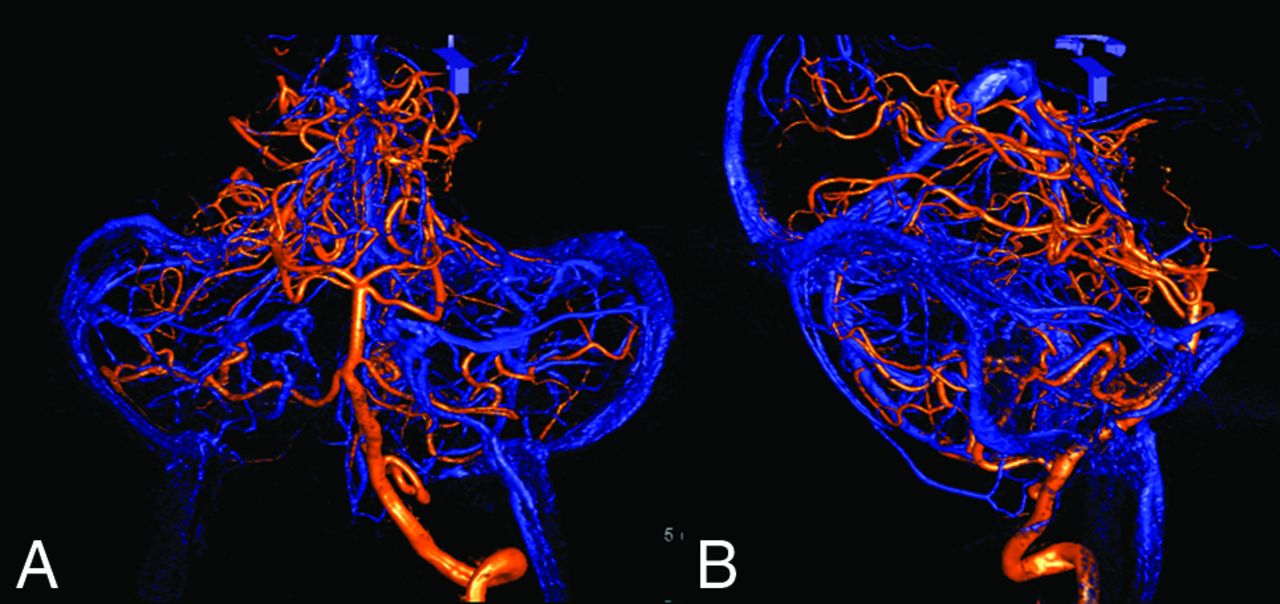
1) In a woman presenting with subarachnoid hemorrhage, DSA revealed a right carotid terminus aneurysm. Rotational angiography of the right ICA was performed as part of routine preoperative evaluation of the aneurysm (Fig 1A). Given the patient’s preference to be treated with clipping, venous 3D also was obtained (Fig 1B) to demonstrate the venous anatomy, especially around the Sylvian fissure, for surgical planning. The 2 subtracted volumes were then fused to show concomitant representation of the arteries and veins (Fig 1C).
2) In a woman presenting with small centrum semiovale hemorrhage, arterial DSA with rotational angiography revealed no evidence of aneurysm or AVM. Rather, it showed a large developmental venous anomaly and, for better visualization, a venous 3D DSA was also obtained. The 2 subtracted volumes were then fused to show concomitant representation of the arteries and veins (Fig 2).
3) In a man presenting with Moyamoya disease, full arterial and venous angiography was performed for surgical planning in anticipation of a possible direct bypass. As part of the cerebral angiogram, a vertebrobasilar AV-3D-DSA was obtained (Fig 3).
DISCUSSION
In this technical note, we describehow to obtain simultaneously merged AV-3D-DSA visualization by using 2 separate, appropriately timed, rotational acquisitions with postprocessing. While there has been description of concomitant arterial and venous-3D reconstruction in cross-sectional imaging by using 2 separate CTA acquisitions, this has not previously been done by using DSA to our knowledge.5,6 Our protocol can be considered a variant of the 2-color 3D-3D fusion of selective rotational cerebral angiograms used to visualize concurrently 2 different arterial territories.7 Notably, this technique can be applied to any volume of interest as demonstrated by the insert in the case 1 illustration.
Other DSA methods to examine concomitant vessels in different phases are, for example, the acquisition of 4D-DSA, which is excellent to analyze the fast inflow and outflow of AVMs, but has the limitation of not distinctly separating arteries and veins in the same volume.4 Another method is the so-called “sequential subtraction,” which uses 2 different masks, one for the bone and a negative one for the veins. Described in 1974, this is a useful technique to show the relationships between the arteries and veins but is limited only to 2D-DSA.8 With another technique, cone beam CT, the 20sDSA DynaCT (Siemens) protocol is long enough to depict both arteries and veins in the same volume, but the technique is not able to separate them for differential visualization.
Obtaining AV-3D-DSA has multiple potential purposes. Because it allows depiction of the relationship between the arteries and the veins in a 3D environment, the technique can be useful in planning open cerebrovascular surgery. It can help the surgeon understand the relevant “angioarchitecture” of a particular situation (depicting arteries and veins in their relationship to one another from the perspective of the surgical window) to better prepare for surgical exploration of cerebrovascular lesions, such as dissection and a clip reconstruction of a complex cerebral aneurysm. Other potential specific applications are its use to distinguish the venous drainage of an AVM or dural AVF from the venous drainage of the adjacent normal parenchyma. Moreover, it has also been extremely useful as a teaching tool for residents and fellows allowing a better understanding of neurovascular anatomy.
In this report, we demonstrate the technique used for the internal carotid and vertebrobasilar territories, but theoretically this can be applied to any vascular territory, granted that the arterial and venous injection protocols are appropriately modified to better set the pertinent flow, volume, and delay of the contrast injection. 2D-DSA images are used to establish appropriate rate, volume, and injection delay settings.
While volumetric MR imaging with 3D-T1 and 3D-T2 can demonstrate the vasculature, both sequences are poor in terms of differentiating arteries compared with veins, and the differentiation may be important for the surgical planning or other purposes.
The method is simple and the main disadvantage is the need to obtain 2 separate 3D-DSAs to obtain the dataset, which means double the amount of radiation and double the contrast volume. For this reason, we recommend strategic use of this technique when clinically necessary to answer specific questions. Theoretically, to obtain the dataset, the 2 phases may potentially be acquired with the same contrast injection, but that is not possible with the software/hardware capability of current Siemens angiographic equipment. Current protocol requires the subject not to move between and during acquisitions. We have obtained excellent quality images in cooperative subjects who were awake (images from Fig 2 were obtained in an awake patient) and patients under general anesthesia.

A, Frontal view arterial 3D-DSA of the right internal carotid artery injection demonstrating a multilobulated carotid terminus aneurysm. B, Frontal view venous 3D-DSA of the right internal carotid artery injection showing the venous outflow of the right hemisphere using mainly a deep middle cerebral vein draining toward the pterygoid plexus through the foramen ovale. C, Fused AV-3D-DSA demonstrating the relationship between the arteries and the veins. Specifically, note the presence of a vein running adjacent to the anterior aneurysm dome (arrow) better demonstrated in the insert (D).

Left lateral (A), right anterior oblique (B), and right lateral (C) views of AV-3D-DSA of the left internal carotid artery injection demonstrating a large developmental venous anomaly draining most of the left frontal hemisphere toward the left internal cerebral vein, which likely developed in association with atretic anterior superior sagittal sinus. Notice also the hypertrophied right inferior petrosal sinus and the plexus of Rektorzik surrounding the left ICA.

Frontal (A) and lateral (B) fused AV-3D-DSA of left vertebral artery injection demonstrating well the relationship between the petrosal veins and the vertebrobasilar system.
To conclude, in this technical note we describe how to obtain a simultaneous arterial and venous 3D-DSA visualization by using 2 separate, appropriately timed rotational acquisitions followed by dedicated, straightforward postprocessing, the results of which may be useful for different clinical and educational purposes.
Acknowledgments
We thank our lead tech Safia Syed for her invaluable relentless support. We also want to acknowledge Zia Shamsi, Osiris Galan, and Duane Thomas from Siemens AG for their 24/7 across campus availability to assist with image quality.
Footnotes
Disclosures: Eytan Raz—UNRELATED: Expert Testimony: Various law firms; Royalties: Springer; Stock/Stock Options: Siemens stocks; Comments: personal portfolio; Travel/Accommodations/Meeting Expenses Unrelated to Activities Listed: MicroVention, Rapid Medical, Stryker. Erez Nossek—UNRELATED: Consultancy: Rapid Medical; Stock/Stock Options: Rapid Medical.
References
- Received December 6, 2020.
- Accepted after revision February 5, 2021.
- © 2021 by American Journal of Neuroradiology


