Article Text

Abstract
Objectives To describe a new technique to obtain minimally invasive but efficient vertebral body (VB) reconstruction, augmentation, and stabilization in severe osteoporotic and neoplastic fractures, combining two pre-existing procedures. The implant of vertebral body stents (VBS) is followed by insertion of percutaneous, fenestrated, cement-augmented pedicular screws that act as anchors to the posterior elements for the cement/stent complex. The screws reduce the risk of stent mobilization in a non-intact VB cortical shell and bridge middle column and pedicular fractures. This procedure results in a 360° non-fusion form of vertebral internal fixation that may empower vertebral augmentation and potentially avoid corpectomy in challenging fractures.
Procedure details This report provides step-by-step procedural details, rationale, and proposed indications for this procedure. The procedure is entirely percutaneous under fluoroscopic guidance. Through transpedicular trocars the VBS are inserted, balloon-expanded and implanted in the VB. Over k-wire exchange the transpedicular screws are inserted inside the lumen of the stents and cement is injected through the screws to augment the stents and fuse the screws to the stents.
Applications This technique may find appropriate applications for the most severe osteoporotic fractures with large clefts, high-degree fragmentation and collapse, middle column and pedicular involvement, and in extensive neoplastic lytic lesions.
Conclusions Stent-Screw-Assisted Internal Fixation (SAIF) might represent a minimally invasive option to obtain VB reconstruction and restoration of axial load capability in severe osteoporotic and neoplastic fractures, potentially obviating the need for more invasive surgical interventions in situations that would pose significant challenges to standard vertebroplasty or balloon kyphoplasty.
- stent
- neoplasm
- metastatic
- spine
- technique
Statistics from Altmetric.com
Introduction
Vertebral augmentation (VA) has been extensively used for pain palliation and stabilization of vertebral body (VB) fractures due to trauma, osteoporosis, and tumors.1–3 The introduction of the Vertebral Body Stent (VBS) (DePuySynthes-Johnson & Johnson) further empowers these techniques. VBS is a balloon-expandable barrel-shaped metallic cage which is percutaneously inserted via uni- or bi-pedicular access. On expansion, the VBS maintains the cavity through balloon deflation and subsequent cement injection. Introduced for the treatment of vertebral compression fractures,4–10 VBS has also been used in neoplastic fractures.11 Most recently it has been tested in cases of extensive osteolysis of the VB to reconstruct the anterior column.12
VBS has several potential advantages over traditional non-instrumented augmentation in that the rigid stent remains expanded after balloon deflation, thus maintaining the restored VB height.7 The VBS metallic mesh virtually guarantees a predictable and reasonably uniform barrel-shaped balloon expansion whereas a compliant balloon often follows the path of least resistance. The barrel shape of the VBS, with its large support surface, provides mechanical support, scaffolds the VB from within and, where necessary, recreates VB walls such as in dehiscent or fragmented cortical boundaries. Furthermore, the metallic mesh helps contain the injected cement in the created cavity. These characteristics potentially favor the use of VBS in the most severe vertebral fractures such as highly fragmented osteoporotic fractures or neoplastic fractures with prominent cortical osteolysis.
Despite these advantages, in the most severe osteolytic or neoplastic fractures the implanted VBS may only be partially contained by the non-intact cortical shell. In that situation, the VBS could potentially be expected to mobilize,6 12 leading to adverse events. Other potential issues with VBS treatment of these lesions is the frequent association with middle column and pedicular fractures, in the face of which VBS augmentation might represent undertreatment.
The aim of this paper is to describe a new technique combining the VBS implant with the insertion of percutaneous transpedicular cannulated fenestrated screws followed by cement deposition through the screw, with the intention to anchor the VBS/cement implant to the posterior elements, reduce the risk of VBS mobilization, and bridge middle column and pedicular fractures.
This Stent-Screw-Assisted Internal Fixation (SAIF) technique, as opposed to the standard surgical external fixation achieved with screws and rods bypassing the index level, might represent a minimally invasive image-guided 360° non-fusion form of vertebral reconstruction and stabilization in severe osteoporotic and neoplastic thoracolumbar vertebral fractures.
Procedural details
This is a technical note describing the procedural details and potential applications of a new technique, combining the use of two established and already reported procedures and devices.7 13 14 The Institutional Review Board approved this investigation and the patients signed a required informed consent to undergo the procedure.
Figure 1 shows the SAIF model, instrumentarium, main procedural steps, and schematic drawings, while figures 2–4 show the SAIF in three different clinical scenarios, representing examples of potential clinical applications of the technique.

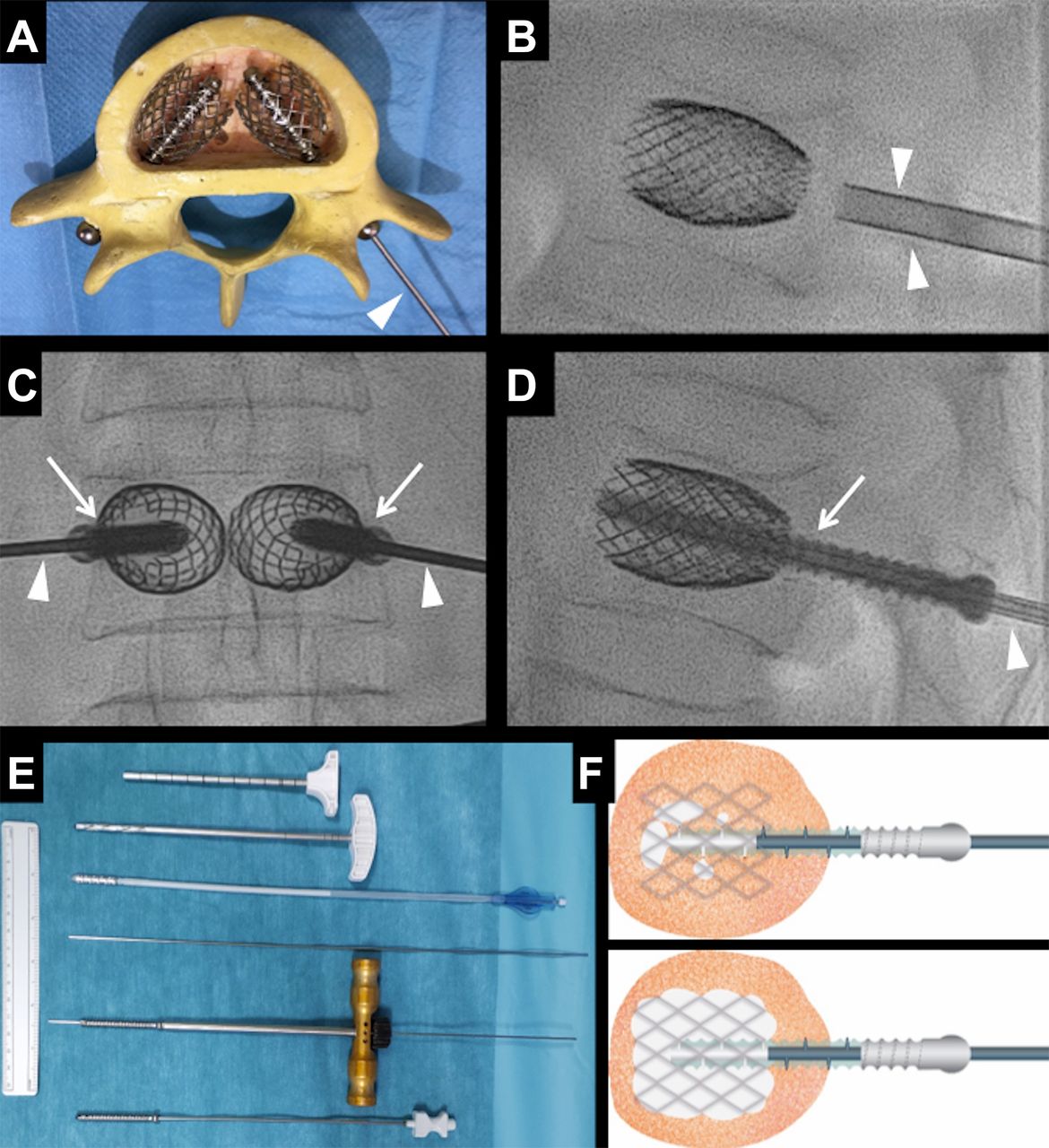
Stent-Screw-Assisted Internal Fixation (SAIF) technique. (A) Model representation of the SAIF implants in a lumbar vertebra. Bilateral vertebral body stents are expanded and implanted, with transpedicular fenestrated screws inserted in their lumen. Cement injection through the screws inside the stents would follow via the injection cannula (arrowhead), but is not shown to maintain visibility of the implants. (B–D) Main fluoroscopy-guided procedural steps of SAIF before cement injection. (B) Lateral fluoroscopic view showing the stents that have been inserted and expanded in the vertebral body through the bilateral transpedicular access cannulae (arrowheads). (C, D) Anteroposterior and lateral views respectively showing that the cannulae have been replaced via k-wire exchange with transpedicular cannulated fenestrated screws (arrows) inside the stents. Over the same k-wire, a cannula for cement injection is inserted inside each screw (arrowheads). (E) Main instrumentarium for the SAIF technique showing, from top to bottom: the 7G trocar, manual coaxial drill, balloon-mounted stent, k-wire, screw mounted on a screwdriver over a k-wire, and the 14G cement injection luer lock cannula inserted in the screw. (F) Schematic drawing showing how the fitting cannula (in blue) can be positioned along the screw stem so that cement (in white) flows through the screw fenestrations distal to the cannula tip and fills the stent lumen.

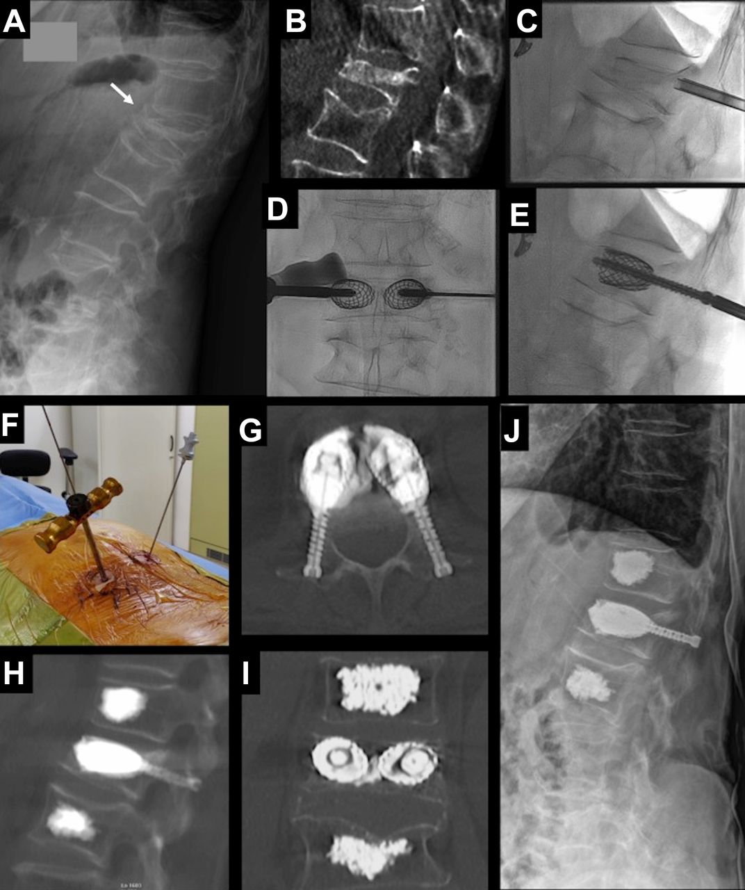
Stent-Screw-Assisted Internal Fixation (SAIF) technique in a L2 hepatocarcinoma metastatic fracture. Adult patient affected by metastatic cancer with disabling drug-resistant mechanical back pain and evidence on (A) MRI and (B, C) CT of a severely collapsed and fragmented L2 vertebral body fracture with involvement of the middle column and posterior wall retropulsion but without neurological deficit. (D) Lateral fluoroscopic view in the prone position showing the result of SAIF with pedicular screws inside the stents filled by PMMA cement augmentation. There is residual intradural contrast agent from intraoperative myelographic control (arrowheads). There is significant height restoration of the vertebral body and optimal stent cement augmentation in the absence of extravertebral leaks. (E–H) Post-procedure CT shows the vertebral body reconstruction by the stents, the cement bridge along the anterior aspect of the stents (arrowheads on E), and the screws cemented inside the stents, anchoring to the posterior elements. Prophylactic vertebral augmentation was performed at L1. The patient reported rapid and sustained pain relief and was discharged the same day after 6 hours of observation. He died 5 months later but until his death he had no further back pain or issues.

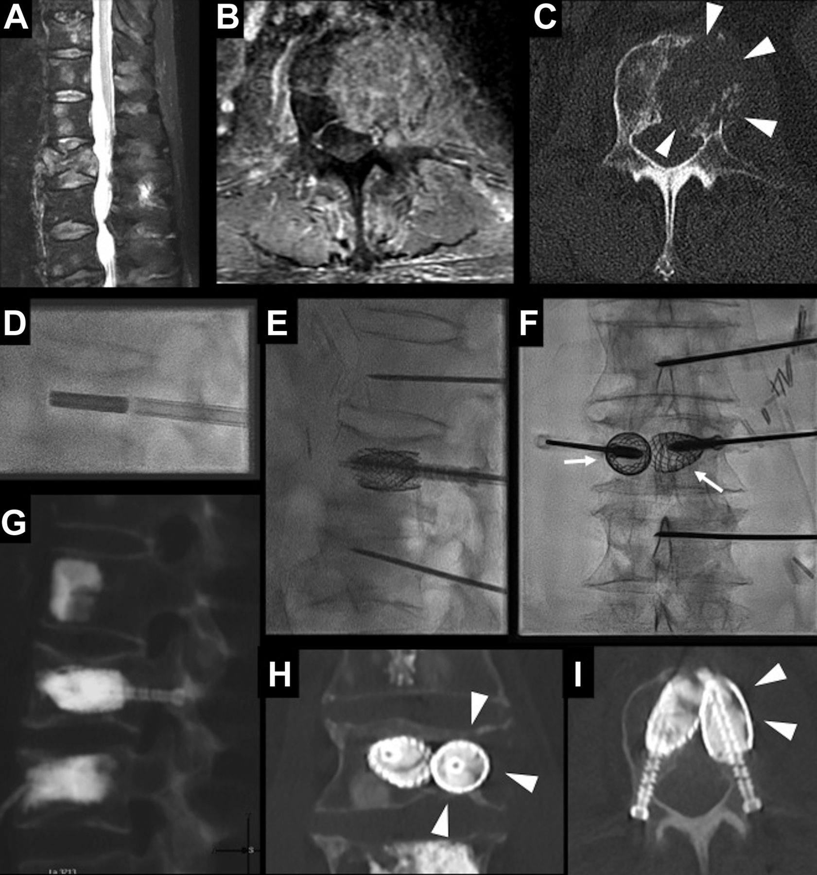
Stent-Screw-Assisted Internal Fixation (SAIF) technique in a L2 renal cell cancer large lytic lesion in a 64-year-old man with renal cell cancer with severe mechanical axial back pain. MRI with sagittal fat-suppressed T2-weighted images (A) and axial fat-suppressed contrast-enhanced T1-weighted images (B) show a pathological fracture with partial collapse of the L2 vertebral body on a large vertebral and extracompartmental enhancing soft tissue mass, centered in the left side of the vertebral body, crossing midline, extending to the left pedicle, ventral epidural space and perivertebral space on the left, towards the psoas. Multilevel metastatic spinal involvement is also noted. (C) Corresponding axial CT image shows the lytic nature of the lesion with largely dehiscent cortical borders at the posterior and left anterolateral walls (arrowheads). Standard vertebral augmentation would pose a significant risk of early extravertebral cement leak and insufficient stabilization due to the extensive cortical osteolysis. (D–F) SAIF fluoroscopic intraprocedural images show insertion of stents, their balloon expansion, and insertion of screws; additional 14G needles are inserted at L1 and L3 for standard augmentation. Note the slight left-sided lateralization of the stents (arrows in F) to obtain maximum protection of the left hemibody, predominantly involved by the lytic lesion. (G–I) Post-procedure CT after cement augmentation shows how the stents with cement have reconstructed the destroyed portion of the vertebral body and now offer support to bear the axial load. The walls of the stents recreate vertebral body walls along the disc endplates and lateral aspect of the vertebral body (arrowheads in H and I) and limit the risk of cement leak. The screws anchor the stent/cement complex to the posterior elements, minimizing the risk of displacement. The patient was able to stand and walk the same day and could undergo radiation treatment for local disease control a few days later.
Stent-Screw-Assisted Internal Fixation (SAIF) technique in an osteoporotic fracture with vertebra plana deformity. A 75-year-old woman with known osteoporosis reported severe back pain following a fall from a height. Imaging revealed a mild L1 compression fracture. Despite best medical conservative treatment, disabling pain persisted and at 14 days follow-up standing plain films revealed progression to vertebra plana deformity (arrow on A) with severe junctional kyphosis (Cobb angle 28°), which was keeping the patient bedridden. (B) CT confirmed the fracture, slightly reduced in the supine position, suggesting a mobile fracture, and milder compression fractures at T12 and L2. Procedural fluoroscopic images show the trocar access parallel to the anticipated alignment of the original pre-fracture endplates (C), to allow the most efficient vertebral height restoration during vertebral stent expansion and screw insertion (D–E). (F) Operation field corresponding to the fluoroscopic image in (D) with the low-profile screwdriver over the k-wire on the left and the cement injection cannula already inserted in the screw on the right. (G–I) Post-procedure CT images show the obtained height restoration and internal vertebral body reconstruction and fixation of L1, and standard cement augmentation at T12 and L2. The stents are interconnected by a ’bridge' of cement and are anchored to the posterior elements by the screws. The patient was then able to stand and walk with significantly reduced pain, and standing plain films at 1-month follow-up (J) showed stability of the implants and of the height restoration, with markedly reduced junctional kyphosis (Cobb angle 15°).
Procedural instructions for bilateral SAIF
The patient is placed under general anesthesia and turned prone (general anesthesia is used based on the institutional preference of the anesthesiologists, but we speculate that SAIF could be performed under moderate sedation of monitored anesthesia care). Intravenous antibiotic prophylaxis is administered.
Under fluoroscopic guidance, a 4.5 mm (7G) caliber trocar, included in the VBS access kit, is positioned via transpedicular access at the pediculo-somatic junction, bilaterally, as per a standard balloon kyphoplasty (BKP), with an oblique lateromedial orientation15 (figure 1). Particular care is taken to insert the trocar parallel to the anticipated alignment of the original pre-fracture endplates to allow the most efficient vertebral height restoration during VBS expansion (figure 4). Manual drills are placed coaxially through trocars to create the necessary space for the balloon-mounted VBS, which are then inserted on each side in the VB. The stents are expanded, as desired and possible, by balloon inflation with a manual hydraulic pump using saline or contrast to try to obtain fracture reduction and height restoration. The balloons are deflated and removed while the expanded stents remain in place (figure 1). At this point the trocars are removed leaving a k-wire (1.4×350 mm, blunt tip) in place. The tract is not dilated. Over the k-wire, through the same 6–8 mm skin stab incision, a low-profile manual screwdriver (figure 4) is used to place a percutaneous transpedicular fenestrated cannulated screw (injection pin, 2B1, Milan, Italy) of desired length (34–55 mm) and caliber (5 or 6 mm) as planned on the basis of the pre-procedure CT axial images. The screw is inserted into the lumen of the stent until the bulbous head reaches the dorsal cortex of the posterior elements. The screwdriver is removed and, over the same k-wire, a 14G 210 mm long cannula with a luer lock hub is inserted in the screw for cement injection (figure 1). The screws have multiple fenestrations at the distal tip and along their entire threaded stem to allow and optimize cement dispersion. The cannula fits within the screw lumen and the position of the injection cannula can be adjusted along the entire stem, from the distal tip to its proximal end, to manage the desired site of cement injection (figure 1). The injection cannula is compatible with all commercially available PMMA cements and with any luer lock injection system. PMMA injection is monitored with real-time fluoroscopy in the lateral view with intermittent anteroposterior checks. Most commonly, cement is injected in the distal third of the screw and is seen permeating from the screw fenestrations inside the lumen of the stent and, after filling the stent, either overflowing to the adjacent stent or to the anterior open end of the stent, or interdigitating through the mesh into the adjacent trabecular spaces. Cement injection is halted if extravertebral leaks occur or if cement approaches the posterior wall. When cement injection is deemed complete, the injection cannula is retracted and the screw left in place. When the PMMA hardens, the screws are fixed in the VBS/cement complex implant and anchor it to the pedicles and posterior element cortex. There is no need to apply suture stitches since the stab incisions are very small.
As with standard vertebral augmentation, the patient is mobilized from the prone position after a time interval that allows the PMMA to cure. The patients are allowed to stand and walk as early as 3 hours after the procedure and, if clinical conditions allow, may be discharged the same day. Clinical and radiological follow-up is similar to that used for patients who have undergone standard vertebral augmentation. This includes a limited C-arm CT at the end of the procedure as well as standing spinal plain films once the patient can mobilize. Patients are routinely followed up at 1 and 6 months, when repeat standing radiographs are obtained.
Applications
We propose that the SAIF technique could be used to perform vertebral augmentation in severe osteoporotic fractures such as those characterized by crush deformity, advanced collapse (Genant grade 3),16 high degree of osseous fragmentation (McCormack comminution grade 2 and 3),17 large osteonecrotic cleft, middle column and pedicular fractures or, more generally, in vertebral fractures with advanced loss of integrity and quality of trabecular and cortical bone (figure 4). Similarly, in neoplastic lesions (figures 2 and 3), the use of SAIF might be considered to augment extensive osteolytic lesions with dehiscent cortical boundaries (Tomita extracompartmental lesion type 4–6)18 that are fractured or at risk of impending collapse.
Discussion
SAIF is a previously undescribed technique, combining the implant of VBS and cement augmentation through percutaneous pedicular screws. In the aggregate the VBS and screws become a solid construct; the screws anchor the VBS to the posterior elements and potentially bridge and stabilize the middle column and pedicular fractures. This represents an advance over traditional augmentation in that the middle column is also stabilized.
Vertebral augmentation, performed with vertebroplasty or BKP, has limitations in complex osteoporotic fractures with advanced bone loss and in extensive lytic lesions.19–21 Cement distribution in these highly destroyed VBs might be unpredictable, uneven, or even result in early extravertebral leak, leading to insufficient augmentation and stabilization or to clinical complications if central vascular migration or neural compression occur. Middle column fractures characterize potentially unstable fractures, and augmentation of the anterior column alone might represent undertreatment. In addition, frequently associated pedicular fractures represent a treatment dilemma, and cement augmentation is biomechanically inefficient on structures that bear tensile forces.22
These injuries are commonly considered for surgical treatment of stabilization that may include corpectomy performed through an anterior or anterolateral approach. At the same time, this approach carries significant invasiveness and morbidity risk,23 24 especially in elderly patients with osteoporosis or neoplastic diseases. Therefore, a minimally invasive treatment option is desirable.
In vertebral lesions with severely altered osseous structure, the SAIF technique offers a VB reconstruction using VBS metallic cages and cement. These bilateral implants scaffold the VB, offering a large support surface along the disc endplates, help to contain cement along dehiscent cortical boundaries (figure 3) and, filled with cement, become a structure of ‘armed concrete’, able to bear the axial load in a solid and uniform manner in the VB. Cement flowing from one stent to the other, either interdigitating through the mesh or passing through the anterior open ends, creates a solid bridge between the two VBS (figures 2 and 4), and the screws that are cemented inside the stents represent the anchors to the most commonly intact posterior elements, largely limiting the risk of VBS mobilization. The whole construct, with the two stents interconnected by the bridge and the two screws as anchors, might be regarded as an internal VB prosthesis fixed to the neural arch (figure 2). Insertion of a unilateral screw can also be considered, especially in neoplastic cases when one of the two pedicles is involved by lytic destruction and would not offer a good anchoring.
Unlike surgical corpectomy, grafting and posterior instrumentation, the disc spaces and the mobility of the spinal functional units are spared. Nevertheless, when indicated, SAIF can be combined with posterior surgical instrumentation. Inserting screws with a rod connecting system, the screws at the target level might even be connected to the bars of a posterior instrumentation. The whole SAIF procedure is image-guided, percutaneous, and minimally invasive, being performed through two small skin stab incisions. The addition of the screws to the VBS is entirely performed over a k-wire, enhancing its safety and rapidity. The cement injection through the screws into the VB can be adjusted according to the preference of the operator, sliding the injection cannula along the screw stem. Recovery is more typical of a percutaneous procedure, so most patients are allowed to stand after a few hours and return to their normal activities within 24 hours after the procedure. Moreover, in neoplastic patients, SAIF does not interfere with chemotherapy or radiation treatment.25
At follow-up thus far we have not encountered VBS/cement mobilization or significant spinal alignment issues at the SAIF-treated levels. Subtle VB bone subsidence and remodeling may occur, although these have tended not to require any treatment.
The risks and contraindications for the SAIF procedure are the same as those for vertebroplasty, BKP or VBS.
The procedure has to be halted if the VBS does not expand due to sclerotic changes within the VB or because of premature rupture of the balloon inside the stent; in these cases the procedure is converted to a standard VBS augmentation. We do not recommend insertion of percutaneous screws if a safe transpedicular approach cannot be obtained.
The additional procedure time and material costs that SAIF requires are justified in consideration of the patient selection, challenging features of the treated lesions, short hospitalization, and fast patient recovery, especially if compared with a standard surgical procedure.
We do not use or recommend VBS and SAIF in young patients with traumatic fractures, in which bone quality is supposed to be good, and large volume implants in the VB with VBS and PMMA may halt spontaneous osseous healing.
In addition, it should be stressed that we propose the SAIF procedure in a neoplastic vertebra as a stabilization procedure, with no intent of local control of disease for which standard oncological treatments should be used.
In conclusion, the SAIF technique might represent a minimally invasive treatment option to reconstruct the VB and restore axial load capability in patients with severe osteoporotic or neoplastic fractures, potentially obviating the need for a more invasive surgical procedure of corpectomy and grafting. Biomechanical and clinical studies are needed to further prove this concept.
References
Footnotes
Contributors All authors contributed to the presented work by substantial contributions to the conception or design of the work; or the acquisition, analysis, or interpretation of data for the work and drafting the work or revising it critically for important intellectual content and final approval of the version to be published, and agree to be accountable for all aspects of the work in ensuring that questions related to the accuracy or integrity of any part of the work are appropriately investigated and resolved.
Funding The authors have not declared a specific grant for this research from any funding agency in the public, commercial or not-for-profit sectors.
Competing interests None declared.
Ethics approval Ethics Committee of Canton Ticino.
Provenance and peer review Not commissioned; externally peer reviewed.
Patient consent for publication Obtained.