
Abstract
BACKGROUND AND PURPOSE: Flow diversion with the Pipeline Embolization Device is increasingly used for endovascular treatment of intracranial aneurysms due to high reported obliteration rates and low associated morbidity. While obliteration of covered branches in the anterior circulation is generally asymptomatic, this has not been studied within the posterior circulation. The aim of this study was to evaluate the association between branch coverage and occlusion, as well as associated ischemic events in a cohort of patients with posterior circulation aneurysms treated with the Pipeline Embolization Device.
MATERIALS AND METHODS: A retrospective review of prospectively maintained databases at 8 academic institutions from 2009 to 2016 was performed to identify patients with posterior circulation aneurysms treated with the Pipeline Embolization Device. Branch coverage following placement was evaluated, including the posterior inferior cerebellar artery, anterior inferior cerebellar artery, superior cerebellar artery, and posterior cerebral artery. If the Pipeline Embolization Device crossed the ostia of the contralateral vertebral artery, its long-term patency was assessed as well.
RESULTS: A cohort of 129 consecutive patients underwent treatment of 131 posterior circulation aneurysms with the Pipeline Embolization Device. Adjunctive coiling was used in 40 (31.0%) procedures. One or more branches were covered in 103 (79.8%) procedures. At a median follow-up of 11 months, 11% were occluded, most frequently the vertebral artery (34.8%). Branch obliteration was most common among asymptomatic aneurysms (P < .001). Ischemic complications occurred in 29 (22.5%) procedures. On multivariable analysis, there was no significant difference in ischemic complications in cases in which a branch was covered (P = .24) or occluded (P = .16).
CONCLUSIONS: There was a low occlusion incidence in end arteries following branch coverage at last follow-up. The incidence was higher in the posterior cerebral artery and vertebral artery where collateral supply is high. Branch occlusion was not associated with a significant increase in ischemic complications.
ABBREVIATIONS:
- PCA
- posterior cerebral artery
- PED
- Pipeline Embolization Device
- VA
- vertebral artery
Flow diversion using the Pipeline Embolization Device (PED; Covidien, Irvine, California) has become an important treatment technique for intracranial aneurysms. Although the mechanism of action of this device is still not completely understood, it is expected to initially reduce the intra-aneurysmal flow leading to aneurysm thrombosis and seal off the aneurysm from circulation by inducing neointimal coverage of the PED surface at the aneurysm neck.1 When the PED spans an arterial branch, there is concern for branch occlusion and subsequent ischemic stroke. Among others, this concern had limited the Food and Drug Administration approval of PED use to the internal carotid artery below the level of the posterior communicating artery.2 Since this decision, the PED has only been used reluctantly in the treatment of posterior circulation aneurysms where multiple branches supply the brain stem. A handful of studies have evaluated the off-label use of the PED in the treatment of posterior circulation aneurysms and reported a relatively high risk of ischemic complications, particularly in the treatment of fusiform aneurysms.3⇓⇓⇓⇓⇓–9 In this study, we aimed to assess the association between branch coverage and occlusion incidence and its impact on ischemic complications.
Materials and Methods
A retrospective review of prospectively maintained databases at 8 academic institutions in North America and Europe was performed from 2009 to 2016 to identify adult patients (18 years of age or older) with posterior circulation aneurysms treated consecutively with the PED. Both ruptured and unruptured aneurysms and all aneurysm shapes were included. Patients younger than 18 years of age were excluded. The following information was collected prospectively by the treating neurointerventionalists: patient demographics, aneurysm characteristics, platelet function test results, antiplatelet regimen, procedural details, angiographic and functional outcomes, and complications. Aneurysm measurements were obtained using digital subtraction angiography, except in cases of partially thrombosed aneurysms in which axial images were also analyzed. Institutional review board approval was obtained at all centers. The primary end point for the study was the occurrence of branch occlusion after placement of the PED across major posterior circulation branches. The secondary end point was the occurrence of ischemic complications following branch occlusion.
Procedure Details
Patients received aspirin, 325 mg daily, and clopidogrel, 75 mg daily, for 3–14 days before the intervention. Platelet function testing was routinely performed by some of the centers using whole-blood lumiaggregometry, light transmission aggregometry, or the VerifyNow P2Y12 assay (Accumetrics, San Diego, California). In those centers, clopidogrel nonresponders were identified on the basis of established cutoff values at the individual institutions and were guided by manufacturer's recommendations. If a patient was identified as a clopidogrel nonresponder, the choice to continue the same dose of clopidogrel, administer a 1-time 600-mg clopidogrel boost within 24 hours preprocedure, or switch to an alternative antiplatelet agent was at the discretion of the interventionalist performing the procedure. All patients were anticoagulated with heparin throughout the procedure. Activated clotting time was used in most cases to guide heparin administration intraprocedurally, with a target of 250–300 seconds; typical dosing consisted of a 3000- to 5000-U bolus at the beginning of the procedure, with hourly dosing of 1000 U. The guide catheter and microcatheter used for PED deployment were at the discretion of the individual interventionalists. The deployment and apposition of the PED to the vessel wall were documented using fluoroscopy. Dual antiplatelet therapy was continued for at least 3 months after the procedure.
Branch Coverage and Obliteration
Branch coverage and occlusion following PED placement were assessed on procedural DSA and last follow-up imaging, respectively. Only complete branch occlusion was considered. MR angiography was used as a follow-up technique in 1 center, but DSA was used to confirm branch occlusion if suspected. Vessels assessed included the posterior inferior cerebellar artery, anterior inferior cerebellar artery, superior cerebellar artery, and posterior cerebral artery (PCA). In addition, if the PED crossed the ostia of the contralateral vertebral artery (VA), the long-term patency of the covered VA was also assessed. Because brain stem perforator vessels are too small to resolve on DSA, we did not attempt to assess them. However, if brain stem infarct occurred, the possibility of perforator infracts was considered. When we assessed predictors of branch occlusion following coverage, each covered branch was considered as a separate entity.
Ischemic Complications
Ischemic complications occurring from the date of the procedure to last follow-up were included. Intraprocedural thromboembolic complications were identified on DSA as either thrombus formation, slow filling of a previously normally filling vessel, or vessel drop-out. Postprocedural ischemic complications were identified using a combination of clinical and radiographic findings. Postprocedural imaging was performed at the discretion of the treating physician and was only obtained due to clinical concern. Routine screening for clinically silent ischemic strokes was not performed. Only ischemic strokes in the territory of the treated vessel were included. An ischemic complication was considered symptomatic if the patient reported symptoms or signs attributable to an ischemic event; this included transient or resolving signs and symptoms.
Statistical Analysis
Statistical analysis was performed using SPSS 21.0 (IBM, Armonk, New York). In univariable analysis, variables were compared between groups with the Mann-Whitney test for numeric variables and the χ2 test for categoric variables. Statistical significance was defined as P < .05. Multivariable logistic regression was performed on candidate predictor variables to identify variables independently associated with branch occlusion following coverage and thromboembolic complications following PED placement after controlling for potential confounders. Accounting for interactions and collinearity among variables was undertaken.
Results
Patient and Aneurysm Characteristics
One-hundred twenty-nine consecutive patients (median age, 58 years; male/female ratio, 1:1.7) underwent 129 procedures to treat 131 posterior circulation aneurysms with the PED. Smoking and multiple aneurysms were encountered in 33.3% and 24.8% of procedures, respectively. Most patients presented with neurologic deficits caused by aneurysm rupture or direct neurovascular compression (55%). Incidental aneurysms were identified in 21.7% of procedures. Treatment of immediate (<24 hours) or acute (>24 hours and <2 weeks) aneurysmal SAH occurred in 14.0% and 5.4% of procedures, respectively. Most aneurysms were fusiform (40.5%) or saccular (37.4%) and were commonly located along the intracranial segment of the VA (35.1%) or the basilar artery (34.5%). The median maximum diameter was 12 mm, and a daughter sac was present in 19.8% of aneurysms. Platelet function testing was performed in 59.7% of procedures, and the incidence of clopidogrel nonresponders was 18.2% of those tested (Table 1).
Baseline characteristics
Treatment Outcome
The median number of PEDs deployed was 1 (range, 1–14). Adjunctive coiling was used in 31% of procedures. Following PED placement, ≥1 branch was covered in 79.8% of procedures. At a median follow-up of 11 months (range, 172 months), complete or near-complete aneurysm occlusion (≥90%) was achieved in 78.1% of aneurysms. Retreatment with endovascular techniques was necessary in 8.4% of aneurysms. At last follow-up, the mRS improved in 34.4% and worsened in 22.4%, inclusive of patients presenting with aneurysmal SAH. The overall mortality rate was 11.2% (Table 2).
Outcome measures
Predictors of Branch Occlusion and Ischemic Stroke following PED Placement
The rate of aneurysm occlusion following coverage was 11% (25/228). There was a higher branch occlusion incidence when the PED covered posterior circulation branches in asymptomatic patients with incidental aneurysms (P < .001). The VA (34.8%) and PCA (23.5%) were associated with a significantly higher incidence of occlusion compared with other posterior circulation branches (P = .001). There was an increased incidence of branch occlusion when ≥2 branches were covered in the same procedure, but this was not statistically significant (P = .07).
Ischemic complications occurred in 22.5% of procedures, while permanent symptomatic ischemic complications occurred in 8.5%. On univariable analysis, the risk of ischemic complications was significantly higher when vascular branches were covered with the PED compared with cases with no branch coverage (26.2% versus 7.7%, P = .04). The territory of ischemia was the same as the covered branch in 72.4% of cases. Among the cases with covered branches, there was no significant difference in the incidence of ischemic complications regarding branch occlusion (P = .16) (Fig 1). All 3 cases in which branch occlusion was associated with an ischemic complication in the same territory involved the AICA (Fig 2). There was no significant difference in branch occlusion (P = .018) or ischemic complications (P = .7) per aneurysm morphology (Table 3 and On-line Table).

Digital subtraction angiography shows a basilar tip saccular aneurysm before treatment with the Pipeline Embolization Device (A). The PED was placed spanning the lower part of the basilar truck into the left posterior cerebral artery. On 4-month follow-up (B), DSA shows complete aneurysm occlusion, along with complete occlusion of the right PCA. The anterior inferior cerebral arteries and superior cerebellar arteries remained patent. The patient remained neurologically intact.

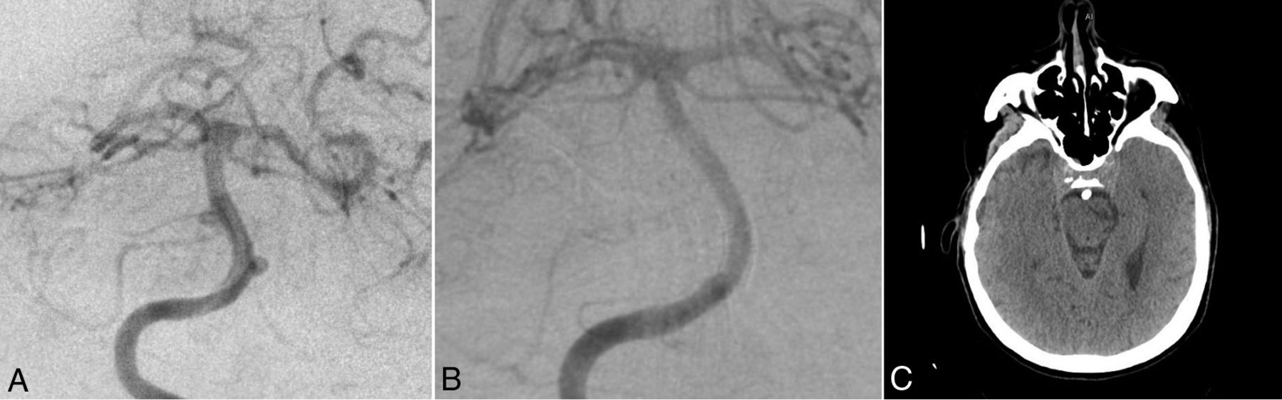
Digital subtraction angiography shows 2 basilar trunk saccular aneurysms before treatment with the Pipeline Embolization Device (A). The PED was placed spanning the AICA bilaterally. On follow-up DSA (B), there was complete occlusion of the left AICA. The patient had a symptomatic left-sided pontine stroke that remained symptomatic at 10-month follow-up (C).
Predictors of branch occlusion following coverage
Discussion
In this multicenter study, we report the fate of major posterior circulation branches after placement of PEDs in the treatment of intracranial aneurysms. In our study, 25 of 228 covered branches were obliterated following coverage (11%). The VA and PCA were associated with the highest incidence of vascular obliteration, while the superior cerebellar artery was associated with the lowest incidence. Despite this variability, there was no significant difference in the incidence of ischemic complications based on the covered branch. The presence of a covered branch was associated with a significant increase in the incidence of ischemia on univariable analysis. However, it was not significant as an independent predictor, regardless of the number of covered branches. Intramural thrombosis was associated with a significant increase in ischemic complication incidence on multivariable analysis.
Treatment of Posterior Circulation Aneurysms Using the PED
Posterior circulation aneurysms are often associated with a higher incidence of morbidity and mortality compared with their anterior circulation counterparts; this is mainly related to higher rates of aneurysm rupture and neurovascular compression caused by large dolichoectatic aneurysms.10,11 Flow diversion using the PED has gained popularity as an off-label treatment option.3⇓⇓⇓⇓⇓–9
Flow diverters are designed to seal aneurysms from the circulation by diverting blood flow away from the aneurysm, allowing intra-aneurysm thrombus formation followed by neointimal growth across the neck of the aneurysm.1 Intimal growth over the luminal surface of a flow-diversion device is expected to be seen as a tissue layer consisting of smooth muscle cells covered by endothelium (endothelization), while thrombus organization is expected to be visualized in the form of smooth-muscle cell invasion and connective tissue formation within the clot.1
Despite widespread reluctance to use the PED for treatment of posterior circulation aneurysms, a handful of studies have attempted to evaluate the safety and efficacy in this high-risk group of aneurysms. The incidence of thromboembolic complications in these studies ranged between 0% and 22.5%3⇓⇓⇓⇓⇓–9 and was particularly high in fusiform aneurysms. A possible explanation for this finding was the higher likelihood of branch coverage when fusiform aneurysms were treated with the PED. Moreover, fusiform aneurysms can often be associated with intramural thrombosis, and placement of the PED is particularly hazardous because critical perforators may only be supplied through tenuous channels crossing the thrombus.9 However, in this study, most ischemic complications were either temporary or asymptomatic, and only 8.5% of procedures were affected by permanent symptomatic ischemic complications.
Branch Coverage and Occlusion following PED Placement
Initial evaluation of the PED by Kallmes et al12,13 showed that despite placement of multiple overlapping flow-diversion devices in the rabbit aorta, lumbar branch vessels remained patent at follow-up. In an assessment of the patency of anterior circulation branches following PED placement, Rangel-Castilla et al14 reported a 15.8% branch occlusion following coverage. The occlusion rate was lowest in the anterior choroidal artery (0%) and almost equal in the ophthalmic artery (10.5%) and posterior communicating artery (10.7%). Both cases of anterior cerebral artery coverage ended with occlusion on last follow-up. Despite branch obliteration, the authors did not identify any clinical sequelae.14
Similar results were reported by separate studies on the fates of the ophthalmic artery,15⇓⇓–18 posterior communicating artery,19 and anterior choroidal artery.19,20 Brinjikji et al21 reported a 45% incidence of posterior communicating artery occlusion or diminished flow at last follow-up, but none of the patients showed clinical symptoms related to vessel obliteration. Most of these patients demonstrated diminished blood flow immediately following PED placement; this finding was significantly associated with a higher occlusion incidence at follow-up. Gawlitza et al22 reported using flow diverters to treat aneurysms in the middle cerebral artery bifurcation and anterior communicating artery complex. Of the covered branches, 10.5% (2/19) had completely occluded on last follow-up. Temporary symptomatic ischemic events in perforator territories occurred in 17.6% of cases, which were reversible in all cases within 24 hours. Follow-up MR imaging disclosed asymptomatic lacunar defects corresponding to covered perforating artery territories in 29.4%. The lack of symptoms was attributed to the supply of respective cortical territories by leptomeningeal collaterals in all cases.22
The high incidence of occlusion of the ophthalmic artery and posterior communicating artery following flow-diverter coverage is attributed to the rich collateral supply from the external carotid artery and PCA, respectively. These collateral vessels might increase the tendency for proximal occlusion if a flow diverter causes some diminution of inflow and the distal anastomosis takes over the end-organ arterial supply.14,15 This collateral supply also explains the absence of clinical symptoms. On the contrary, terminal arteries without significant collateral supply, such as the anterior choroidal artery or middle cerebral artery perforators, are more likely to remain patent after coverage by a flow diverter. The pressure gradient across these arteries is more than that across vessels with a rich collateral supply, thus increasing the threshold for branch occlusion by a flow diverter.14,20
Other considerations regarding the use of the PED for treatment of aneurysms near large branches include the branch that is incorporated into the neck of an aneurysm. In these cases, the use of the PED is controversial and often does not result in aneurysm occlusion. This outcome can be seen with flow diversion of posterior communicating artery aneurysms and may be related to retrograde aneurysm filling.18,21
In this series, the fate of major posterior circulation branches has been assessed for the first time in a large and diverse group of patients. Similar to the anterior circulation branches, major branching arteries including the PICA, AICA, and superior cerebellar artery had a low incidence of branch occlusion after coverage with the PED. However, following occlusion of these branches, the risk of ischemic complications might be high. In our series, all 3 cases in which branch occlusion was associated with ischemic complications in the same territory involved the AICA. The relatively high incidence of VA and PCA occlusion was thought to result from a rich collateral arterial supply and was not significantly associated with ischemic complications. Like the study of Puffer et al,15 the number of PEDs deployed was not found to have any significant relation to branch occlusion incidence. Moreover, there was no significant correlation between branch occlusion and thromboembolic events.
Limitations
The primary limitations are the retrospective study design and variability in the management and follow-up protocols of patients across centers. The inclusion of multiple institutions, however, improves the generalizability of the findings. Indications for using the PED for posterior circulation aneurysms were at the discretion of the participating institution. Immediate branch flow following PED placement was not assessed. The time of branch occlusion and the status of perforators, which is difficult to precisely detect on DSA, was also not assessed. Lack of consistent follow-up brain images could result in missed silent strokes. Moreover, MRA was used to assess the branch occlusion in a subset of patients, along with the limitations related to this imaging technique. The lack of significant association between branch coverage and end points of interest may be influenced by type II error.
Conclusions
This is the first study to evaluate the fate of posterior circulation branches covered by a PED with attention to the risk of ischemic complications. There was a low incidence of branch occlusion following coverage in most vessels. Moreover, there was no significant increase in the incidence of ischemic complications following branch occlusion compared with covered branches that remained patent. Intramural thrombosis was an independent predictor of ischemic complications.
Footnotes
Disclosures: Christophe Cognard—UNRELATED: Consultancy: Stryker, Medtronic, MicroVention. Elad I. Levy—UNRELATED: Board Membership: Acute Ischemic Stroke Clinical Advisory Board (Stryker), Advisory Board (NeXtGen Biologics), Advisory Board (MEDX), Advisory Board (Cognition Medical); Consultancy: Pulsar Vascular; Expert Testimony: Medical Malpractice, Comments: renders medical/legal opinion as an expert witness; Payment for Lectures Including Service on Speakers Bureaus: Medtronic, Abbott Vascular, Comments: honorarium for training and lectures (Medtronic), carotid training sessions for physicians (Abbott Vascular); Stock/Stock Options: Intratech Medical Ltd, NeXtGen Biologics, Neuravi/now Codman Neuro (Johnson & Johnson), sold April 2017. Thomas R. Marotta—UNRELATED: Consultancy: proctor for PED Medtronic; Patents (Planned, Pending or Issued): no money for eCLIPS/Evasc; Stock/Stock Options: eCLIPS/Evasc. Ajith J. Thomas—UNRELATED: Expert Testimony: CRICO, Comments: cerebrovascular disorders; OTHER RELATIONSHIPS: I am on the Data Safety Monitoring Board of the SCENT trial, a flow-diverter trial.* Adnan H. Siddiqui—UNRELATED: Consultancy: Amnis Therapeutics, Boston Scientific, Canon Medical Systems USA, Cerebrotech Medical Systems, Cerenovus, Claret Medical, Corindus Vascular Robotics, EndoStream Medical, Guidepoint Global consulting, Imperative Care, Integra, MicroVention, Penumbra, Rapid Medical, Rebound Therapeutics Corp, Silk Road Medical, StimMed, Stryker, Three Rivers Medical Center, VasSol, W.L. Gore & Associates, Medtronic; Stock/Stock Options: Amnis Therapeutics, Apama Medical, Blinktbi, Buffalo Technology partners, Cardinal Health, Cerebrotech Medical Systems, Claret Medical, Cognition Medical, EndoStream Medical, Imperative Care, International Medical Distribution Partners, Rebound Therapeutics Corp, Silk Road Medical, StimMed, Synchron, Three Rivers Medical, Viseon Spine; Other: Cerenovus LARGE trial and ARISE II trial, MicroVention FRED Trial and CONFIDENCE study, MUSC POSITIVE trial, Penumbra Separator 3D trial, COMPASS trial, INVEST trial, Comments: National Principal Investigator/Steering Committees. *Money paid to the institution.
References
- Received November 21, 2017.
- Accepted after revision April 10, 2018.
- © 2018 by American Journal of Neuroradiology


Jump to section
Related Articles
Cited By...
- Learning Curve for Flow Diversion of Posterior Circulation Aneurysms: A Long-Term International Multicenter Cohort Study
- Treatment of posterior circulation non-saccular aneurysms with flow diversion versus stent-assisted coiling: a systematic review and meta-analysis
- Periprocedural to 1-year safety and efficacy outcomes with the Pipeline Embolization Device with Shield technology for intracranial aneurysms: a prospective, post-market, multi-center study
- The FRED for Cerebral Aneurysms of the Posterior Circulation: A Subgroup Analysis of the EuFRED Registry