Advertisement

Main menu

  • Home
  • Content
    • Current Issue
    • Publication Preview--Ahead of Print
    • Past Issue Archive
    • Case of the Week Archive
    • Classic Case Archive
    • Case of the Month Archive
    • COVID-19 Content and Resources
  • For Authors
    • Author Policies
    • Manuscript Submission Guidelines
  • About Us
    • About AJNR
    • American Society of Neuroradiology
  • Submit a Manuscript
  • Podcasts
    • Podcasts
    • Subscribe on iTunes
  • More
    • Subscribers
    • Permissions
    • Advertisers
    • Alerts
    • Feedback

User menu

  • Subscribe
  • Alerts
  • Log in

Search

  • Advanced search
American Journal of Neuroradiology

American Journal of Neuroradiology

  • Subscribe
  • Alerts
  • Log in

Advanced Search

  • Home
  • Content
    • Current Issue
    • Publication Preview--Ahead of Print
    • Past Issue Archive
    • Case of the Week Archive
    • Classic Case Archive
    • Case of the Month Archive
    • COVID-19 Content and Resources
  • For Authors
    • Author Policies
    • Manuscript Submission Guidelines
  • About Us
    • About AJNR
    • American Society of Neuroradiology
  • Submit a Manuscript
  • Podcasts
    • Podcasts
    • Subscribe on iTunes
  • More
    • Subscribers
    • Permissions
    • Advertisers
    • Alerts
    • Feedback
  • Follow AJNR on Twitter
  • Visit AJNR on Facebook
  • Follow AJNR on Instagram
  • Join AJNR on LinkedIn
  • RSS Feeds
LetterLETTER

Medullary Edema and Enhancement with a Straight Upper Border in Cases of Dural Arteriovenous Fistulas

D. Byrne, J.P. Walsh, T. Lynch and E.C. Kavanagh
American Journal of Neuroradiology August 2018, 39 (8) E90-E91; DOI: https://doi.org/10.3174/ajnr.A5681
D. Byrne
aDepartment of Radiology Mater Misericordiae University Hospital Dublin, Ireland
  • Find this author on Google Scholar
  • Find this author on PubMed
  • Search for this author on this site
  • ORCID record for D. Byrne
J.P. Walsh
aDepartment of Radiology Mater Misericordiae University Hospital Dublin, Ireland
  • Find this author on Google Scholar
  • Find this author on PubMed
  • Search for this author on this site
  • ORCID record for J.P. Walsh
T. Lynch
bDublin Neurologic Institute, Department of Neurology Mater Misericordiae University Hospital Dublin, Ireland
  • Find this author on Google Scholar
  • Find this author on PubMed
  • Search for this author on this site
  • ORCID record for T. Lynch
E.C. Kavanagh
cDepartment of Radiology Mater Misericordiae University Hospital Dublin, Ireland
dSchool of Medicine University College Dublin, Ireland
  • Find this author on Google Scholar
  • Find this author on PubMed
  • Search for this author on this site
  • ORCID record for E.C. Kavanagh
  • Article
  • Figures & Data
  • Info & Metrics
  • References
  • PDF
Loading

We read the recent article by Copelan et al,1 “Dural Arteriovenous Fistulas: A Characteristic Pattern of Edema and Enhancement of the Medulla on MRI” with great interest. The authors describe an unusual but characteristic imaging appearance of patchy medullary edema and enhancement with areas of sparing in cases of dural arteriovenous (AV) fistulas.

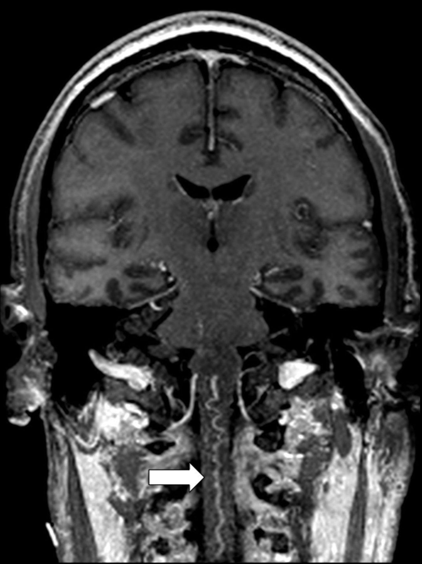
We previously encountered a similar case of a patient with edema and abnormal patchy enhancement in the medulla on MR imaging with initial misdiagnosis and treatment for neuromyelitis optica. Subsequent DSA demonstrated an arteriovenous fistula with an arterial feeder from the posterior division of the right middle meningeal artery draining into a tortuous transcerebellar vein into a mildly distended varicose anterior spinal vein (Figs 1 and 2). We also note that similar to the cases described by Copelan et al, the edema and enhancement pattern in the medulla demonstrate a characteristically straight upper border (Fig 3). It has previously been reported that dural AV shunts develop at the site where the bridging veins penetrate the dura and then reflux into the pontomesencephalic and anterior medullary systems, potentially resulting in brain stem or cerebellar venous ischemia2 and likely accounting for the straight upper border of edema due to a vascular etiology. We believe that the presence of medullary edema and enhancement with a straight upper border should alert the radiologist to the possibility of an underlying dural AV fistula.

Fig 1.
  • Download figure
  • Open in new tab
  • Download powerpoint
Fig 1.

Coronal T1 postcontrast MIP image demonstrates the anterior spinal vein, which is distended and mildly varicose.

Fig 2.
  • Download figure
  • Open in new tab
  • Download powerpoint
Fig 2.

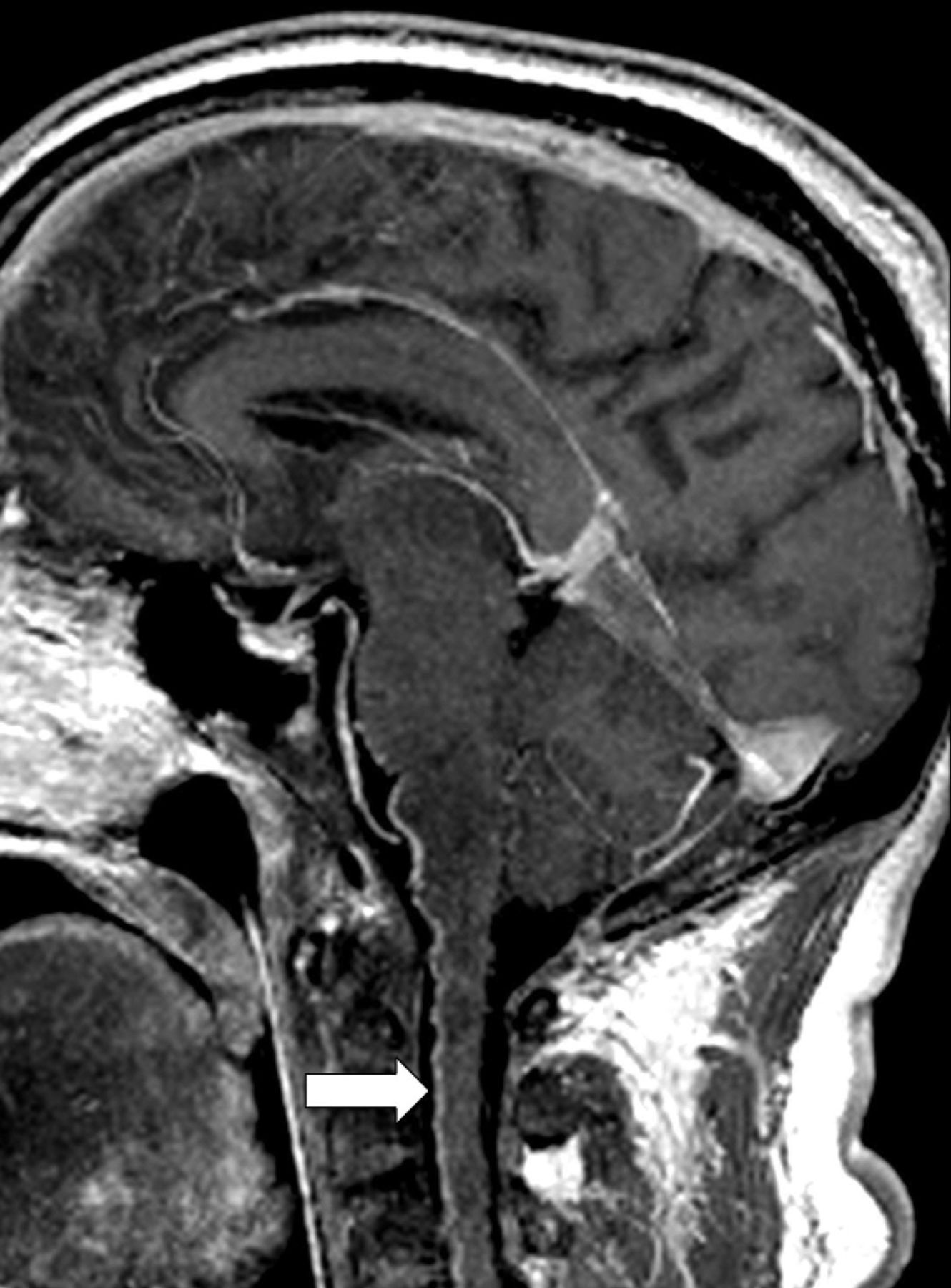
Sagittal T1 postcontrast MIP image demonstrates the anterior spinal vein, which is distended and mildly varicose.

Fig 3.
  • Download figure
  • Open in new tab
  • Download powerpoint
Fig 3.

Sagittal FLAIR demonstrates medullary edema with a straight upper border.

References

  1. 1.↵
    1. Copelan AZ,
    2. Krishnan A,
    3. Marin H, et al
    . Dural arteriovenous fistulas: a characteristic pattern of edema and enhancement of the medulla on MRI. AJNR Am J Neuroradiol 2018;39:238–44 doi:10.3174/ajnr.A5460 pmid:29217743
    Abstract/FREE Full Text
  2. 2.↵
    1. Alvarez H,
    2. Sasaki-Adams D,
    3. Castillo M
    . Resolution of brainstem edema after treatment of a dural tentorial arteriovenous fistula. Interv Neuroradiol 2015;21:603–08 doi:10.1177/1591019915591741 pmid:26116648
    CrossRefPubMed
  • © 2018 by American Journal of Neuroradiology
PreviousNext
Back to top

In this issue

American Journal of Neuroradiology: 39 (8)
American Journal of Neuroradiology
Vol. 39, Issue 8
1 Aug 2018
  • Table of Contents
  • Index by author
  • Complete Issue (PDF)
Advertisement
Print
Download PDF
Email Article

Thank you for your interest in spreading the word on American Journal of Neuroradiology.

NOTE: We only request your email address so that the person you are recommending the page to knows that you wanted them to see it, and that it is not junk mail. We do not capture any email address.

Enter multiple addresses on separate lines or separate them with commas.
Medullary Edema and Enhancement with a Straight Upper Border in Cases of Dural Arteriovenous Fistulas
(Your Name) has sent you a message from American Journal of Neuroradiology
(Your Name) thought you would like to see the American Journal of Neuroradiology web site.
CAPTCHA
This question is for testing whether or not you are a human visitor and to prevent automated spam submissions.
Citation Tools
Medullary Edema and Enhancement with a Straight Upper Border in Cases of Dural Arteriovenous Fistulas
D. Byrne, J.P. Walsh, T. Lynch, E.C. Kavanagh
American Journal of Neuroradiology Aug 2018, 39 (8) E90-E91; DOI: 10.3174/ajnr.A5681

Citation Manager Formats

  • BibTeX
  • Bookends
  • EasyBib
  • EndNote (tagged)
  • EndNote 8 (xml)
  • Medlars
  • Mendeley
  • Papers
  • RefWorks Tagged
  • Ref Manager
  • RIS
  • Zotero
Share
Medullary Edema and Enhancement with a Straight Upper Border in Cases of Dural Arteriovenous Fistulas
D. Byrne, J.P. Walsh, T. Lynch, E.C. Kavanagh
American Journal of Neuroradiology Aug 2018, 39 (8) E90-E91; DOI: 10.3174/ajnr.A5681
del.icio.us logo Digg logo Reddit logo Twitter logo CiteULike logo Facebook logo Google logo Mendeley logo
  • Tweet Widget
  • Facebook Like
  • Google Plus One
Purchase

Jump to section

  • Article
    • References
  • Figures & Data
  • Info & Metrics
  • References
  • PDF

Related Articles

  • No related articles found.
  • PubMed
  • Google Scholar

Cited By...

  • No citing articles found.
  • Crossref
  • Google Scholar

This article has not yet been cited by articles in journals that are participating in Crossref Cited-by Linking.

More in this TOC Section

  • Fair Performance of CT in Diagnosing Unilateral Vocal Fold Paralysis
  • Reply:
  • Regarding “Altered Blood Flow in the Ophthalmic and Internal Carotid Arteries in Patients with Age-Related Macular Degeneration Measured Using Noncontrast MR Angiography at 7T”
Show more LETTERS

Similar Articles

Advertisement

News and Updates

  • Lucien Levy Best Research Article Award
  • Thanks to our 2022 Distinguished Reviewers

Resources

  • Evidence-Based Medicine Level Guide
  • AJNR Podcast Archive
  • Librarian Resources
  • Terms and Conditions

Opportunities

  • Get Peer Review Credit from Publons

American Society of Neuroradiology

  • Neurographics
  • ASNR Annual Meeting
  • Fellowship Portal

© 2023 by the American Society of Neuroradiology | Print ISSN: 0195-6108 Online ISSN: 1936-959X

Powered by HighWire