
Abstract
BACKGROUND AND PURPOSE: Our purpose was to determine the characteristic MR features of early-onset (before age 2 years) versus late-onset (after age 2 years) globoid cell leukodystrophy (GLD).
METHODS: Thirty-four brain MR images in 22 patients with GLD were reviewed. A severity score (0 to 32), based on a point system derived from the location and extent of disease and the presence of focal and/or global atrophy, was calculated for each examination.
RESULTS: Of the 22 patients, three were asymptomatic and 19 were symptomatic. Ten patients had early-onset disease, whereas nine had late-onset disease. MR images of all patients showed abnormalities. In the early-onset group (n = 10; mean maximum MR score, 8.1; range, 3–18), 90% had pyramidal tract involvement, 80% had cerebellar white matter involvement, 70% had deep gray matter involvement, 60% had posterior corpus callosal involvement, 50% had parietooccipital white matter involvement, and 40% had cerebral atrophy. Serial MR imaging in four of these patients revealed progressive disease. In the late-onset group (n = 9; mean maximum MR score, 5.6; range, 4–10), 100% had pyramidal tract involvement, 100% had parietooccipital white matter involvement, 89% had posterior corpus callosal involvement, and none had cerebellar white matter involvement, deep gray matter involvement, or cerebral atrophy. Serial MR imaging in one patient with late-onset GLD did not reveal any change. A spectrum of findings was observed in the three patients who were asymptomatic.
CONCLUSION: Cerebellar white matter and deep gray matter involvement are present only in early-onset GLD. Pyramidal tract involvement is a characteristic finding in both early- and late-onset GLD. This scoring method for brain MR observations will assist in the objective assessment of the impact of hematopoietic stem cell transplantation in patients with GLD.
Globoid cell leukodystrophy (GLD), an autosomal recessive disorder caused by a deficiency of galactocerebrosidase, results in white matter degeneration associated with large macrophages. The clinical expression of the disease varies with age of onset. The early-onset form presents with fisting and hypertonicity and progresses rapidly to seizures, tonic spasms, and incapacitation. It culminates in death, often by 2 years of age. The late-onset form has a more protracted course. Hematopoietic stem cell (HSC) transplantation has been reported to halt disease progression in GLD by post-HSC transplantation brain MR observation (1). MR imaging readily provides early evidence of brain involvement. In this report, we present the MR imaging results of 22 patients with biochemically proved GLD and indicate that characteristic MR findings distinguish early- from late-onset disease.
Methods
The patient group was selected from families seen for consideration of HSC transplantation. Twenty-two patients with the biochemical defect for GLD were divided into early-onset (clinical symptoms before or at the age of 2 years), late-onset (after the age of 2 years), and asymptomatic groups. MR imaging of the brain was performed on a variety of imaging units from many institutions. Imaging at the University of Minnesota was performed on 1.5-T units. All MR images included T1-weighted sagittal images and T2-weighted axial images. Five of these patients have undergone successful HSC transplantation. The final population evaluated included 22 patients with 34 MR examinations.
A severity score (0 to 32) was calculated for each MR image according to a point system derived from the location and extent of involvement and the presence of focal and/or global atrophy by one neuroradiologist (Table 1). Each image was evaluated at least twice in separate reading sessions. The scoring system was based on a methodology used for evaluating adrenoleukodystrophy (ALD) (2, 3). In the GLD scoring system, the corticospinal projection fibers (pyramidal tract) were compartmentalized into three locations (supratentorial, internal capsule, and brain stem) instead of two compartments. The cerebellum was scored into two separate compartments (white matter and deep gray nuclei). The auditory pathway was not scored. The scoring system was otherwise identical to that used with ALD.
GLD scoring system
MR involvement referred to any signal changes within the brain parenchyma that could be explained by GLD. Involvement could be seen as T1 hyperintensity or hypointensity, T2 hyperintensity or hypointensity, or enhancement. MR severity scores and major areas of MR involvement were plotted or charted with respect to the patient's age at the time of the examination. Patients with serial MR studies were examined for changes over time.
Results
The age range of the 22 patients with GLD was 1 month to 18.5 years at the time of the MR examinations. All MR examinations revealed abnormalities, including images in three asymptomatic patients and three images in a patient before he became overtly symptomatic.
The severity scores of these patients were plotted versus age, as shown in Figure 1. The major areas of involvement on each MR study regarding clinical onset and age at the time of MR examination are outlined in Table 2.
GLD MR results regarding age

GLD MR severity score versus age. Connecting lines indicate patients with serial examinations
The mean maximum MR severity score for the early-onset group was 8.1 (range, 3–18). The mean maximum MR severity score for the late-onset group was 5.6 (range, 4–10). The mean MR severity score for the asymptomatic group was 3.2 (range, 1.5–5.0).
In the early-onset group of 10 patients, the MR findings, in order of declining percentages, were as follows: pyramidal tract, 90%; cerebellar white matter, 80%; deep gray matter, 70%; posterior corpus callosum, 60%; parietooccipital white matter, 50%; and atrophy, 40%. In the late-onset group of nine patients, MR findings were as follows: pyramidal tract, 100%; parietooccipital white matter, 100%; posterior corpus callosum, 89%; cerebellar white matter, 0%; deep gray matter, 0%; and atrophy, 0%.
Serial brain MR studies were available in five patients. On the basis of the severity scores, overall progression of MR disease was found in the four patients with early-onset GLD. A late-onset GLD patient with two MR studies was stable over a 6-month period.
Discussion
Our results (Table 2) and a review of the literature (4–21) (Table 3) indicate that GLD has a characteristic MR pattern that depends on the age of onset. We divided GLD into early-onset (clinical presentation before or at the age of 2 years) and late-onset forms (clinical presentation after the age of 2 years). The early-onset group included patients classified in the literature as infantile or late infantile onset, whereas the late-onset group included patients classified as early juvenile, late juvenile, and adult onset. While these categories can be clinically useful with regard to disease course and prognosis, the only successful form of therapy in both early- and late-onset GLD is HSC transplantation (1).
Review of literature: MR findings in 27 patients with GLD ordered by age of onset
In patients with clinical symptoms before or at the age of 2 years, GLD usually involved the cerebellar white matter, the deep gray nuclei (dentate, basal ganglia, and/or thalamic), and the pyramidal tract (corticospinal tract), with later atrophy and involvement of the corpus callosum (posterior body and/or splenium) and supratentorial parietooccipital white matter (Figs 2 and 3). Finelli et al (9) and others have emphasized the difficulty in identifying brain involvement with MR imaging in early-onset GLD because of subtle deep gray nuclear abnormalities. The deep gray nuclear involvement may be subtle in the basal ganglia region and thalamus and can usually be better detected on CT than on MR imaging (Fig 3). The MR signal abnormalities in the basal ganglia and thalamic nuclei were variable on T1- and T2-weighted sequences, but usually decreased on T1-weighted sequences and increased on T2-weighted sequences. The cause of the basal ganglia and thalamic abnormalities remains unknown. The findings may be related to transient calcifications or to alterations in lipids, water, and proteins attributable to myelin breakdown (6).

Patient 1: early-onset GLD.
A–D, Axial T2-weighted images (4000/100/2 [TR/TE/excitations]) at 4 months of age show increased signal intensity in the dentate nuclei (curved arrows in A, target lesions), in cerebellar white matter (arrowheads in A), the brain stem pyramidal tract (arrows in B), the pyramidal tract within the posterior limb of the internal capsule (arrows in C), and the pyramidal tract in the corona radiata (arrows in D), resulting in a GLD MR severity score of 5.
E–H, Axial T2-weighted images (3000/80/1) at 10 months of age show stable dentate nuclear involvement (curved arrows in E), progressive cerebellar white matter (arrowheads in E), and progressive pyramidal tract involvement (arrows in F, G, and H). The development of moderate global atrophy, as evidenced by a widened third ventricle width (8 mm), widened frontal horn ratio (41%), and widened subarachnoid spaces, results in an MR severity score of 7.
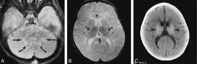
Patient 5: early-onset GLD.
A and B, Axial T2-weighted images (3000/80/1) show subtle increased signal intensity within the cerebellar white matter (straight arrows in A), dentate nuclei (curved arrows in A), and posterior limbs of the internal capsule (arrows in B). There is relatively normal signal intensity within the basal ganglia and thalami. These are the characteristic MR findings in early-onset GLD. Because of the coexistent involvement of the corona radiata pyramidal tract, the MR severity score was 4.
C, Axial noncontrast CT scan at 7 months of age (2 weeks after MR imaging in B) shows abnormal increased density within the basal ganglia and thalamic nuclei (arrowheads).
In comparison with frequent subtle basal ganglia and thalamic findings, signal abnormalities in cerebellar gray and white matter were readily visible on MR images and were the earliest findings, along with pyramidal tract involvement, in the group with early-onset GLD. The dentate nuclei often had a target appearance with a high signal intensity center (8) (Fig 2). Barone et al (6) and others have reported cerebellar white matter involvement to be a primary finding in early-onset GLD. Abnormal findings in the cerebellar white matter were present in one of our patients at 1 month of age. Vanhanen et al (13) have emphasized the importance of cerebellar findings in differentiating infantile GLD from Rett syndrome and infantile neuronal ceroid lipofuscinosis.
Early diagnosis of GLD brain involvement can be determined with MR imaging if the cerebellar signal abnormalities and pyramidal tract involvement are detected in the developing brain, which is incompletely myelinated before 18 months of age. Posterior corpus callosal involvement, supratentorial white matter involvement, and atrophy often develop within 2 months to 1 year of age in early-onset GLD if not present on the initial MR image.
The most common imaging findings in late-onset GLD included involvement of the pyramidal tract, corpus callosum, and parietooccipital white matter (Fig 4). None of the patients in our series examined clinically after the age of 2 years had cerebellar white matter or deep gray matter involvement. Although Barone et al (6) described a patient with basal ganglia involvement at age 12 years, this patient's clinical age of onset was 2 years, classifying this patient with the early-onset form of GLD. Cerebral atrophy was also not characteristically observed in the late-onset group, both in our series and in the literature.

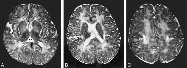
Patient 17: late-onset GLD.
A, Axial fluid-attenuated inversion recovery MR image (6500/105, 180/1) shows symmetric increased signal intensity within the pyramidal tract (arrows) and parietooccipital white matter (arrowheads).
B, Axial T2-weighted MR image (3400/90/1) shows increased signal intensity in the posterior body of the corpus callosum (curved arrows) as well as the pyramidal tract (straight arrows).
Our group of three asymptomatic patients consisted of a 16-month-old patient with cerebellar white matter, basal ganglia, and posterior corpus callosal involvement; a 2-year 7-month-old patient with isolated parietooccipital involvement; and an 8-year-old patient with posterior corpus callosal and parietooccipital involvement. Interestingly, none of our asymptomatic patients had pyramidal tract involvement. Since the pyramidal tract involves the motor pathways, it was not surprising that its involvement by GLD was clinically apparent.
Pyramidal pathway involvement was the most characteristic finding in both infantile- and late-onset GLD. Of our symptomatic patients and the patients from the literature review, 96% had pyramidal tract abnormalities revealed by initial MR imaging. Of our four patients without pyramidal tract involvement, three were asymptomatic. Barone et al (6) emphasized that pyramidal tract involvement was a leading radiologic sign in the late-onset form of GLD. Our data indicated that this involvement was a leading radiologic sign in all forms of GLD. In patients of all ages, pyramidal pathway involvement from the motor cortex through the corona radiata, internal capsule, and brain stem was rather striking, unless the white matter had not yet myelinated. In infants with immature white matter, pyramidal tract involvement can be identified by a higher signal intensity than the normal high signal intensity of immature white matter on T2-weighted pulse sequences (Fig 5). The MR studies in two of our patients with early-onset GLD were initially mistakenly interpreted as showing normal findings because of the failure to recognize pyramidal tract and cerebellar abnormalities.

Patient 6: early-onset GLD.
A–C, Axial T2-weighted images (2800/80/1) at 7 months of age show increased signal intensity in the pyramidal tract (arrows in A–C). Pyramidal tract involvement in children with white matter that has not yet myelinated can be identified by higher than normal high signal intensity of immature white matter on T2-weighted images.
Corpus callosal involvement was also a characteristic MR finding in both early- and late-onset GLD. Six of our 10 patients with early-onset GLD had corpus callosal involvement detected on MR studies, although only four of these patients had corpus callosal involvement detected on their initial MR study. Our four patients with early-onset GLD who did not have corpus callosal involvement did not undergo MR imaging after 10 months of age. A review of the literature indicated similar findings regarding corpus callosal involvement in the patients with early-onset GLD, with 10 of 11 patients having involvement detected at the time of their last MR study, although only seven of these patients had documented corpus callosal involvement on their initial examination. In late-onset GLD, eight of our nine patients and all of the patients reviewed in the literature had corpus callosal involvement. In all cases in which the corpus callosum was involved, the site of abnormality was within the posterior body or splenium of the corpus callosum.
Because of the usual involvement of the posterior portion of the corpus callosum, pyramidal tract, and parietal white matter, the late-onset form of GLD can mimic ALD (2). Where pyramidal tract involvement tends to be a key feature in GLD, it is more of a secondary feature of ALD. Most cases of ALD have dramatic visual and auditory pathway involvement. In comparison, auditory pathway involvement is not seen in GLD, and visual pathway involvement in GLD is more anterior than ALD. Optic hypertrophy and optic atrophy may occur in GLD (7, 22). With MR imaging, the 15% of patients with the pyramidal form of ALD may appear similar to patients with late-onset GLD (2). Other diseases with pyramidal tract involvement, such as amyotrophic lateral sclerosis, should also be included in the differential diagnosis of late-onset GLD. Late-onset GLD can also mimic an infiltrating glioma (14).
MR imaging revealed symmetric findings, with two exceptions. The only patients in this series with asymmetric disease were monochorionic monoamniotic twins, whose MR images showed nearly mirror image involvement (23) (Fig 6).

Patients 18 and 19: late-onset GLD.
A, Axial T2-weighted image (2700/90/1) in patient 19 shows asymmetric increased signal intensity in the right coronal radiata pyramidal tract (straight arrow) and the right parietooccipital white matter (curved arrow).
B, Axial T2-weighted image (2700/90/1) in patient 18 (monozygotic twin of patient 19) shows asymmetric increased signal in the left corona radiata pyramidal tract (straight arrow) and bilateral increased signal in the parietooccipital white matter (curved arrows).
Our data and the findings in the literature showed progressive disease by MR imaging in early-onset GLD (5–8, 11, 19). Results of serial examinations in our four patients with early-onset GLD indicated an overall progression of MR observations (on the basis of the final MR severity scores being higher than the initial MR severity score), with one exception. In one of these patients, the last MR severity score actually improved after treatment of severe hydrocephalus, resulting in less white matter signal abnormalities because of the resolution of hydrocephalus-induced transependymal flow. This was a limitation of the scoring system, in that it was often not possible to prospectively distinguish abnormalities caused directly by GLD from those caused indirectly by other processes.
Late-onset GLD may also have progressive MR findings, although at a slower time interval (6, 15, 17, 22). Our single patient with a serial examination was stable only over a 6-month interval. One of our late-onset patients was previously reported to have a progression of MR findings (22).
The major use of the GLD MR severity score is to provide a measure for brain involvement. It was designed to be applicable to standard MR examinations and reproducible by any neuroradiologist or other individual familiar with MR neuroanatomy of the pediatric brain. Both T1- and T2-weighted images are useful in determining a severity score. Besides determining global or focal atrophy, T1-weighted images are also useful in determining basal ganglia involvement and supratentorial white matter involvement in the immature white matter in early-onset GLD. The scoring system has limitations in the early-onset form of GLD. Hydrocephalus and its transependymal flow-induced white matter signal abnormalities can mimic the GLD white matter involvement. Determining whether ventricular enlargement is attributable to atrophy or hydrocephalus can also be problematic. Along with neuropsychological testing and clinical examination, this MR scoring system is being used as a tool to evaluate asymptomatic and symptomatic patients with GLD and to monitor their responses to therapy. On the basis of the MR severity score, a group of selected GLD patients who have undergone successful HSC transplantation have experienced either stabilization or improvement in MR observations (1).
Conclusion
GLD has characteristic findings on brain MR imaging according to age of clinical onset. In both the early- and late-onset forms of the disease, pyramidal tract involvement was a characteristic imaging finding. In early-onset GLD, the leading imaging findings were signal abnormalities within cerebellar white matter, the deep gray nuclei, and the pyramidal tract. Brain involvement in the parietooccipital white matter and corpus callosum developed rapidly, along with global atrophy. In late-onset GLD, MR brain involvement was characterized by signal abnormalities within the pyramidal tract, posterior corpus callosum, and parietooccipital white matter. Knowledge of these characteristic brain MR findings contribute to rapid and specific diagnosis of GLD in the proper clinical setting.
Footnotes
↵1 Address reprint requests to Daniel J. Loes, MD, Suburban Radiologic Consultants, Ltd, 6401 France Ave South, Edina, MN 55435.
References
- Received March 10, 1998.
- Copyright © American Society of Neuroradiology
In this issue


Jump to section
Related Articles
Cited By...
- Novel approaches to quantify CNS involvement in children with Pompe disease
- Brain MRI features and scoring of leukodystrophy in adult-onset Krabbe disease
- MRI characteristics and scoring in HDLS due to CSF1R gene mutations
- Teaching NeuroImages: Selective corticospinal tract involvement in late-onset Krabbe disease
- Metachromatic Leukodystrophy: A Scoring System for Brain MR Imaging Observations
- Peripheral nerve involvement in Krabbe disease: a guide to therapy selection and evaluation.
- Krabbe disease: Neurophysiologic studies and MRI correlations
- MR Imaging and Proton MR Spectroscopy in Adult Krabbe Disease