
Abstract
Summary: Benign ductal cysts of the accessory lacrimal glands are uncommon lesions of the orbit, arising from the glands of Wolfring and Krause. We report two patients with histopathologically proved cysts in whom CT scans revealed well-circumscribed extraconal cystic lesions adjacent to the globe, involving both eyelids. Radiologists should be aware of these rare lesions so as to include them in the differential diagnosis of orbital cysts.
Tear secretion is a function of both the major and the accessory lacrimal glands. The major glands are located in the anterior superolateral part of the orbit and are responsible for reflex tear secretion. The accessory lacrimal glands (of Wolfring and Krause) are dispersed among the conjunctival fornices and the upper tarsal border and are responsible for basal tear secretion. Obstruction of the excretory duct of any lacrimal gland results in the entity called dacryops, or simple ductal cyst. We describe the CT appearance of dacryops of the accessory lacrimal glands in two patients.
Case Reports
Case 1
A 4-year-old boy presented with a 3-month history of a bulge in his right lower lid, which had been increasing progressively in size. No history of orbital trauma or inflammation could be elicited. Ophthalmologic examination was normal. Exophthalmometry revealed only a 1-mm difference between the right and left side. There was no increase in the lid bulge or in the exophthalmometric measurement upon Valsalva maneuver. A CT scan of the orbits revealed a 1 × 0.8-cm well-circumscribed rounded extraconal cystic lesion near the right orbital floor, abutting the inferior aspect of the globe and in continuity with the lower eyelid (Fig 1). Complete excision of the lesion was carried out through a transconjunctival lower lid approach. Histologic examination revealed a cyst lined by a layer of cuboidal epithelium surrounded by a layer of spindled myoepithelial cells. In areas, the epithelium was flattened. The cyst wall was thin and consisted of chronically inflamed vascularized fibrous tissue.

4-year-old boy with right lower lid bulge.
A and B, Axial (A) and coronal (B) unenhanced CT scans of the orbits show a well-defined water-density rounded lesion compatible with a cyst in the orbital floor abutting the inferior aspect of the globe and in continuity with the mid aspect of the lower eyelid (arrows, A). A pathologic specimen confirmed a simple ductal cyst of the accessory lacrimal glands.
Case 2
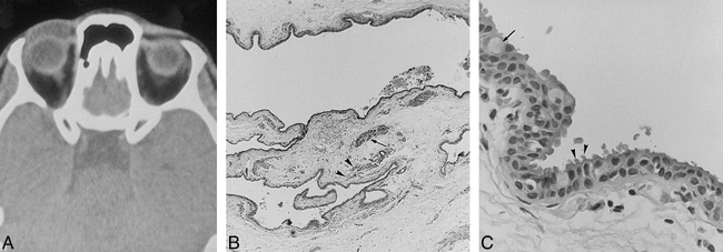
A 37-year-old woman presented with a mass in the right upper eyelid. The lesion had appeared 1 year earlier and had been growing in size intermittently. No history of trauma or prior inflammatory conditions was present. Physical examination revealed a 2.5 × 2-cm movable soft-tissue lesion in the medial aspect of the left upper lid. A CT scan of the orbits showed a 2-cm well-circumscribed cystic lesion in the central and medial aspect of the left upper lid, adjacent to the anterior aspect of the globe (Fig 2A). The lesion was completely excised. Histopathologic examination showed a collapsed cyst lined by two distinct cell layers. The luminal cell layer showed cytoplasmic outpouchings as well as interspersed goblet cells. The cyst wall showed patchy infiltrates of lymphocytes and plasma cells as well as a few glands (Fig 2B and C).

37-year-old woman with right upper lid lesion of 1 year's duration, located mainly in its medial aspect.
A, Axial CT scan of the superior aspect of the orbits reveals a cystic lesion of the right upper eyelid, inseparable from the anterior aspect of the globe. Histopathologic findings (see B and C) were consistent with a benign ductal cyst of the accessory lacrimal glands. The cyst probably arose from the glands of Wolfring.
B, Low-power view shows a collapsed cyst with patches of chronic inflammatory cells (arrowheads) and a few glands (arrow) in its wall (H and E, original magnification ×30).
C, High-power view of the cyst lining shows a double cell layer, confirming the ductal origin of the cyst. Cytoplasmic apical microvillous processes (arrowheads) from many of the luminal cells are noted, consistent with decapitation secretion. Occasional goblet cells are also seen (arrow) (H and E, original magnification ×400).
Discussion
Lacrimal glandular tissue is present in both the major and the accessory lacrimal glands, and, rarely, in ectopic locations within the orbit (1). Each of these glands is partly responsible for tear secretion. The major lacrimal gland is divided into two lobes, the orbital and the palpebral. The major lacrimal gland is located in the anterior superotemporal quadrant of the orbit and is mainly responsible for reflex tear secretion through ductules that empty directly into the upper outer fornix. The accessory lacrimal glands of Wolfring and Krause are responsible for basal tear secretion and also differ from the major gland with respect to their location. Wolfring glands are located in and around the upper tarsal border and, to a lesser extent, in the lower tarsal border (2). Krause glands are located within the conjunctiva of both upper and lower fornices (2) (Fig 3). The sole duct of each accessory gland empties directly onto the adjacent conjunctival surface.

Drawing (sagittal view) of the upper eyelid and fornix shows the location of the accessory lacrimal glands of Wolfring and Krause. The former are close to the tarsus, while the latter are located at the fornix. The ciliary glands (of Zeis and Moll), and the tarsal Meibomian glands are also illustrated
A ductal cyst of the major lacrimal gland, or dacryops, is a rare clinical entity, encountered mainly in its palpebral lobe (1, 3, 4). Ductal cysts of the accessory lacrimal glands of Wolfring and Krause are even more uncommon than those of the major lacrimal gland and are rarely reported in the ophthalmologic literature (1, 2, 5–7). Clinically, cysts of the accessory gland develop insidiously and present as painless cystic masses of the lids. They are of variable size and are located in the previously described anatomic locations of the accessory glands. Most frequently, these cysts occur in patients with previous traumatic or inflammatory conditions of the conjunctiva, particularly in patients with trachoma (2, 5). Less commonly, a congenital anomaly of an excretory duct or alteration in the composition of secretions may be the cause of cyst formation (1). Histopathologically, these lesions are simple benign cysts lined by a layer of nonciliated cuboidal epithelial cells and occasional goblet cells, with an underlying layer of myoepithelial cells (Fig 2C). The cyst wall consists of fibrous tissue of variable thickness, often infiltrated by lymphocytes and plasma cells (Fig 2B) (1, 2, 4). Decapitation secretion has been described in many cysts (Fig 2C). The double-layered lining of these cysts supports their ductal origin. These histologic features differentiate the accessory lacrimal gland cysts from the more common dermoid and epidermoid cysts of the orbit. These latter cysts are in fact lined by stratified squamous epithelial cysts and do not contain goblet cells (1).
CT findings of ductal cysts involving the major lacrimal glands have been reported previously, and consist of fluid CT-density lesions in the region of the lacrimal glands, lateral to the globe (3, 8). In a few cases, the lacrimal cysts had soft-tissue density on CT scans (1, 4).
To our knowledge, CT findings of ductal cysts of the accessory lacrimal glands have not been reported previously. In our patients, the CT scans showed the cystic nature of the lesions and their location in the upper and lower eyelids without evidence of a bony abnormality. The location of the lesion in case 2 denotes most probably a Wolfring ductal cyst. In case 1, the cyst may have arisen from either Wolfring or Krause glands.
The main differential diagnosis of these benign ductal cysts is dermoid or epidermoid cysts (1, 4, 8). Histopathologic examination can help one readily differentiate these entities. Nevertheless, CT scans may also be helpful in the diagnosis by showing the fat-density characteristic of dermoid and epidermoid cysts, as well as a bony fossa in the adjacent orbit, because these lesions arise within the diploe of bone. Another important differential diagnosis is echinococcus, or hydatid, cysts of the orbit, which is a rare entity (1, 2, 4). Subconjunctival cysticercosal cyst is also a consideration (2).
The management of choice of ductal cysts of the accessory lacrimal glands is total surgical excision of the intact cyst through a conjunctival route (1, 2). Failure to do this may lead to recurrence or fistula formation.
Conclusion
We have reported the CT findings of accessory lacrimal glands ductal cysts, a rare disease not previously reported in the radiologic literature. These cysts should be included in the differential diagnosis of cystic disease of the orbit. CT is helpful in delineating the cystic nature of the lesion as well as its location, size, and relationship to the globe and bony orbit.
Footnotes
↵1 Address reprint requests to Nabil J. Khoury, MD, American University of Beirut, 850 Third Ave, 18th Floor, New York NY, 10022.
- Received October 6, 1998.
- Accepted after revision February 1, 1999.
- Copyright © American Society of Neuroradiology


