
Article Figures & Data
Figures
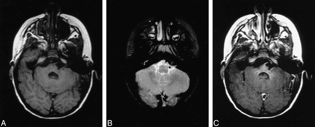
- fig 1.
A, Axial T1-weighted (700/17/1) MR image shows mild asymmetry of the lower basis pontis and middle cerebellar peduncle on the left. Note the geographic region of subtle hypointensity centrally and to the left of midline and questionable punctate hyperintense foci near the midline.
B, Axial T2-weighted (2200/90/1) image showing abnormal hyperintensity within the left ventral portion of the brain stem at the level of the inferior cerebellar peduncle/medulla. This focus is slightly caudal to the region of hypointensity seen in figure 1B.
C, Post-gadolinium T1-weighted (700/19/1) axial image reveals homogeneous enhancement within the rostral pons and adjacent cerebellar peduncles. The magnitude of contrast enhancement is disproportionately greater than the focal signal changes seen on the T2-weighted images (see figure 1B).
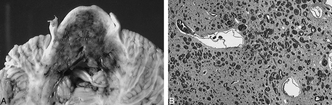
- fig 2.
A, Gross anatomic specimen showing axial section through mid-pontine level. The capillary malformation is clearly demarcated by the extensive bluish discoloration involving nearly the entire cross-sectional area of the basis pons and tegmentum with extension into the middle cerebellar peduncles. There are patchy areas of relatively more dense discoloration representing increased density of telangiectasia.
B, Microscopic specimen showing the basis pontis. Vast numbers of blood-filled endothelium-lined vascular channels measuring 50–150 microns in diameter have replaced much of the brain parenchyma. There is also gliosis and neuronal drop-out, but no evidence of hemorrhage.


