
Article Figures & Data
Figures
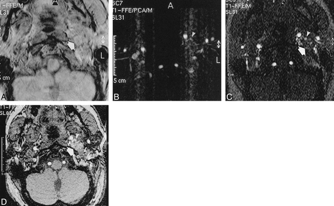
- fig 1.
A 38-year-old-man with a left-sided vagal paraganglioma.
A, PC magnitude image (26/9/2/20° [TR/TE/excitations/flip angle]) shows good tumor visualization (large arrow).
B, PC image shows feeding vessels (arrowheads), but ghost artifacts from pulsatility of vessels degrades the image.
C, 2D TOF axial source image (28/5.1/2/60° [TR/TE/excitations/flip angle]) only shows feeding branches from the ascending pharyngeal artery (arrowheads), but the margins of the tumor are only poorly visualized (large arrow).
D, 3D TOF axial source image (25/6.9/1/20° [TR/TE/excitations/flip angle]) shows good visualization of the tumor (large arrow) and also nicely depicts the feeding branches from the ascending pharyngeal artery (arrowheads).
- fig 2.
Graph of ROC curves of reader 1 for the three MR angiography techniques shows the highest area under the curve for 3D TOF MR angiography. Areas under the curves (AUC) for both readers are given
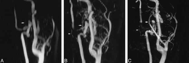
- fig 3.
A 38-year-old-man with a left-sided vagal paraganglioma. Slightly anteroposterior, oblique MIPs are presented. A muscular branch from the vertebral artery adds to the extensive vascularization of this tumor.
A, 3D PCA MIP (26/9/2/20° [TR/TE/excitations/flip angle]) hardly shows a branch from the occipital artery (arrow) as tumor feeder. The muscular branch from the vertebral artery is not at all visible.
B, 2D TOF MIP (28/5.1/2/60° [TR/TE/excitations/flip angle]) not only shows supply from the occipital artery (arrow), but a small feeding branch from the vertebral artery (arrowhead) also is depicted.
C, 3D TOF MIP (25/6.9/1/20° [TR/TE/excitations/flip angle]) shows more clearly than the 2D TOF MIP vascularization from the occipital artery (arrow) as well as from the vertebral artery (arrowhead).
- fig 4.
A 53-year-old-man with bilateral carotid body tumors.
A, Anteroposterior view of the 3D TOF MIP (25/6.9/1/20° [TR/TE/excitations/flip angle]) shows the thyrocervical trunk (arrowheads), adding to the vascularization of the carotid body tumor.
B, On the aortic arch angiogram, showing both carotid body tumors, the vascularization from the thyrocervical trunk (arrowheads) was confirmed.
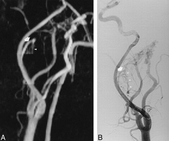
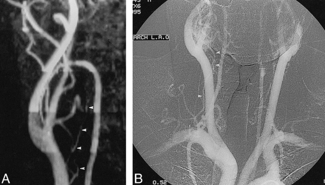
- fig 5.
A 49-year-old-man with a vagal paraganglioma (lateral view).
A, 3D TOF MIP (25/6.9/1/20° [TR/TE/excitations/flip angle]) shows enlarged ascending pharyngeal artery (large arrow) as a feeder despite the fact that the cranially placed slab for saturation of venous flow obscures the descending trunk of this artery (arrowheads).
B, On the digital subtraction angiogram, the complete course of the ascending pharyngeal artery (large arrow), including its descending trunk (arrowheads), is visible.


Tables
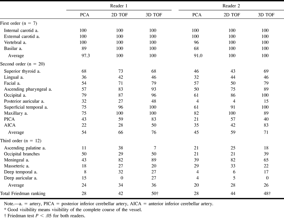
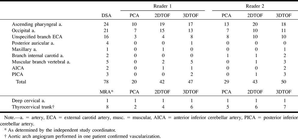
 TABLE 1:
Percentage of good visibility* of 39 normal neck arteries as determined by the three different MR angiography techniques for both readers
- TABLE 2:
Number of feeding arteries as detected by the three different MR angiography techniques for both readers as compared with DSA, the standard of reference. Results were divided by putting the threshold between probably and equivocally present rankings
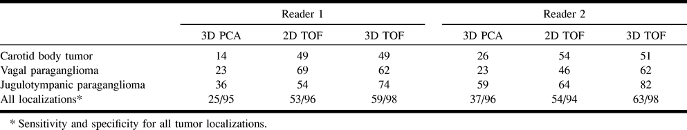
- TABLE 3:
Sensitivity percentage for detection of feeding arteries per tumor localization for reader 1 and 2. Results were divided by putting the threshold between probably and equivocally present rankings


