Advertisement

Main menu

  • Home
  • Content
    • Current Issue
    • Publication Preview--Ahead of Print
    • Past Issue Archive
    • Case of the Week Archive
    • Classic Case Archive
    • Case of the Month Archive
    • COVID-19 Content and Resources
  • For Authors
  • About Us
    • About AJNR
    • Editors
    • American Society of Neuroradiology
  • Submit a Manuscript
  • Podcasts
    • Subscribe on iTunes
    • Subscribe on Stitcher
  • More
    • Subscribers
    • Permissions
    • Advertisers
    • Alerts
    • Feedback
  • Other Publications
    • ajnr

User menu

  • Subscribe
  • Alerts
  • Log in

Search

  • Advanced search
American Journal of Neuroradiology

American Journal of Neuroradiology

  • Subscribe
  • Alerts
  • Log in

Advanced Search

  • Home
  • Content
    • Current Issue
    • Publication Preview--Ahead of Print
    • Past Issue Archive
    • Case of the Week Archive
    • Classic Case Archive
    • Case of the Month Archive
    • COVID-19 Content and Resources
  • For Authors
  • About Us
    • About AJNR
    • Editors
    • American Society of Neuroradiology
  • Submit a Manuscript
  • Podcasts
    • Subscribe on iTunes
    • Subscribe on Stitcher
  • More
    • Subscribers
    • Permissions
    • Advertisers
    • Alerts
    • Feedback
  • Follow AJNR on Twitter
  • Visit AJNR on Facebook
  • Follow AJNR on Instagram
  • Join AJNR on LinkedIn
  • RSS Feeds
Research ArticleBRAIN

Intraventricular CSF Pulsation Artifact on Fast Fluid-Attenuated Inversion-Recovery MR Images: Analysis of 100 Consecutive Normal Studies

Rohit Bakshi, Shelton D. Caruthers, Vallabh Janardhan and Mohammad Wasay
American Journal of Neuroradiology March 2000, 21 (3) 503-508;
Rohit Bakshi
aFrom the Dent Neurologic Institute, Lucy Dent Imaging Center, Kaleida Health-Millard Fillmore Hospital, Buffalo, NY (R.B., V.J., M.W.); and Philips Medical Systems, Magnetic Resonance Division, Boston, MA (S.D.C.).
  • Find this author on Google Scholar
  • Find this author on PubMed
  • Search for this author on this site
Shelton D. Caruthers
aFrom the Dent Neurologic Institute, Lucy Dent Imaging Center, Kaleida Health-Millard Fillmore Hospital, Buffalo, NY (R.B., V.J., M.W.); and Philips Medical Systems, Magnetic Resonance Division, Boston, MA (S.D.C.).
  • Find this author on Google Scholar
  • Find this author on PubMed
  • Search for this author on this site
Vallabh Janardhan
aFrom the Dent Neurologic Institute, Lucy Dent Imaging Center, Kaleida Health-Millard Fillmore Hospital, Buffalo, NY (R.B., V.J., M.W.); and Philips Medical Systems, Magnetic Resonance Division, Boston, MA (S.D.C.).
  • Find this author on Google Scholar
  • Find this author on PubMed
  • Search for this author on this site
Mohammad Wasay
aFrom the Dent Neurologic Institute, Lucy Dent Imaging Center, Kaleida Health-Millard Fillmore Hospital, Buffalo, NY (R.B., V.J., M.W.); and Philips Medical Systems, Magnetic Resonance Division, Boston, MA (S.D.C.).
  • Find this author on Google Scholar
  • Find this author on PubMed
  • Search for this author on this site
  • Article
  • Figures & Data
  • Info & Metrics
  • References
  • PDF
Loading

Article Figures & Data

Figures

  • Tables
  • fig 1.
    • Download figure
    • Open in new tab
    • Download powerpoint
    fig 1.

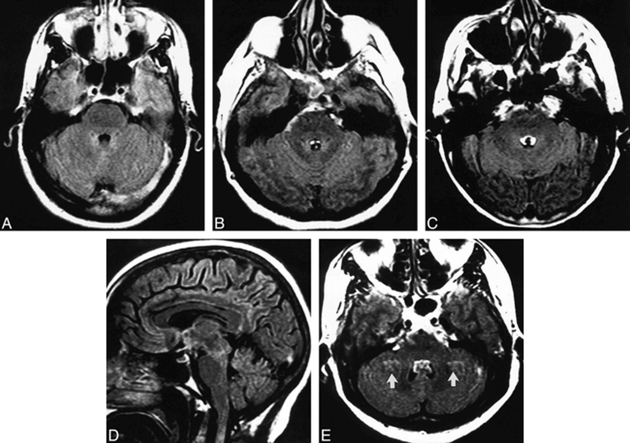
    Fourth ventricular CSF pulsation artifact in four subjects.

    A–C, FLAIR axial images of three patients show increasing severity of fourth ventricular CSF pulsation artifact with increasing age: grade 0 in a 32-year-old (A); grade 1 in a 43-year-old (B); and grade 2 in a 71-year-old (C). Subarachnoid CSF artifact is also present in the basal cisterns in B and C.

    D, The fourth ventricular CSF pulsation artifact that was present on the axial FLAIR image (C) is not visible on this sagittal FLAIR image.

    E, FLAIR axial image of a subject with fourth ventricular CSF pulsation artifact also shows ghost pulsation artifacts (arrows) in the phase-encoding axis (left to right), causing superimposition of hyperintensities on the bilateral cerebellar parenchyma.

  • fig 2.
    • Download figure
    • Open in new tab
    • Download powerpoint
    fig 2.

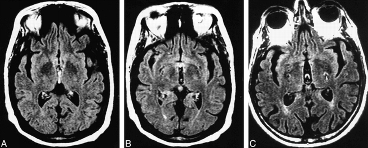
    Third ventricular CSF pulsation artifact in two subjects.

    A and B, Contiguous FLAIR axial images of a 67-year-old man referred for dizziness. The patient has a grade 2 third ventricular VCSFA in the inferior aspect of the third ventricle (A) and a large third ventricle.

    C, FLAIR axial image of a subject with third ventricular CSF pulsation artifact also has bilateral ghost pulsation artifacts in the phase-encoding axis (left to right), causing superimposition of hyperintensities on the bilateral adjacent brain. The most prominent ghost artifact (arrow) is in the left basal ganglia.

  • fig 3.
    • Download figure
    • Open in new tab
    • Download powerpoint
    fig 3.

    Lateral ventricular CSF pulsation artifact in two subjects.

    A and B, Contiguous FLAIR axial images of a 70-year-old man show focal lateral ventricular CSF pulsation artifacts bilaterally, more prominent on the right, in the region of the foramen of Monro (arrows).

    C, A subject with lateral ventricular CSF pulsation artifact (open arrow) also has ghost pulsation artifacts in the phase-encoding axis (left to right), causing superimposition of hyperintensities on the bilateral adjacent brain parenchyma.

  • fig 4.
    • Download figure
    • Open in new tab
    • Download powerpoint
    fig 4.

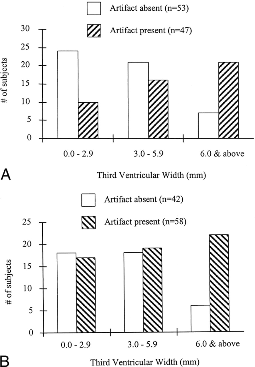
    A, The group with third ventricular VCSFA had significantly larger third ventricles (P < .0001) than did those without third ventricular VCSFA. For illustrative purposes, the patients are divided into categories based on small, medium, and large third ventricles.

    B, The group with fourth ventricular VCSFA had significantly larger third ventricles (P < .01) than did those without fourth ventricular VCSFA. For illustrative purposes, the patients are divided into categories based on small, medium, and large third ventricles.

Tables

  • Figures
  • Table1

    Ventricular CSF pulsation artifact on fast FLAIR MR images as related to sex, age, and size of third ventricle

PreviousNext
Back to top

In this issue

American Journal of Neuroradiology
Vol. 21, Issue 3
1 Mar 2000
  • Table of Contents
  • Index by author
Advertisement
Print
Download PDF
Email Article

Thank you for your interest in spreading the word on American Journal of Neuroradiology.

NOTE: We only request your email address so that the person you are recommending the page to knows that you wanted them to see it, and that it is not junk mail. We do not capture any email address.

Enter multiple addresses on separate lines or separate them with commas.
Intraventricular CSF Pulsation Artifact on Fast Fluid-Attenuated Inversion-Recovery MR Images: Analysis of 100 Consecutive Normal Studies
(Your Name) has sent you a message from American Journal of Neuroradiology
(Your Name) thought you would like to see the American Journal of Neuroradiology web site.
CAPTCHA
This question is for testing whether or not you are a human visitor and to prevent automated spam submissions.
Citation Tools
Intraventricular CSF Pulsation Artifact on Fast Fluid-Attenuated Inversion-Recovery MR Images: Analysis of 100 Consecutive Normal Studies
Rohit Bakshi, Shelton D. Caruthers, Vallabh Janardhan, Mohammad Wasay
American Journal of Neuroradiology Mar 2000, 21 (3) 503-508;

Citation Manager Formats

  • BibTeX
  • Bookends
  • EasyBib
  • EndNote (tagged)
  • EndNote 8 (xml)
  • Medlars
  • Mendeley
  • Papers
  • RefWorks Tagged
  • Ref Manager
  • RIS
  • Zotero
Share
Intraventricular CSF Pulsation Artifact on Fast Fluid-Attenuated Inversion-Recovery MR Images: Analysis of 100 Consecutive Normal Studies
Rohit Bakshi, Shelton D. Caruthers, Vallabh Janardhan, Mohammad Wasay
American Journal of Neuroradiology Mar 2000, 21 (3) 503-508;
Reddit logo Twitter logo Facebook logo Mendeley logo
  • Tweet Widget
  • Facebook Like
  • Google Plus One

Jump to section

  • Article
    • Abstract
    • Methods
    • Results
    • Discussion
    • Conclusion
    • Footnotes
    • References
  • Figures & Data
  • Info & Metrics
  • References
  • PDF

Related Articles

  • No related articles found.
  • PubMed
  • Google Scholar

Cited By...

  • What are White Matter Hyperintensities Made of? Relevance to Vascular Cognitive Impairment
  • Double Inversion Recovery MR Sequence for the Detection of Subacute Subarachnoid Hemorrhage
  • Morphologic, Distributional, Volumetric, and Intensity Characterization of Periventricular Hyperintensities
  • White matter hyperintensities on MRI in high-altitude U-2 pilots
  • 3D Fluid-Attenuated Inversion Recovery Imaging: Reduced CSF Artifacts and Enhanced Sensitivity and Specificity for Subarachnoid Hemorrhage
  • Analysis of Genetic Variability and Whole Genome Linkage of Whole-Brain, Subcortical, and Ependymal Hyperintense White Matter Volume
  • Normal Findings on Brain Fluid-Attenuated Inversion Recovery MR Images at 3T
  • Reduction of CSF and Blood Flow Artifacts on FLAIR Images of the Brain with k-Space Reordered by Inversion Time at each Slice Position (KRISP)
  • Diffusion-weighted MRI as an evolving standard of care in acute stroke
  • Crossref
  • Google Scholar

This article has not yet been cited by articles in journals that are participating in Crossref Cited-by Linking.

More in this TOC Section

  • SWI or T2*: Which MRI Sequence to Use in the Detection of Cerebral Microbleeds? The Karolinska Imaging Dementia Study
  • Progression of Microstructural Damage in Spinocerebellar Ataxia Type 2: A Longitudinal DTI Study
  • Statin Therapy Does Not Affect the Radiographic and Clinical Profile of Patients with TIA and Minor Stroke
Show more BRAIN

Similar Articles

Advertisement

News and Updates

  • Lucien Levy Best Research Article Award
  • Thanks to our 2022 Distinguished Reviewers
  • Press Releases

Resources

  • Evidence-Based Medicine Level Guide
  • How to Participate in a Tweet Chat
  • AJNR Podcast Archive
  • Ideas for Publicizing Your Research
  • Librarian Resources
  • Terms and Conditions

Opportunities

  • Share Your Art in Perspectives
  • Get Peer Review Credit from Publons
  • Moderate a Tweet Chat

American Society of Neuroradiology

  • Neurographics
  • ASNR Annual Meeting
  • Fellowship Portal
  • Position Statements

© 2023 by the American Society of Neuroradiology | Print ISSN: 0195-6108 Online ISSN: 1936-959X

Powered by HighWire