
Abstract
BACKGROUND AND PURPOSE: Lesions associated with acute stroke are often missed by diffusion-weighted imaging (DWI), suggesting that the sensitivity of this technique for detecting acute ischemic stroke may not be as high as initially thought. Our aim was to estimate the rate of false-negative DWI studies in patients with persistent neurologic deficit due to an ischemic stroke and to identify which stroke lesions are most likely to be missed by DWI.
METHODS: We reviewed MR images obtained within 48 hours after stroke onset in 139 patients admitted for symptoms consistent with ischemic stroke in whom the deficit lasted more than 24 hours. Cases of negative initial DWI findings with an ischemic lesion visible on follow-up MR studies and a final diagnosis of arterial ischemic stroke were analyzed in terms of delay between onset of symptoms and initial DWI (MR latency), size and vascular distribution of the lesions, and relationship to findings in patients with positive initial DWI results.
RESULTS: We found eight cases (5.8%) of false-negative initial DWI studies, of which four were positive on initial fluid-attenuated inversion recovery (FLAIR) imaging. Follow-up FLAIR/DWI showed a hyperintensity matching clinical presentation in all eight patients. The mean size of the lesion was 0.19 ± 0.16 cm3. False-negative studies occurred more often in cases of stroke in the posterior (19%) than in the anterior (2%) circulation or when DWI was obtained within 24 hours after symptom onset. Of the six false-negative vertebrobasilar stroke lesions, five were located in the brain stem. In all, 31% of patients with vertebrobasilar ischemic stroke had a false-negative initial DWI study during the first 24 hours.
CONCLUSION: A false-negative DWI study is not uncommon during the first 24 hours of ischemic stroke. Vertebrobasilar stroke should therefore not be ruled out on the basis of early negative DWI, especially when symptoms persist and are suggestive of this diagnosis.
During the last few years, diffusion-weighted MR imaging (DWI) has been shown to be an excellent tool for the detection of acute stroke (1–8), although its sensitivity in this regard would appear to have limitations, as witnessed by an increasing number of reports of false-negative cases (1, 4, 5–8). The rate of negative DWI studies in patients with acute ischemic stroke is highly variable, however, ranging from 0% (2) to 21% (1, 4, 8, 9), notwithstanding that the stroke lesions in these studies were not always confirmed radiologically. In light of these findings, the reliability of negative DWI studies for excluding ischemic stroke seems questionable. This is a clinically relevant problem, since DWI is routinely used for diagnostic purposes and is now proposed for guiding aggressive therapy during the first few hours after the onset of neurologic symptoms (2, 10, 11).
In our stroke center, DWI is used as a first-line diagnostic tool for patients with sudden onset of neurologic symptoms who are admitted for a suspected ischemic event within 48 hours of onset. In this study, our aim was to estimate the rate of false-negative DWI studies in patients with persistent neurologic deficit due to an ischemic stroke and to identify which stroke lesions were most likely to be missed by DWI.
Methods
From the overall population who underwent DWI studies for a suspected stroke at our center between January 1998 and August 1999, we retrospectively selected those who met the following criteria: patients who were admitted to the stroke center with signs and symptoms highly suggestive of arterial ischemic stroke in whom the onset time could be determined, in whom DWI was performed within 48 hours after symptom onset, and in whom neurologic symptoms were still present 24 hours after onset. One hundred thirty-nine patients fulfilled these criteria.
Before MR imaging, all patients underwent an emergency clinical evaluation by a stroke neurologist and had an unenhanced CT scan to exclude intracranial hemorrhage. All initial MR examinations were performed immediately after admission on a 1.5-T MR unit and included two sequences: a fast-FLAIR sequence with 4 minutes imaging time, 5-mm axial interleaved slices, 256 × 256 matrix, 24 × 24-cm field of view, 10002/148 TR/TEeff, 2200 inversion time (TI), and 32 kHz bandwidth, and a DWI sequence. For the latter, the first 50 patients were studied with an “experimental” multislice single-shot spin-echo diffusion echo-planar sequence with a pair of diffusion gradients centered on the 180° pulse with 6-mm axial slice thickness, 1.5-mm gap, 96 × 64 matrix, 28 × 21-cm field of view, and 4000/120 TR/TEeff. Five sets of 17 slices were acquired with five b values (δ = 25 ms, Δ = 28 ms), starting from b = 0 s/mm2 up to 800 s/mm2, with diffusion gradients applied simultaneously in three orthogonal spatial directions. For the remaining 89 patients, the DWI sequence was modified as follows: spin-echo multislice single-shot echo-planar imaging sequences with a pair of diffusion gradients centered on the 180° pulse with 6-mm axial slice thickness, 1.5-mm gap, 96 × 64 matrix, 28 × 21-cm field of view, and 2825/92.6 TR/TEeff. Sixteen slices were acquired with a baseline T2 acquisition (b = 0 s/mm2) and a b = 1000 s/mm2 (diffusion gradient G = 22 mT/m, active during 31 ms). The diffusion gradients were successively and separately set in the three orthogonal directions for a total DWI acquisition time of 11 seconds to cover the whole brain. Trace images were then generated and apparent diffusion coefficient (ADC) maps calculated with a dedicated software program (Functool, General Electric, Buc, France).
Initial MR imaging was performed within 48 hours after stroke onset in all patients and within 24 hours in 93 patients. In each case, the final diagnosis was made by a stroke neurologist, who reviewed all available clinical data, paraclinical studies, including etiologic cardiac and arterial work-up, and radiologic follow-up. All initial MR images were reviewed retrospectively by a neuroradiologist who was aware of the acute neurologic symptoms but blinded to the clinical and imaging follow-up. Initial DWI was considered positive for the diagnosis of recent arterial stroke whenever hyperintensities with reduced ADC values were observed in clinically relevant brain regions. Because ADC maps may not be reliable in small lesions, ADC values were not taken into account for lesions smaller than 5 mm. The result of this review was compared with the report made at the time of initial MR imaging. In case of discrepancy, a second neuroradiologist was asked to determine whether an acute stroke lesion was visible on DWI. In patients with negative initial MR findings, a follow-up MR study, including FLAIR and DWI, was systematically performed before hospital discharge. All patients with normal initial and follow-up MR imaging had a clinical examination at 3 months to confirm the diagnosis made at hospital discharge.
Among the 139 patients in our study group, the final diagnosis was arterial ischemic stroke in 130 (94%). Stroke lesions were located in the anterior circulation in 99 patients (69 imaged within 24 hours) and in the posterior circulation in 31 patients (19 imaged within 24 hours). The remaining nine patients had a final diagnosis other than arterial stroke: two cases each of aura migrainous and nonorganic deficit and one case each of venous stroke, multiple sclerosis, postictal deficit, toxic encephalitis, and peripheral vertigo.
False-negative initial DWI studies corresponded to normal initial DWI findings, a stroke lesion clearly visible on follow-up MR studies, and a final diagnosis of arterial stroke. Clinical data, lesion size, findings on FLAIR images, delay between onset of symptoms and initial MR (MR latency), and vascular distribution (ie, anterior or posterior circulation) of the stroke lesions in patients with false-negative DWI findings were analyzed. The statistical analysis compared the patients who had a false-negative initial DWI with those who had a positive initial DWI in terms of MR latency and vascular distribution. The χ2-test was used to study these two variables. The influence of MR latency on the probability of false-negative DWI findings was further analyzed for lesions located in the anterior or posterior circulation using logistic regression (GNU Octave software, Eaton et al, University of Wisconsin): the probability of false-negative DWI was graphically plotted against time (from initial onset to MR imaging) for lesions located in the anterior circulation and for those of the posterior circulation separately. The true-positive and false-negative DWI lesions in patients with vertebrobasilar stroke imaged within 24 hours were compared in terms of spatial distribution and size using the Fisher's exact or Mann-Whitney U-test. For all statistical analyses, a difference was considered significant at P < .05. All calculations were expressed as mean ± SD. Lesion size was estimated from follow-up FLAIR images. The abnormality was manually outlined on each image using an image analysis package (Functool). The area of abnormality on each section was multiplied by the section thickness (5 mm, no gap) to obtain a volume.
Results
Of the 139 patients in our study group, eight (5.8%) presented with a sudden acute neurologic deficit and had a negative initial DWI study but a final diagnosis of arterial stroke on the basis of follow-up MR imaging findings and clinical data. The Table 1 summarizes the demographic data, stroke risk factors, neurologic onset, and radiologic findings in these eight patients, who included seven men and one woman with a mean age of 58 years (range, 44–74 years).
Findings in eight patients with sudden acute neurologic deficit and negative initial findings on diffusion-weighted MR imaging study
Initial MR imaging, including DWI and FLAIR, was performed between 30 minutes and 22 hours (mean, 8.8 ± 8.5 hours) after the acute onset of clinical symptoms. Two of the eight patients with false-negative initial DWI findings were studied with the experimental DWI pulse sequence and have been reported previously (6). There was no statistical difference in the false-negative rate between the patients imaged with the experimental DWI sequence (two of 50 patients) and those imaged with the modified DWI sequence (six of 89 patients, Fisher's exact test: P = .7). Of the eight patients with false-negative initial DWI findings, four (cases 1, 2, 4, and 6) had concomitant FLAIR images showing a focal hyperintensity in clinically relevant brain regions (Table).
Follow-up MR examinations were performed between 8 hours and 7 days (mean, 88 ± 59 hours) after clinical onset in these eight patients. Follow-up MR imaging revealed a clear-cut hyperintensity in all eight patients on both FLAIR and DWI studies (Figs 1–3). In the four patients with negative initial DWI but positive FLAIR studies, the hyperintensity on follow-up DWI matched the location of the initial FLAIR abnormalities (Figs 1 and 2). The mean lesion size was 0.19 ± 0.16 cm3. Lesions were located in the posterior circulation in six patients (five in the brain stem and one in the subthalamic area) and in the anterior circulation in the other two patients (one in the middle cerebral artery and on in the anterior cerebral artery). Of the 31 patients with vertebrobasilar stroke, six (19%) had a false-negative initial DWI study. When only the 19 patients imaged within 24 hours after stroke onset were considered, six (31%) with a vertebrobasilar stroke had a false-negative initial DWI study. In contrast, the two patients with a false-negative initial DWI study who had a stroke lesion in the anterior circulation accounted for only 2% of the 99 patients with a definite anterior stroke lesion. No false-negative initial DWI study was observed in patients imaged after 24 hours.
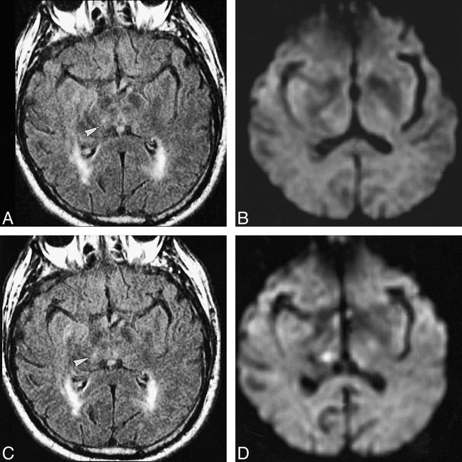
Case 4: 74-year-old man with sudden left paresthesia.
A–D, Seven hours after the onset of symptoms, FLAIR image (10002/148/1, TI = 2200) (A) shows multiple diffuse areas of periventricular hyperintensity with a small hyperintensity in the right subthalamic area (arrowhead, A and C), whereas DWI (2825/92.6/1) (B) is considered normal. Forty-eight hours after symptom onset, FLAIR image remains unchanged (C) while DWI shows a clear hyperintensity in the right subthalamic area (D), matching clinical presentation.
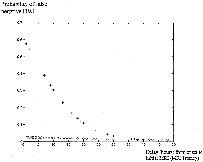
Statistical analysis confirmed that false-negative initial DWI findings occurred more frequently when the stroke lesion was located in the vertebrobasilar territory (χ2 = 12.3, P = .0004, 1 df), and when imaging was performed within 24 hours after symptom onset (χ2 = 4.07, P = .04, 1 df). The results of the logistic regression analysis within each vascular topography subgroup are presented in Figure 4. This analysis showed that the occurrence of false-negative DWI findings decreased significantly when MR latency increased (𝛉 = 0.438, β = 0.127, P = .04) for vertebrobasilar stroke lesions. This link was no longer significant when lesions were located in the anterior circulation (𝛉 = −3.6, β = 0.018, P > .05). Among the 19 patients with a vertebrobasilar stroke imaged within 24 hours after onset, false-negative DWI lesions were significantly smaller (final volume: median, 0.18 cm3; range, 0.05–0.5 cm3) than true-positive DWI lesions (final volume: median, 0.6 cm3; range, 0.2–23 cm3) by Mann Whitney U-test (P = .04). Moreover, false-negative DWI lesions were located significantly more often in the brain stem (five of the six vertebrobasilar stroke lesions) than were the true-positive DWI lesions (three of the 10 vertebrobasilar stroke lesions) by Fisher's exact test (P = .04).

Case 6: 61-year-old man with sudden right crural hemiparesis.
A–D, Twenty-two hours after onset of symptoms, FLAIR image (10002/148/1, TI = 2200) (A) shows a small cortical hyperintensity in the left paracentral lobule (arrowhead, A and C), whereas DWI (2825/92.6/1) (B) fails to show a stroke lesion. Four days later, the lesion is still visible on FLAIR image (C), and DWI (D) displays a hyperintensity in the paracentral lobule, consistent with a recent stroke lesion and matching clinical presentation.

Case 7: 45-year-old man with sudden onset of vertigo, dysmetria, and somnolence.
A–D, On initial MR examination, performed 5.5 hours after onset, MR angiogram (not shown) displayed a partially thrombosed dolichobasilar artery, which was responsible for the mass effect on the brain stem and heterogeneous signal anterior to the pons on FLAIR images. Initial FLAIR image (A) (10002/148/1, TI = 2200) and DWI (B) fail to reveal a stroke lesion. On follow-up MR examination, performed 15 hours after onset, FLAIR image shows a small right-sided brain stem hyperintensity (arrowhead) (C), which is more clearly visible on DWI (D).

Estimation of the probability of false-negative DWI findings by means of a logistic regression. The probability of false-negative DWI findings is plotted against time for stroke lesions located in the anterior (circles) and posterior (crosses) circulation. For vertebrobasilar stroke lesions (crosses), the probability of false-negative DWI findings diminishes when MR latency increases (𝛉 = 0.438, β = 0.127, P = .04). For lesions located in the anterior circulation (circles), this relation is no longer significant
Discussion
Although it has been documented that DWI is highly sensitive for the diagnosis of acute ischemic stroke, there is increasing evidence that it may fail to detect acute stroke lesions (1, 4–6, 7–9). Our results emphasize the fact that false-negative DWI findings in acute stroke are not rare events. However, a controversy exists, as the rate of negative DWI studies in acute stroke varies among studies, ranging from 0% (2) to 21% (1, 4, 8, 9). A large proportion of the reported negative DWI studies occurred in patients with transient ischemic attack (TIA); that is, a neurologic deficit resolving completely within 24 hours. Indeed, TIA accounted for 40% of false-negative DWI findings reported by Lövblad et al (4), for 18% in the study by Ay et al (1), and for all the cases reported in the study by Lutsep et al (5). This is in agreement with the observation that DWI findings are often negative in patients with short-lasting TIA, and remain negative on follow-up studies (12).
Our aim was to identify which stroke lesions were most likely to be missed by DWI. Since clinical and extensive paraclinical examinations are often insufficient to reliably discriminate between TIA and other causes of reversible neurologic deficit, we excluded all patients with a deficit resolving within 24 hours and focused our attention on patients with a signature of stroke on follow-up MR examinations. Similarly, Ay et al (1) selected patients for whom “the deficit was still present at the time of MR imaging” and thus excluded most cases of short-lasting TIA.
Two large studies reported false-negative DWI findings in the acute phase of ischemic stroke lesions (1, 4). Apart from TIA, Ay et al (1) reported 12 patients with false-negative DWI studies in whom the best final diagnosis was a cerebral ischemic event. Of these, only half had definite stroke lesions on follow-up MR images. Lövblad et al (4) reported 18 patients with false-negative DWI findings. In their study, the final diagnosis of stroke was based on clinical data and, except for one patient, follow-up MR imaging was not reported. Since follow-up MR imaging was not systematically performed in either study, the precise rate and features of the false-negative DWI population are unknown.
Our data describe a group of eight patients with negative initial DWI studies and formally identified stroke lesions on follow-up MR images. The rate of false-negative DWI (5.8%) in our study population is much higher than that reported by Ay et al (1.5%) but lower than that reported by Lövblad et al (9.3%).
In our study, all false-negative cases occurred in patients imaged within the first 24 hours after the onset of symptoms. False-negative DWI studies have also been reported in patients imaged more than 24 hours after stroke onset (1, 7). These cases are, however, questionable, since in one study (1) MR confirmation was lacking and in the other (7) the duration of symptoms was unknown (7). Most of the false-negative findings reported by Lövblad et al (4) were in patients who “seemed to have [a] small lesion localized to the brain stem.” Our study confirms that small lesions located in the posterior vascular territory may be missed by DWI. Although very few cases have been reported (1, 9, 11), we did not observe any large hemispheric infarct with negative initial DWI findings.
Although several reports have suggested that false-negative DWI results depend on the location of the lesion and MR latency (1, 4), our study is the first to ascertain the proportion of false-negative studies in relation to these parameters. Within the first 24 hours, the rate of false-negative DWI studies was much higher when the stroke occurred in the posterior circulation (31%) than when it occurred in the anterior circulation (2%), and brain stem lesions were at high risk of being missed by DWI.
Overall, patients presenting with small vertebrobasilar stroke lesions imaged soon after the onset of symptoms were those most likely to have negative DWI findings. Several reasons can be advanced. First, the lesions could be too small for the resolution of the DWI echo-planar sequence. This hypothesis is supported by the fact that FLAIR images, with a higher spatial resolution, displayed a signal abnormality in half the lesions that were not visible on DWI. Second, the signal-to-noise ratio could be insufficient in the first few hours after onset. And, last, magnetic susceptibility artifacts occurring in echo-planar imaging cause brain stem distortions that could blur image analysis. This hypothesis is supported by the high rate of brain stem lesions missed by DWI in this study. DWI performed with turbo spin-echo sequences provides high-resolution images with few susceptibility artifacts and might overcome these technical limitations, particularly in the posterior fossa (13). It has been stated that normal DWI findings coupled with normal perfusion MR findings can identify all patients who present with stroke symptoms but do not have cerebral ischemia (11). However, bolus-tracking perfusion MR imaging also uses echo-planar pulse sequences, and thus has the same drawbacks as echo-planar DWI (limited spatial resolution and strong susceptibility artifacts). In addition, perfusion maps are often normal in lacunar infarcts (11). Thus, one can hypothesize that most echo-planar perfusion maps would have remained negative given the small size and the vertebrobasilar location of the ischemic stroke lesions missed on DWI examinations.
Conclusion
Our data suggest that false-negative DWI studies are not uncommon during the first 24 hours of a stroke; that negative DWI findings obtained in the first 24 hours after onset are not a reliable indicator by which to rule out a stroke lesion, especially if symptoms are suggestive of a stroke in the posterior circulation; and that false-negative DWI findings are no longer observed after 24 hours. These are clinically important results, since they indicate that the diagnosis of stroke should not be ruled out on the basis of early negative DWI studies. Moreover, DWI should be repeated more than 24 hours after onset in the event of a negative initial DWI study in a patient with long-lasting clinical symptoms consistent with ischemic stroke.
Footnotes
1 Supported in part by a research grant (P980101, CRC 97120) from Assistance Publique Hôpitaux de Paris.
↵2 Address reprint requests to Catherine Oppenheim, MD, Department of Neuroradiology, Groupe Hospitalier Pitié-Salpêtrière, Paris VI University, 47 Boulevard de l'Hôpital, 75651 Paris, Cedex 13, France.
References
- Received November 29, 1999.
- Accepted after revision February 21, 2000.
- Copyright © American Society of Neuroradiology
In this issue


Jump to section
Related Articles
Cited By...
- Acute peripheral facial paralysis caused by tegmental pontine infarction
- Cerebrovascular disease in sickle cell disease
- Time Course and Clinical Correlates of Retinal Diffusion Restrictions in Acute Central Retinal Artery Occlusion
- Early detection and monitoring of cerebral ischemia using calcium-responsive MRI probes
- Current endovascular strategies for posterior circulation large vessel occlusion stroke: report of the Society of NeuroInterventional Surgery Standards and Guidelines Committee
- Use of HINTS in the acute vestibular syndrome. An Overview
- Diagnosis of DWI-negative acute ischemic stroke: A meta-analysis
- Optimal Timing of Diffusion-Weighted Imaging to Avoid False-Negative Findings in Patients With Transient Ischemic Attack
- Proposed Standardized Neurological Endpoints for Cardiovascular Clinical Trials: An Academic Research Consortium Initiative
- Acute Ischemic Stroke Infarct Topology: Association with Lesion Volume and Severity of Symptoms at Admission and Discharge
- Stroke risk stratification in acute dizziness presentations: A prospective imaging-based study
- How I treat and manage strokes in sickle cell disease
- Sensitivity of Diffusion- and Perfusion-Weighted Imaging for Diagnosing Acute Ischemic Stroke Is 97.5%
- Head impulse gain and saccade analysis in pontine-cerebellar stroke and vestibular neuritis
- Characteristics of central lesions in patients with dizziness determined by diffusion MRI in the emergency department
- Posterior circulation ischaemic stroke
- The Use of Diffusion Imaging to Detect Microstructure in the Preterm Brain
- Validity of Negative High-Resolution Diffusion-Weighted Imaging in Transient Acute Cerebrovascular Events
- An Updated Definition of Stroke for the 21st Century: A Statement for Healthcare Professionals From the American Heart Association/American Stroke Association
- Stroke syndromes associated with DWI-negative MRI include ataxic hemiparesis and isolated internuclear ophthalmoplegia
- Accuracy of Diffusion-Weighted Imaging in the Diagnosis of Stroke in Patients With Suspected Cerebral Infarct
- Clinical Reasoning: A 54-year-old man with unilateral weakness and vascular risk factors
- Secondary Prevention of Overt Strokes in Sickle Cell Disease: Therapeutic Strategies and Efficacy
- Does my dizzy patient have a stroke? A systematic review of bedside diagnosis in acute vestibular syndrome
- 1H-NMR-Based Metabolomics Study of Cerebral Infarction
- Characteristics of patients with minor ischaemic strokes and negative MRI: a cross-sectional study
- Posterior versus anterior circulation strokes: comparison of clinical, radiological and outcome characteristics
- Diffusion-weighted MRI in acute stroke within the first 6 hours: 1.5 or 3.0 Tesla?
- Safety of tPA in stroke mimics and neuroimaging-negative cerebral ischemia
- HINTS to Diagnose Stroke in the Acute Vestibular Syndrome: Three-Step Bedside Oculomotor Examination More Sensitive Than Early MRI Diffusion-Weighted Imaging
- Change in Diffusion-Weighted Imaging Infarct Volume Predicts Neurologic Outcome at 90 Days: Results of the Acute Stroke Accurate Prediction (ASAP) Trial Serial Imaging Substudy
- The California, ABCD, and Unified ABCD2 Risk Scores and the Presence of Acute Ischemic Lesions on Diffusion-Weighted Imaging in TIA Patients
- Normal head impulse test differentiates acute cerebellar strokes from vestibular neuritis
- When to Expect Negative Diffusion-Weighted Images in Stroke and Transient Ischemic Attack
- False-negative diffusion-weighted imaging with lateral medullary infarction
- Recommendations for Comprehensive Stroke Centers: A Consensus Statement From the Brain Attack Coalition
- Diffusion-Weighted MRI in 300 Patients Presenting Late With Subacute Transient Ischemic Attack or Minor Stroke
- Repetitive TMS temporarily alters brain diffusion
- The clinical significance of diffusion-weighted MR imaging in infratentorial strokes
- Evaluation of Hyperintense Vessels on FLAIR MRI for the Diagnosis of Multiple Intracerebral Arterial Stenoses
- Diffusion- and Perfusion-Weighted Magnetic Resonance Imaging of the Brain Before and After Coronary Artery Bypass Grafting Surgery
- A prospective, longitudinal diffusion tensor imaging study of brain injury in newborns
- Benefits of Perfusion MR Imaging Relative to Diffusion MR Imaging in the Diagnosis and Treatment of Hyperacute Stroke