Advertisement

Main menu

  • Home
  • Content
    • Current Issue
    • Publication Preview--Ahead of Print
    • Past Issue Archive
    • Case of the Week Archive
    • Classic Case Archive
    • Case of the Month Archive
    • COVID-19 Content and Resources
  • For Authors
  • About Us
    • About AJNR
    • Editors
    • American Society of Neuroradiology
  • Submit a Manuscript
  • Podcasts
    • Subscribe on iTunes
    • Subscribe on Stitcher
  • More
    • Subscribers
    • Permissions
    • Advertisers
    • Alerts
    • Feedback
  • Other Publications
    • ajnr

User menu

  • Subscribe
  • Alerts
  • Log in

Search

  • Advanced search
American Journal of Neuroradiology

American Journal of Neuroradiology

  • Subscribe
  • Alerts
  • Log in

Advanced Search

  • Home
  • Content
    • Current Issue
    • Publication Preview--Ahead of Print
    • Past Issue Archive
    • Case of the Week Archive
    • Classic Case Archive
    • Case of the Month Archive
    • COVID-19 Content and Resources
  • For Authors
  • About Us
    • About AJNR
    • Editors
    • American Society of Neuroradiology
  • Submit a Manuscript
  • Podcasts
    • Subscribe on iTunes
    • Subscribe on Stitcher
  • More
    • Subscribers
    • Permissions
    • Advertisers
    • Alerts
    • Feedback
  • Follow AJNR on Twitter
  • Visit AJNR on Facebook
  • Follow AJNR on Instagram
  • Join AJNR on LinkedIn
  • RSS Feeds
Research ArticleBRAIN

Persistence of Gadolinium in CSF: A Diagnostic Pitfall in Patients with End-stage Renal Disease

Ansaar T. Rai and Jeffery P. Hogg
American Journal of Neuroradiology August 2001, 22 (7) 1357-1361;
Ansaar T. Rai
  • Find this author on Google Scholar
  • Find this author on PubMed
  • Search for this author on this site
Jeffery P. Hogg
  • Find this author on Google Scholar
  • Find this author on PubMed
  • Search for this author on this site
  • Article
  • Figures & Data
  • Info & Metrics
  • References
  • PDF
Loading

Article Figures & Data

Figures

  • fig 1.
    • Download figure
    • Open in new tab
    • Download powerpoint
    fig 1.

    Sagittal (A) 500/8/1 (TR/TE/excitation) and axial (B) 566/12/1 precontrast T1-weighted and axial FLAIR (C) 10002/97.5/1 images of the brain demonstrate normal signal intensity of the CSF with normal contrast enhancement between the CSF and adjacent brain.

    Sagittal (D) 500/8/1 and axial (E) 566/12/1 precontrast T1-weighted images 10 days later, demonstrate increased signal intensity within the subarachnoid space, manifested by an isointense appearance of the sulci as compared with the adjacent brain. Additionally, there is increased signal intensity within the ocular globes on the axial image. The axial FLAIR (F) 10002/97.5/1 image demonstrates diffuse significant increased signal intensity within the subarachnoid space and the ventricles. A–F were filmed at the same values for window and level, and were performed on the same scanner.
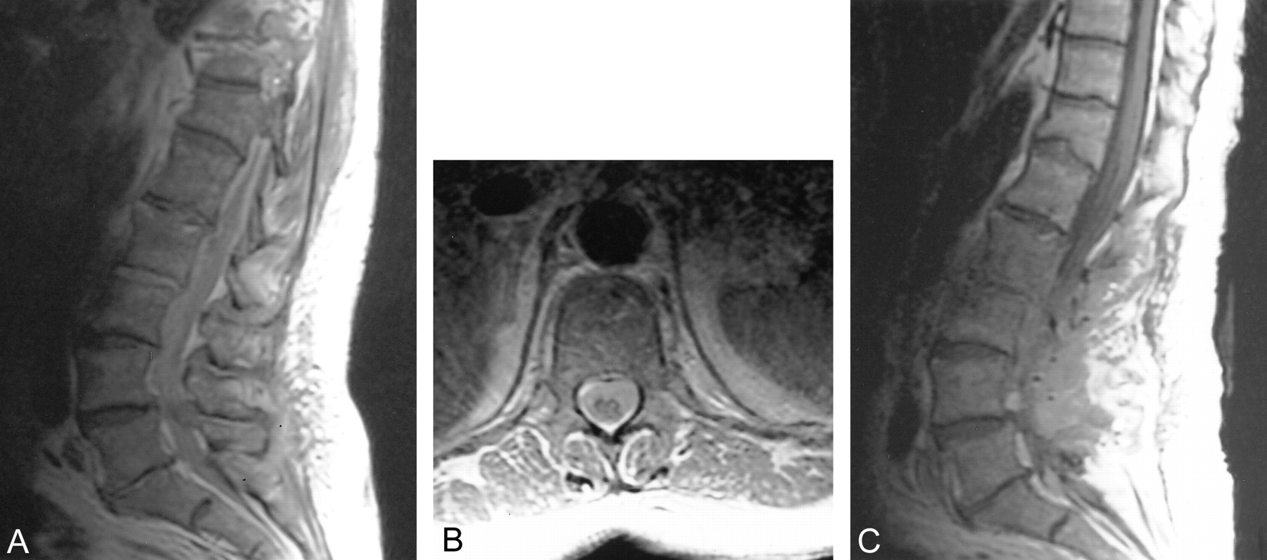
  • fig 2.
    • Download figure
    • Open in new tab
    • Download powerpoint
    fig 2.

    Sagittal (A) 516/12/2 and axial (B) 686/11.6/2 precontrast T1-weighted images of the spine demonstrate diffuse increased signal intensity within the subarachnoid space. Sagittal precontrast T1-weighted (C) 550/12/2 image of the lumbar spine performed 2 weeks later shows resolution of the previously seen increased signal intensity and a normal signal within the CSF. A–C were filmed at the same values for window and level, and were performed on the same scanner

PreviousNext
Back to top

In this issue

American Journal of Neuroradiology: 22 (7)
American Journal of Neuroradiology
Vol. 22, Issue 7
1 Aug 2001
  • Table of Contents
  • Index by author
Advertisement
Print
Download PDF
Email Article

Thank you for your interest in spreading the word on American Journal of Neuroradiology.

NOTE: We only request your email address so that the person you are recommending the page to knows that you wanted them to see it, and that it is not junk mail. We do not capture any email address.

Enter multiple addresses on separate lines or separate them with commas.
Persistence of Gadolinium in CSF: A Diagnostic Pitfall in Patients with End-stage Renal Disease
(Your Name) has sent you a message from American Journal of Neuroradiology
(Your Name) thought you would like to see the American Journal of Neuroradiology web site.
CAPTCHA
This question is for testing whether or not you are a human visitor and to prevent automated spam submissions.
Citation Tools
Persistence of Gadolinium in CSF: A Diagnostic Pitfall in Patients with End-stage Renal Disease
Ansaar T. Rai, Jeffery P. Hogg
American Journal of Neuroradiology Aug 2001, 22 (7) 1357-1361;

Citation Manager Formats

  • BibTeX
  • Bookends
  • EasyBib
  • EndNote (tagged)
  • EndNote 8 (xml)
  • Medlars
  • Mendeley
  • Papers
  • RefWorks Tagged
  • Ref Manager
  • RIS
  • Zotero
Share
Persistence of Gadolinium in CSF: A Diagnostic Pitfall in Patients with End-stage Renal Disease
Ansaar T. Rai, Jeffery P. Hogg
American Journal of Neuroradiology Aug 2001, 22 (7) 1357-1361;
Reddit logo Twitter logo Facebook logo Mendeley logo
  • Tweet Widget
  • Facebook Like
  • Google Plus One

Jump to section

  • Article
    • Abstract
    • Case Reports
    • Discussion
    • Footnotes
    • References
  • Figures & Data
  • Info & Metrics
  • References
  • PDF

Related Articles

  • No related articles found.
  • PubMed
  • Google Scholar

Cited By...

  • Differential diagnosis of hyperintense cerebrospinal fluid on fluid-attenuated inversion recovery images of the brain. Part I: pathological conditions
  • Persistence of Gadolinium Contrast Enhancement in CSF: A Possible Harbinger of Gadolinium Neurotoxicity?
  • Gadolinium encephalopathy in a patient with renal failure
  • Crossref
  • Google Scholar

This article has not yet been cited by articles in journals that are participating in Crossref Cited-by Linking.

More in this TOC Section

  • Predictors of Reperfusion in Patients with Acute Ischemic Stroke
  • Enhanced Axonal Metabolism during Early Natalizumab Treatment in Relapsing-Remitting Multiple Sclerosis
  • Progression of Microstructural Damage in Spinocerebellar Ataxia Type 2: A Longitudinal DTI Study
Show more BRAIN

Similar Articles

Advertisement

News and Updates

  • Lucien Levy Best Research Article Award
  • Thanks to our 2022 Distinguished Reviewers
  • Press Releases

Resources

  • Evidence-Based Medicine Level Guide
  • How to Participate in a Tweet Chat
  • AJNR Podcast Archive
  • Ideas for Publicizing Your Research
  • Librarian Resources
  • Terms and Conditions

Opportunities

  • Share Your Art in Perspectives
  • Get Peer Review Credit from Publons
  • Moderate a Tweet Chat

American Society of Neuroradiology

  • Neurographics
  • ASNR Annual Meeting
  • Fellowship Portal
  • Position Statements

© 2023 by the American Society of Neuroradiology | Print ISSN: 0195-6108 Online ISSN: 1936-959X

Powered by HighWire