
Abstract
BACKGROUND AND PURPOSE: The MR appearance of white matter tracts in the hypothalamus and the role of the hypothalamus as a memory mechanism have not been sufficiently described in clinical settings. Heavily T2-weighted black-and-white reversed (T2R) images were assessed to reveal their visualization and clinical significance.
METHODS: One hundred healthy subjects and three patients with hypothalamic lesions underwent fast spin-echo MR imaging to reveal the postcommissural fornix (PF) and mammillothalamic tract (MT).
RESULTS: The PF was identifiable in axial and/or coronal sections in all healthy subjects. No remarkable asymmetry of its size or course was evident. Both anteroposterior and vertical dimensions ranged from 10.5 to 14 mm. The MT was visible in one or two axial sections above the mammillary body in 64% of healthy subjects and in a coronal section in 36%. Two patients with glioblastoma multiforme and lacunar infarct at the hypothalamus presented with anterograde amnesia; T2R imaging revealed involvement of both the PF and MT. The third patient had a suprasellar craniopharyngioma with PF injury sparing the MT resulting from surgical manipulation and was free of memory deficit. Anterograde amnesia was evident only when both the PF and MT were injured.
CONCLUSION: T2R images have made a high rate of detection of the PF and MT possible and could provide a more detailed correlation of hypothalamic neuroanatomy and memory mechanism in clinical settings.
The advent of superconductive MR imaging has allowed a more detailed normal and pathologic anatomy of central nervous system lesions to be depicted. Standard clinical MR imaging studies most commonly are used to provide T1-weighted, T2-weighted, proton density–weighted, and intravenous contrast-enhanced T1-weighted images. In addition to these common MR sequences, heavily T2-weighted MR images have been highlighted in depicting fine cisternal, as well as neural, structures (1, 2).
It is known that two main white matter tracts are present in the hypothalamus: the postcommissural fornix (PF) and mammillothalamic tract (MT) (3, 4). The MR imaging appearance of the individual white matter tract has not been sufficiently described in clinical settings. Delineation of the anatomic details of these structures will contribute to clarifying the functional role of the hypothalamus as a memory mechanism. In this study, heavily T2-weighted MR imaging sequences were performed to visualize the PF and MT in subjects without neurologic abnormalities as well as patients with a hypothalamic lesion, an endeavor that has yet to be attempted. The clinical significance of these MR images is discussed.
Methods
MR imaging assessment of 100 healthy subjects (72 men, 28 women; age range, 17–72 years; average age, 46 years) was performed. These healthy subjects composed a population presenting with neurologic complaints but no found abnormality.
White matter tracts were identifiable as a result of their relatively higher signal intensity compared with that of the surrounding hypothalamic gray matter (Figs 1–4). The largest perceptual contrast difference between the gray matter and the white matter around the hypothalamus was investigated by changing the window levels. The anatomic details in the axial and coronal sections were studied. Visualization of each white matter tract was determined by three neurosurgeons (N.S., H.M., K.S.) through radiologic evaluation, after which the final decision was made by consensus.

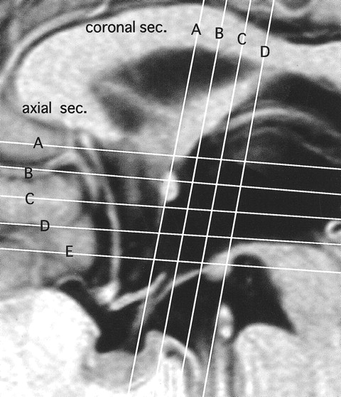
Horizontal and vertical white lines show section planes in axial and coronal views in figs 2 A–E and 3 A–D, respectively
A 1.5-T MR imaging system was used with heavily T2-weighted fast spin-echo and black-and-white reversed imaging (T2R) (5800/220 [TR/TE]; section thickness, 3 mm; intersection gap, 0.5 mm) (2).
Results
PF in Healthy Subjects
The PF was defined as white matter tract extending from each fornical column, running behind the anterior commissure, and terminating at the mammillary body (4, 5).
Axial Section
Fornical columns were observed one section level above the anterior commissure (Fig 2A). The PF was identifiable in all healthy subjects and was spread 10.5 to 14 mm (three to four axial sections) between levels of the anterior commissure and the mammillary body, without motion artifact (Figs 1 and 2B–E). The PF was observed to be approximately round in shape and 2- to 3-mm thick on the axial cut-surface, and it was visualized as a high-signal-intensity spot just behind the anterior commissure and exposed to the cerebrospinal fluid space of the third ventricle (Fig 2B). In the next lower section, the PF was still exposed to the third ventricle (Fig 2C). The next lower section showed ovoid fibers directing posteriorly toward the mammillary body (Fig 2D). At the next level, the PF anteriorly joined to the mammillary body (Fig 2E). In six subjects, the PF was recognizable but not clearly visualized either unilaterally or bilaterally (Fig 2F). No remarkable asymmetry of its size or course was evident.

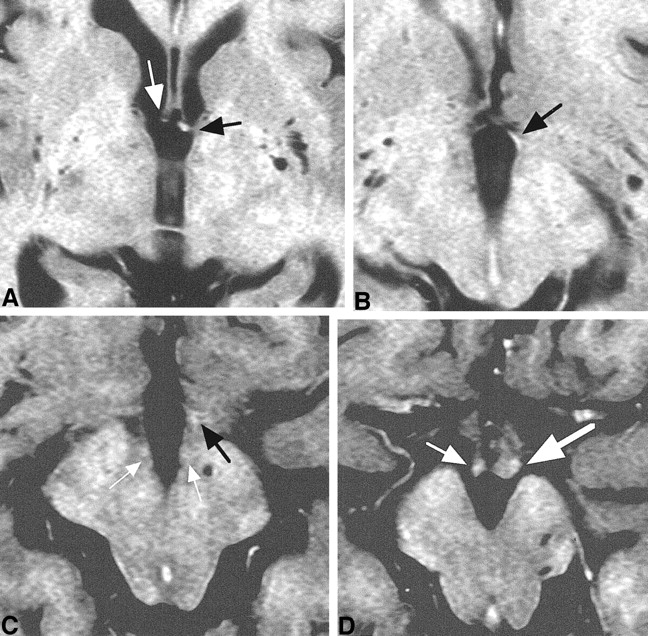
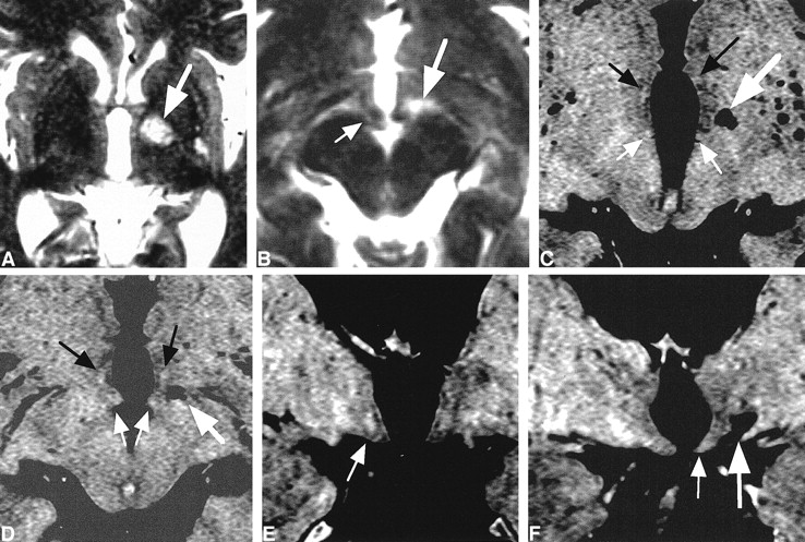
Axial images of consecutive five sections in one healthy subject shown in figure 1 (A–E) and one section in another subject (F).
A, Fornical columns (arrow) were observed in one section level above the anterior commissure.
B, PF (white arrow) was observed as a high-signal-intensity spot just behind the anterior commissure (black arrow) and exposed to the cerebrospinal fluid space of the third ventricle.
C, In the next lower section, PF (arrow) was still exposed to the third ventricle.
D, In the next lower section, ovoid PF (white arrow) directing posteriorly toward the mammillary body was seen. Ill-defined high-signal-intensity spot (MT; black arrow) was visible 4 mm posterior to PF.
E, At the next level, PF (white arrow) joined to the mammillary body (black arrow) anteriorly and laterally.
F, PF (white arrow) was visible but ill-defined bilaterally. Obscure PF was seen in 6% of healthy subjects. MT (black arrow) was identifiable in this case.
Coronal Section
In the coronal section, the PF was spread 10.5 to 14 mm (three to four sections) in levels between the anterior commissure and the mammillary body (Figs 1 and 3A–D). The fornical column was evident at the anterior commissure section or one section posterior to it (Fig 3A and E). In the next one or two posterior sections, the descending portion of the PF was evident just above the floor of the third ventricle in 78% of the healthy subjects (Fig 3B and C). At the mammillary body level, the entry point of the PF was unidentifiable (Fig 3D).

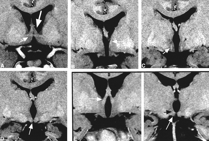
Coronal images of four consecutive sections in one healthy subject (A–D) and two sections in another subject (E and F).
A, The fornical columns (large arrow) were evident at the anterior commissure (small arrow) section.
B, In the next posterior section, the descending portion of PF (arrow) was identifiable, more clearly on the left.
C, In the next posterior section, the descending portion of PF (arrow) was evident bilaterally, above the floor of the third ventricle. It was visible in 80% of the healthy subjects.
D, In the next section, the mammillary body (arrow) was identifiable. At this level, the entry point of the PF was unidentifiable.
E, The fornical columns (arrow) were evident.
F, At the level of mammillary body (white arrow), the origin of MT (black arrow) was visible.
MT in Healthy Subjects
The MT was defined as a white matter tract running 4–8 mm posterior to the PF (5).
Axial Section
The MT was detectable as a high-signal-intensity spot in one or two sections above the mammillary body level (Fig 2D and F). The high signal was often obscure and less evident than that of PF, and it was visualized in 64% of subjects (Fig 2D and F).
Coronal Section
The origin of the MT from the mammillary body was evident in 36% of healthy subjects (Fig 3D and F).
Case Reports
Case 1: Hypothalamic Glioblastoma Multiforme
A right-handed 34-year-old man reported progressive forgetfulness spanning 6 months. His family and professional colleagues noted that he could not remember where he had placed objects, recent points of conversation, and events of the day. Symptoms and signs associated with endocrinology or ophthalmology were absent. No confabulation was noted. MR images showed a large left hypothalamic mass lesion (Fig 4). A coronal image showed that the right fornical column and PF were displaced off the midline, and the left fornical column and PF were not visible (Fig 4B). Axial sections showed that the right PF and MT were displaced but visible, although the left PF and MT were relocated inferiorly and deformed by the mass (Fig 4C–E). The left mammillary body and PF were displaced and deformed inferiorly (Fig 4F). The main lesion was located at the PF and MT on the left side, based on their deformity, displacement, and surrounding brain edema. Such involvement of both white matter tracts was suspected to cause this patient's memory impairment. Stereotactic surgery for the biopsy yielded histologic results compatible with glioblastoma multiforme. The patient was transferred to another institution for radiation and chemotherapy.

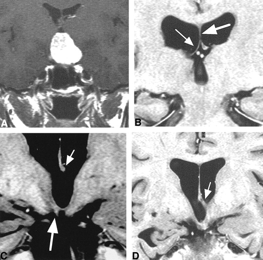
Case 1.
A, Sagittal image shows a huge left hypothalamic mass lesion.
B, Coronal image shows that the right fornical column (small arrow) and PF (large arrow) were displaced off the midline, and the left PF was not visible.
C, Right fornical column or PF (arrow) was identifiable as a slightly high-signal-intensity spot. Left one was unidentifiable because of a huge hypothalamic mass.
D and E, Right PF (small white arrow) and MT (small black arrow) were visible. Left PF (large white arrow) and MT (large black arrow) were displaced inferiorly and deformed by the mass.
F, Right PF (small white arrow) and mammillary body (small black arrow) were displaced laterally. Left mammillary body (large black arrow) was shifted inferiorly, and left PF (large white arrow) was clearly visible as a high-signal-intensity spot.
Case 2: Hypothalamic Infarct
A right-handed 70-year-old man reported recent memory loss spanning 1 week. He could not remember the names of his close friends. He was aware of his memory deficit. He had been mildly hypertensive for 7 years before this consultation, although this condition was untreated. T2-weighted MR images revealed a left hypothalamic high-signal-intensity lesion, 8 mm, that was compatible with the diagnosis of lacunar infarct (Fig 5A and B). The patient was prescribed ticlopidine hydrochloride, 200 mg. His symptoms gradually improved and completely disappeared in 3 months.

Case 2.
A and B, T2-weighted axial images. A high-signal-intensity spot (large arrow), 8 mm in maximum size and compatible with the diagnosis of lacunar infarction, was visible in the left hypothalamus in A and lateral to the superior part of the mammillary body (small arrow) in B.
C and D, Axial T2R images. A lacunar infarct (large white arrow) was visible in the left hypothalamus. PFs (black arrows) and MTs (small white arrows) were visible. Both white matter tracts seemed slightly smaller or less evident in signal intensity on the left.
E, Coronal T2R image. Descending portion of PF (arrow) was more evident on the right than on the left, which is compatible with the axial images in fig 5C and D.
F, Coronal T2R image. Left mammillary body (small arrow) was decreased in size compared with the right. A lacunar infarct (large arrow) was visible a few millimeters lateral to the left third ventricle wall.
A year later, T2R images showed an old infarct of a few millimeters located in the left hypothalamus between the PF and MT (Fig 5C–F). Both white matter tracts were thought to be involved and related to the amnesic symptoms in the acute stage.
Case 3: Fornical Injury from Surgery
A 35-year-old woman reported headache and visual field defect. On admission, bitemporal hemianopia was noted. MR imaging revealed a suprasellar tumor composed of a homogeneous solid component with small cysts (Fig 6A). On the basis of the diagnosis of craniopharyngioma, an anterior interhemispheric approach was performed. The right frontal lobe was laterally retracted during the operation to gain a surgical field in the third ventricle. The tumor was subtotally removed with improvement of visual field defect.

Case 3.
A, T1-weighted coronal image with enhancement. A suprasellar mass with an almost homogeneous enhancement effect was noted.
B-D, T2R coronal MR images.
B, The right half of fornical body (small arrow), attached to the inferior surface of the septum pellucidum (large arrow), was thin and decreased in size.
C, The left half of the fornical body (small arrow) was seen. The right mammillary body was smaller than the left one (large arrow).
D, A fornical column was seen on the left only (arrow).
Two years later, the woman presented with narrowing of her visual field, and evaluation revealed an enlargement of the cystic component. Transsphenoidal surgery and subsequent gamma knife radiosurgery were performed. She recovered without apparent neurologic deficit, including antero- and retrograde amnesia. Follow-up T2R MR images revealed that the size of the right half of the fornical body was remarkably decreased (Fig 6B and C). Further anteriorly, the right fornical column was absent (Figs 6D and 7A). The right PF was not identifiable (Fig 7B and C), and the right mammillary body was decreased in size (Figs 6C and 7D). This patient was suspected to have a unilateral fornical injury from over-retraction of the right frontal lobe during the operation and subsequent decrease in size of the right PF without MT impairment. The memory impairment was not apparent.
Case 3. Axial T2R images.
A, Fornical column was visible on the left side (black arrow). The right fornical column (white arrow) was decreased in size.
B, Only the left PF (arrow) was visible.
C, PF (black arrow) was visible on the left side only. MT (white arrows) was visible bilaterally.
D, Although bilateral mammillary bodies (arrows) were visible, the right side (small arrow) was decreased in size. The size decrease was compatible with the coronal section (fig 6C).
Discussion
Anatomy of PF and MT
Anatomically, there are two major white matter tracts in the hypothalamus: the PF and MT (4–6).
The fornix is composed of arch-shaped white matter fibers connecting the hippocampus to the mammillary body (3, 4). The main parts of the fornix are the crura, commissure, the body, and the two columns (3, 4). Each column further divides into pre- and postcommissural fornices (25% and 75%, respectively) at the level of the anterior commissure (3). The precommissural fibers extend to the septal, lateral preoptic, diagonal, and anterior hypothalamic nuclei. The PF mainly projects to the mammillary body (3, 4).
The MT is a major part of mammillary efferent fibers (4, 5). They primarily arise from the medial mammillary nucleus and initially form a well-defined bundle, the principal mammillary bundle (fasciculus mammillaris princeps) (4, 5). This bundle dorsally passes for a short distance and divides into two components: a larger MT and a smaller mammillotegmental tract. The MT terminates at the anterior thalamic nuclei (4, 5).
MR Study of PF and MT in Healthy Subjects
Although the essential parts of fornical structures are neuroradiologically demonstrated on standard MR images (3, 7), the PF has not been sufficiently described in clinical settings. In our study, the PF was identifiable in 100% of healthy subjects, without motion artifact. The PF is bordered anteriorly and superiorly by the anterior commissure and posteriorly and inferiorly by the mammillary body. Just behind the anterior commissure, the PF forms arches posteriorly and inferiorly in the hypothalamic nuclei toward the mammillary body. Both anteroposterior and vertical dimensions of PF ranged from 10.5 to 14 mm.
The origin of MT was identifiable in only 64% of healthy subjects from axial sections and 36% from coronal sections in our MR parameters (Figs 2D and F, 3D and F). Depiction of the MT was more difficult than that of the PF. Such radiologic ambiguity of MT is speculated to result from its short, well-defined course, its complex origin from trunk fibers of the principal mammillary bundle, and its divergent fiber course terminating in the relatively large anterior thalamic nuclei (4–6). The section direction being nonspecific to the course of MT in this study might be another reason for the radiologic ambiguity of this tract.
MR Study of PF and MT in Clinical Settings
To our knowledge, this is the first report to radiologically delineate the anatomy of the PF and MT in clinical settings.
To date, clinicoradiologic analyses of white matter tracts in the hypothalamus are scarce. Studies have focused mainly on the fornix as it relates to temporal lobe epilepsy or surgical planning of transcallosal or transventricular tumor resection (7–11). One study assessed the volumetry of the limbic system in healthy subjects compared with that in patients with epilepsy (8). Other studies correlated the asymmetry of the crus, body, or mammillary body with temporal lobe epilepsy (7, 9, 10). The fornix at the side of the temporal lobe lesion has been reportedly atrophic and is clinically useful in determining the epileptogenic side (7). Clinical evidence concerning symptomatology from a localized lesion has been limited, although anterograde amnesia resulting from a localized injury of the fornical body and columns was reported (12). Accordingly, the clinical significance of the PF and MT was not sufficiently described in any of these reports.
Functional Consideration of PF and MT
Medial diencephalic injury causes memory impairment similar to that from medial temporal lobe injury, which has been confirmed in animal experiments and human clinical observations (13–16). This type of memory impairment is called diencephalic amnesia and is related to profound impairment of memory for recent events and mild behavioral changes. Memory for remote events is relatively unaffected. Although general intellectual functions might remain at fairly high levels, these patients demonstrate an inability to learn new facts and skills (4, 13). Anatomic sites responsible for this type of amnesia are reported to be the PF, mammillary body, MT, anterior thalamic nuclei, dorsomedial nucleus, and intralaminar nucleus (4, 13). It is known that in animal experiments spatial memory was selectively impaired by the destruction of anterior thalamic nuclei, which has direct fornical and indirect fornix/mammillary body/MT projections. Injury of direct fornical connection produces more severe memory deficit than that of the mammillary body or MT (13). Thus, a site-dependent difference of memory deficit was elucidated to a certain extent in animal experiments. However, a detail of clinical manifestations in humans has not been sufficiently explained, partly because of spatial and contrast limitations of conventional MR sequences (1, 2). In this respect, T2R is expected to overcome such technical limitations of conventional MR images and provide a detailed anatomy of pathologic lesions in the hypothalamus (2, 14, 15).
Case 1 is believed to show unilateral PF and MT involvement, although involvement of anterior thalamic nuclei or contralateral PF was not excluded. Although Case 2 had a localized infarct between the two tracts in the chronic stage, both tracts were speculated to be involved in the acute stage, based on a wider high signal lesion of ischemic and/or edematous area in T2-weighted images. Both patients had recent memory impairment, and both PF and MT were considered to be involved in producing the anterograde amnesia. A dominant-side lesion may be related to the symptomatic appearance in our cases (14–16). It also has been reported that, when it occurs, a lesion on the dominant side was common (6, 14–16). Memory deficit was not evident in Case 3, in which the patient had a unilateral fornical injury without MT involvement.
Considering all these factors, impairment of both the PF and MT might be the cause of recent memory deficits. However, further investigation is needed since causes, lesion sides in respect to hemispheric dominance, or lesion extents in our cases were varied.
Thus, direct visualization of each tract will contribute to a more clear delineation of the functional anatomy of the hypothalamus as a memory mechanism.
Two surgical implications of the MR findings in Case 3 follow. First, excessive, laterally directed brain retraction in the anterior interhemispheric approach for a large suprasellar tumor should be particularly avoided, because such retraction might easily tear the anterior commissure and fornix, which are overstretched by the large tumor and are fragile enough to be torn with even a slight brain retraction (16, 17). Second, the importance of perioperative evaluation of the fornix in large suprasellar tumors is emphasized. When a one-sided fornical decrease in size is noticed, or even suspected, a respective surgical approach and intraoperative manipulation must be chosen to preserve an intact fornix.
Conclusion
Direct visualization of the PF and MT in the hypothalamus by T2R imaging could more precisely delineate the functional neuroanatomy for memory produced by hypothalamic lesions.
Footnotes
1 Address reprint requests to Naokatsu Saeki, Department of Neurological Surgery, Chiba University School of Medicine, Inohana 1–8–1, Chuo-ku, Chiba City, Chiba 260–8670, Japan.
References
- Received December 1, 2000.
- Accepted after revision April 19, 2001.
- Copyright © American Society of Neuroradiology
In this issue


Jump to section
Related Articles
Cited By...
- No citing articles found.