Advertisement

Main menu

  • Home
  • Content
    • Current Issue
    • Accepted Manuscripts
    • Article Preview
    • Past Issue Archive
    • Video Articles
    • AJNR Case Collection
    • Case of the Week Archive
    • Case of the Month Archive
    • Classic Case Archive
  • Special Collections
    • AJNR Awards
    • ASNR Foundation Special Collection
    • Most Impactful AJNR Articles
    • Photon-Counting CT
    • Spinal CSF Leak Articles (Jan 2020-June 2024)
  • Multimedia
    • AJNR Podcasts
    • AJNR SCANtastic
    • Trainee Corner
    • MRI Safety Corner
    • Imaging Protocols
  • For Authors
    • Submit a Manuscript
    • Submit a Video Article
    • Submit an eLetter to the Editor/Response
    • Manuscript Submission Guidelines
    • Statistical Tips
    • Fast Publishing of Accepted Manuscripts
    • Graphical Abstract Preparation
    • Imaging Protocol Submission
    • Author Policies
  • About Us
    • About AJNR
    • Editorial Board
    • Editorial Board Alumni
  • More
    • Become a Reviewer/Academy of Reviewers
    • Subscribers
    • Permissions
    • Alerts
    • Feedback
    • Advertisers
    • ASNR Home

User menu

  • Alerts
  • Log in

Search

  • Advanced search
American Journal of Neuroradiology

American Journal of Neuroradiology

ASHNR American Society of Functional Neuroradiology ASHNR American Society of Pediatric Neuroradiology ASSR
  • Alerts
  • Log in

Advanced Search

  • Home
  • Content
    • Current Issue
    • Accepted Manuscripts
    • Article Preview
    • Past Issue Archive
    • Video Articles
    • AJNR Case Collection
    • Case of the Week Archive
    • Case of the Month Archive
    • Classic Case Archive
  • Special Collections
    • AJNR Awards
    • ASNR Foundation Special Collection
    • Most Impactful AJNR Articles
    • Photon-Counting CT
    • Spinal CSF Leak Articles (Jan 2020-June 2024)
  • Multimedia
    • AJNR Podcasts
    • AJNR SCANtastic
    • Trainee Corner
    • MRI Safety Corner
    • Imaging Protocols
  • For Authors
    • Submit a Manuscript
    • Submit a Video Article
    • Submit an eLetter to the Editor/Response
    • Manuscript Submission Guidelines
    • Statistical Tips
    • Fast Publishing of Accepted Manuscripts
    • Graphical Abstract Preparation
    • Imaging Protocol Submission
    • Author Policies
  • About Us
    • About AJNR
    • Editorial Board
    • Editorial Board Alumni
  • More
    • Become a Reviewer/Academy of Reviewers
    • Subscribers
    • Permissions
    • Alerts
    • Feedback
    • Advertisers
    • ASNR Home
  • Follow AJNR on Twitter
  • Visit AJNR on Facebook
  • Follow AJNR on Instagram
  • Join AJNR on LinkedIn
  • RSS Feeds

AJNR is seeking candidates for the AJNR Podcast Editor. Read the position description.

OtherBRAIN

Angiographic Features of Spontaneous Intracranial Hypotension

Scott A. Koss, John L. Ulmer and Lotfi Hacein-Bey
American Journal of Neuroradiology April 2003, 24 (4) 704-706;
Scott A. Koss
  • Find this author on Google Scholar
  • Find this author on PubMed
  • Search for this author on this site
John L. Ulmer
  • Find this author on Google Scholar
  • Find this author on PubMed
  • Search for this author on this site
Lotfi Hacein-Bey
  • Find this author on Google Scholar
  • Find this author on PubMed
  • Search for this author on this site
  • Article
  • Figures & Data
  • Info & Metrics
  • Responses
  • References
  • PDF
Loading

Abstract

Summary: The imaging characteristics of spontaneous intracranial hypotension have been well described in the clinical and imaging literature. We present a case of spontaneous intracranial hypotension with typical clinical and laboratory features that were thought to be suspicious for a ruptured aneurysm. Blood in the CSF in conjunction with headaches led to cerebral angiography that showed diffuse enlargement of cortical and medullary veins. The angiographic findings were diagnostic of spontaneous intracranial hypotension and consistent with the Monro-Kellie hypothesis.

Spontaneous intracranial hypotension (SIH) was initially described by Schaltenbrand (1) as a spontaneous positional headache, neck stiffness, nausea, tinnitus, and vertigo in patients with low CSF pressure. The diagnosis is based on clinical presentation, CSF evaluation, and diffuse pachymeningeal enhancement on MR images (2, 3). We present a case of spontaneous intracranial hypotension with typical clinical and MR imaging findings and with pertinent findings shown by cerebral angiography.

Case Report

A 50-year-old woman presented with severe headaches that she experienced while in the upright position but that were relieved when she was supine. No abnormal neurologic findings were revealed by physical examination. A lumbar puncture was performed with an undetectable opening pressure. CSF studies revealed 629 red blood cells without evidence of a traumatic tap, elevated protein of 257 mg/dL, and glucose of 89 mg/dL and was positive for lymphocytes and neutrophils. Initial CT showed decreased basal cistern visualization interpreted at an outside institution as possible subacute subarachnoid hemorrhage. Cerebral angiography was requested by the neurosurgery service to identify a possible source of subarachnoid hemorrhage. The angiogram excluded an intracranial aneurysm but showed an enlarged anterior falx artery and enlargement of cortical and medullary veins (Fig 1). MR imaging showed diffuse meningeal enhancement, increased T2 signal intensity within the pachymeninges, decreased size of the prepontine and superior cerebellar cisterns, and downward displacement of hypothalamic structures and the cerebellar tonsils (Fig 2). Myelography and radionuclide cisternography did not reveal a CSF leak. The angiographic and MR imaging findings were indicative of spontaneous intracranial hypotension.

Fig 1.
  • Download figure
  • Open in new tab
  • Download powerpoint
Fig 1.

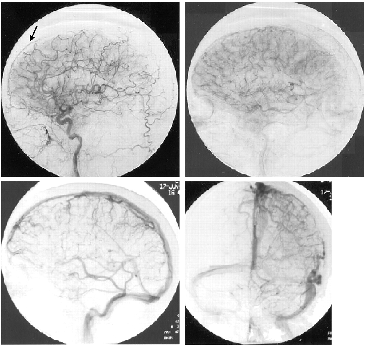
Arteriograms of the left carotid artery. Arterial and venous phases show an enlarged anterior falx artery (arrow) and multiple enlarged cortical and medullary veins, associated with SIH.

Fig 2.
  • Download figure
  • Open in new tab
  • Download powerpoint
Fig 2.

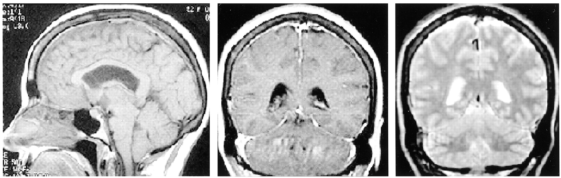
MR images of SIH. Sagittal view T1-weighted image (left) shows depressed cerebellar tonsils, hypothalamic structures, and effacement of the prepontine cistern. Coronal view T1-weighted image (center) shows diffuse dural thickening and enhancement, and coronal view T2-weighted image (right) shows dural high T2 signal intensity. These findings resolved with the symptoms.

The patient received supportive treatment, and her symptoms improved. Follow-up MR imaging performed 1 month later showed near complete resolution of the meningeal enhancement, improved patency of the basal cisterns, and less hypothalamic/tonsilar displacement.

Discussion

Diagnostic imaging of patients with suspected SIH has led to a number of characteristic descriptions. Findings on MR imaging include subdural fluid collections, downward descent of the midbrain and cerebellar tonsils, and a reduction in the size of the prepontine cistern. However, the most characteristic finding on MR imaging is diffuse pachymeningeal enhancement (2).

Many hypothesize that the dural enhancement of SIH is due to venous engorgement as opposed to inflammatory changes (3, 4). This is primarily based on the Monro-Kellie hypothesis, which states that with an intact skull, the sum of the brain volume plus CSF volume plus intracranial blood volume is constant (5). Therefore, a reduction in the CSF volume should result in an increase in the intracranial blood volume. With an increase in venous blood, there is presumably accumulation of contrast material in the pachymeninges due to the lack of a blood-brain barrier. Furthermore, Chung et al (2) have suggested that the increase in dural signal intensity on long TR weighted images may discriminate SIH from pachymeningitis. Meningeal biopsies in cases of SIH lack evidence of inflammation, infection, or neoplasia, further supporting this hypothesis (6).

Visualization of venous engorgement in association with SIH has infrequently been described. The MR imaging literature has described dilated cervical epidural veins in cases of orthostatic headaches (7, 8). Although there are numerous differential considerations, generally, no secondary signs support another cause for cervical epidural venous dilation. These findings will spontaneously resolve with the patient’s symptoms and are thought to be the sequelae of CSF hypotension. A paucity of angiographic descriptions of SIH is available in the literature. The only published data of which we are aware are presented by Christoforidis et al (4), who described an attenuated meningeal blush and an enlarged anterior falx artery. Cerebral angiography in our case of SIH showed an enlarged anterior falx artery and markedly enlarged cortical and medullary veins. The venous engorgement shown is in accordance with the Monro-Kellie rule. This further supports the hypothesis that diffuse pachymeningeal enhancement seen on MR images in cases of SIH is a hydrostatic phenomena due to increased intracranial blood volume.

The presence of >600 red cells in the CSF was the driving force behind the arteriogram obtained in this case. Yet, abnormal amounts of blood and protein in the CSF is a common feature of SIH and is thought to be a result of meningeal hyperemia and red blood cell diapedesis (9, 10). Up to 1100 cells have been seen with nontraumatic lumbar punctures in cases of SIH (2, 4). Furthermore, a traumatic lumbar puncture is more likely with low CSF pressure. Xanthochromia being the hallmark of chronic subarachnoid hemorrhage is a common observation in association with SIH. It has been suggested that SIH may have clinical and diagnostic features that are similar to those of subarachnoid hemorrhage caused by other factors (10). However, the presence of subarachnoid hemorrhage in conjunction with characteristic clinical features of SIH should lead first to MR imaging. Had MR imaging been available in our case, the patient likely would not have undergone arteriography to exclude another source of subarachnoid hemorrhage. Should angiography be required in atypical clinical scenarios, the findings can be diagnostic of SIH and are explained by the Monro-Kellie hypothesis.

References

  1. ↵
    Schaltenbrand G. Normal and pathological physiology of cerebrospinal fluid circulation. Lancet 1953;I:805–809
  2. ↵
    Chung SJ, Kim JS, Lee MC. Syndrome of cerebral spinal fluid hypovolemia: clinical and imaging features and outcome. Neurology 2000;55:1321–1327
    Abstract/FREE Full Text
  3. ↵
    Fishman RA, Dillon WP. Dural enhancement and cerebral displacement secondary to intracranial hypotension. Neurology 1993;43:609–611
    Abstract/FREE Full Text
  4. ↵
    Christoforidis GA, Mehta BA, Landi JL, Czarnecki EJ, Piaskowski RA. Spontaneous intracranial hypotension: report of four cases and review of the literature. Neuroradiology 1998;40:636–643
    CrossRefPubMed
  5. ↵
    Mokri B. The Monro-Kellie hypothesis: applications in CSF volume depletion. Neurology 2001;56:1746–1748
    Abstract/FREE Full Text
  6. ↵
    Mokri B, Parisi JE, Scheithauer BW, Piepgras DG, Miller GM. Meningeal biopsy in intracranial hypotension: meningeal enhancement on MRI. Neurology 1995;45:1801–1807
    Abstract/FREE Full Text
  7. ↵
    Clarot F, Callonnec F, Douvrin F. Giant cervical epidural veins after lumbar puncture in a case of intracranial hypotension. AJNR Am J Neuroradiol 2000;21:787–789
    Abstract/FREE Full Text
  8. ↵
    Forderreuther S, Yousry I, Empl M, Straube A. Dilated cervical epidural veins and extra arachnoid fluid collection in orthostatic headaches. Neurology 2001;57:527–529
    Abstract/FREE Full Text
  9. ↵
    Rando TA, Fishman RA. Spontaneous intracranial hypotension: report of 2 cases and review of the literature. Neurology 1992;42:481–487
    Abstract/FREE Full Text
  10. ↵
    Schievink WI, Wijdicks EF, Meyer FB, Sonntag VK. Spontaneous intracranial hypotension mimicking aneurysmal subarachnoid hemorrhage. Neurosurgery 2001;48:513–517
    CrossRefPubMed
  • Received July 8, 2002.
  • Accepted after revision July 15, 2002.
  • Copyright © American Society of Neuroradiology
View Abstract
PreviousNext
Back to top

In this issue

American Journal of Neuroradiology: 24 (4)
American Journal of Neuroradiology
Vol. 24, Issue 4
1 Apr 2003
  • Table of Contents
  • Index by author
Advertisement
Print
Download PDF
Email Article

Thank you for your interest in spreading the word on American Journal of Neuroradiology.

NOTE: We only request your email address so that the person you are recommending the page to knows that you wanted them to see it, and that it is not junk mail. We do not capture any email address.

Enter multiple addresses on separate lines or separate them with commas.
Angiographic Features of Spontaneous Intracranial Hypotension
(Your Name) has sent you a message from American Journal of Neuroradiology
(Your Name) thought you would like to see the American Journal of Neuroradiology web site.
CAPTCHA
This question is for testing whether or not you are a human visitor and to prevent automated spam submissions.
Cite this article
Scott A. Koss, John L. Ulmer, Lotfi Hacein-Bey
Angiographic Features of Spontaneous Intracranial Hypotension
American Journal of Neuroradiology Apr 2003, 24 (4) 704-706;

Citation Manager Formats

  • BibTeX
  • Bookends
  • EasyBib
  • EndNote (tagged)
  • EndNote 8 (xml)
  • Medlars
  • Mendeley
  • Papers
  • RefWorks Tagged
  • Ref Manager
  • RIS
  • Zotero
0 Responses
Respond to this article
Share
Bookmark this article
Angiographic Features of Spontaneous Intracranial Hypotension
Scott A. Koss, John L. Ulmer, Lotfi Hacein-Bey
American Journal of Neuroradiology Apr 2003, 24 (4) 704-706;
del.icio.us logo Twitter logo Facebook logo Mendeley logo
  • Tweet Widget
  • Facebook Like
  • Google Plus One

Jump to section

  • Article
    • Abstract
    • Case Report
    • Discussion
    • References
  • Figures & Data
  • Info & Metrics
  • Responses
  • References
  • PDF

Related Articles

  • No related articles found.
  • PubMed
  • Google Scholar

Cited By...

  • Dual-Energy CT in Enhancing Subdural Effusions that Masquerade as Subdural Hematomas: Diagnosis with Virtual High-Monochromatic (190-keV) Images
  • CT Myelography for the Planning and Guidance of Targeted Epidural Blood Patches in Patients with Persistent Spinal CSF Leakage
  • MR Imaging of the Optic Nerve Sheath in Patients with Craniospinal Hypotension
  • Spontaneous intracranial hypotension: a cause of severe acute headache
  • Spontaneous intracranial hypotension: a cause of severe acute headache
  • Changes in the appearance of venous sinuses after treatment of disordered intracranial pressure
  • Crossref
  • Google Scholar

This article has not yet been cited by articles in journals that are participating in Crossref Cited-by Linking.

More in this TOC Section

  • Usefulness of Quantitative Susceptibility Mapping for the Diagnosis of Parkinson Disease
  • White Matter Alterations in the Brains of Patients with Active, Remitted, and Cured Cushing Syndrome: A DTI Study
  • Qualitative and Quantitative Analysis of MR Imaging Findings in Patients with Middle Cerebral Artery Stroke Implanted with Mesenchymal Stem Cells
Show more BRAIN

Similar Articles

Advertisement

Indexed Content

  • Current Issue
  • Accepted Manuscripts
  • Article Preview
  • Past Issues
  • Editorials
  • Editor's Choice
  • Fellows' Journal Club
  • Letters to the Editor
  • Video Articles

Cases

  • Case Collection
  • Archive - Case of the Week
  • Archive - Case of the Month
  • Archive - Classic Case

Special Collections

  • AJNR Awards
  • ASNR Foundation Special Collection
  • Most Impactful AJNR Articles
  • Photon-Counting CT
  • Spinal CSF Leak Articles (Jan 2020-June 2024)

More from AJNR

  • Trainee Corner
  • Imaging Protocols
  • MRI Safety Corner

Multimedia

  • AJNR Podcasts
  • AJNR Scantastics

Resources

  • Turnaround Time
  • Submit a Manuscript
  • Submit a Video Article
  • Submit an eLetter to the Editor/Response
  • Manuscript Submission Guidelines
  • Statistical Tips
  • Fast Publishing of Accepted Manuscripts
  • Graphical Abstract Preparation
  • Imaging Protocol Submission
  • Evidence-Based Medicine Level Guide
  • Publishing Checklists
  • Author Policies
  • Become a Reviewer/Academy of Reviewers
  • News and Updates

About Us

  • About AJNR
  • Editorial Board
  • Editorial Board Alumni
  • Alerts
  • Permissions
  • Not an AJNR Subscriber? Join Now
  • Advertise with Us
  • Librarian Resources
  • Feedback
  • Terms and Conditions
  • AJNR Editorial Board Alumni

American Society of Neuroradiology

  • Not an ASNR Member? Join Now

© 2025 by the American Society of Neuroradiology All rights, including for text and data mining, AI training, and similar technologies, are reserved.
Print ISSN: 0195-6108 Online ISSN: 1936-959X

Powered by HighWire