Advertisement

Main menu

  • Home
  • Content
    • Current Issue
    • Accepted Manuscripts
    • Article Preview
    • Past Issue Archive
    • Video Articles
    • AJNR Case Collection
    • Case of the Week Archive
    • Case of the Month Archive
    • Classic Case Archive
  • Special Collections
    • AJNR Awards
    • ASNR Foundation Special Collection
    • Most Impactful AJNR Articles
    • Photon-Counting CT
    • Spinal CSF Leak Articles (Jan 2020-June 2024)
  • Multimedia
    • AJNR Podcasts
    • AJNR SCANtastic
    • Trainee Corner
    • MRI Safety Corner
    • Imaging Protocols
  • For Authors
    • Submit a Manuscript
    • Submit a Video Article
    • Submit an eLetter to the Editor/Response
    • Manuscript Submission Guidelines
    • Statistical Tips
    • Fast Publishing of Accepted Manuscripts
    • Graphical Abstract Preparation
    • Imaging Protocol Submission
    • Author Policies
  • About Us
    • About AJNR
    • Editorial Board
    • Editorial Board Alumni
  • More
    • Become a Reviewer/Academy of Reviewers
    • Subscribers
    • Permissions
    • Alerts
    • Feedback
    • Advertisers
    • ASNR Home

User menu

  • Alerts
  • Log in

Search

  • Advanced search
American Journal of Neuroradiology

American Journal of Neuroradiology

ASHNR American Society of Functional Neuroradiology ASHNR American Society of Pediatric Neuroradiology ASSR
  • Alerts
  • Log in

Advanced Search

  • Home
  • Content
    • Current Issue
    • Accepted Manuscripts
    • Article Preview
    • Past Issue Archive
    • Video Articles
    • AJNR Case Collection
    • Case of the Week Archive
    • Case of the Month Archive
    • Classic Case Archive
  • Special Collections
    • AJNR Awards
    • ASNR Foundation Special Collection
    • Most Impactful AJNR Articles
    • Photon-Counting CT
    • Spinal CSF Leak Articles (Jan 2020-June 2024)
  • Multimedia
    • AJNR Podcasts
    • AJNR SCANtastic
    • Trainee Corner
    • MRI Safety Corner
    • Imaging Protocols
  • For Authors
    • Submit a Manuscript
    • Submit a Video Article
    • Submit an eLetter to the Editor/Response
    • Manuscript Submission Guidelines
    • Statistical Tips
    • Fast Publishing of Accepted Manuscripts
    • Graphical Abstract Preparation
    • Imaging Protocol Submission
    • Author Policies
  • About Us
    • About AJNR
    • Editorial Board
    • Editorial Board Alumni
  • More
    • Become a Reviewer/Academy of Reviewers
    • Subscribers
    • Permissions
    • Alerts
    • Feedback
    • Advertisers
    • ASNR Home
  • Follow AJNR on Twitter
  • Visit AJNR on Facebook
  • Follow AJNR on Instagram
  • Join AJNR on LinkedIn
  • RSS Feeds

AJNR is seeking candidates for the AJNR Podcast Editor. Read the position description.

OtherINTERVENTIONAL

Aneurysm Regression after Coil Embolization of a Concurrent Aneurysm

Michael M. Chow, William E. Thorell and Peter A. Rasmussen
American Journal of Neuroradiology April 2005, 26 (4) 917-921;
Michael M. Chow
  • Find this author on Google Scholar
  • Find this author on PubMed
  • Search for this author on this site
William E. Thorell
  • Find this author on Google Scholar
  • Find this author on PubMed
  • Search for this author on this site
Peter A. Rasmussen
  • Find this author on Google Scholar
  • Find this author on PubMed
  • Search for this author on this site
  • Article
  • Figures & Data
  • Info & Metrics
  • Responses
  • References
  • PDF
Loading

Abstract

Summary: A 43-year-old woman with two incidental paraclinoid internal carotid artery aneurysms underwent coil embolization of the larger superior hypophyseal aneurysm and 10 weeks later underwent follow-up angiography that showed regression of the smaller, more distal paraclinoid aneurysm. We demonstrate that, although it is a rare occurrence, aneurysms can involute. We discuss potential mechanisms of this phenomenon and review the literature on aneurysm regression.

Angiographic regression of an aneurysm is a rare occurrence. Most of the reports in the literature have described aneurysms that have ruptured, and a significant portion of them have also involved vasospasm. In this case report, we present a unique case of an unruptured small paraclinoid internal carotid artery (ICA) aneurysm that disappeared after preceding endovascular treatment of a larger concurrent ICA aneurysm. A mechanism for this extraordinary event is postulated, and the literature on regressing aneurysms is reviewed.

Case Report

A 43-year-old right-handed white woman underwent MR imaging of the brain because of a cerebellar hemorrhage. There was no evidence of subarachnoid (SAH) or intraventricular hemorrhage on MR images or noncontrast CT scans of the head. The hemorrhage was secondary to overdrainage of CSF via a lumbar subarachnoid drain placed because of inadvertent durotomy during lumbar decompression and fusion surgery. A right ICA aneurysm was suspected on the basis of MR imaging findings. Following full recovery from her parenchymal hematoma, she underwent diagnostic cerebral angiography that demonstrated two cerebral aneurysms in the paraclinoid region of the right ICA. The first aneurysm, a superior hypophyseal artery aneurysm, measured 9 mm and projected medially from the ophthalmic segment. The second aneurysm, originating on the ventral ICA wall proximal to the anterior choroidal artery, measured 4 mm and extended laterally and inferiorly (Fig 1A and B).

Fig 1.
  • Download figure
  • Open in new tab
  • Download powerpoint
Fig 1.

AP (A) and lateral (B) right ICA angiograms show a 9-mm superior hypophyseal aneurysm and a 4-mm ventral paraclinoid ICA aneurysm before coil embolization.

The patient underwent coil embolization with the goal of treating both aneurysms at one time. A combination of Guglielmi detachable coils (Boston Scientific, Fremont, CA) and hydrocoils (Microvention, Aliso Viejo, CA) was used to embolize the more proximal and larger aneurysm without complication (Fig 2A and B). Although the microwire and microcatheter were maneuvered inside the second aneurysm, a stable microcatheter position in the second aneurysm was never attained. After a significant number of attempts, coil embolization of the second aneurysm was postponed. Full heparinization, which was begun after placement of the access sheath in the right common femoral artery, was continued overnight. The patient was discharged on aspirin from the hospital on the first postprocedure day.

Fig 2.
  • Download figure
  • Open in new tab
  • Download powerpoint
Fig 2.

AP (A) and lateral (B) right ICA angiograms obtained immediately after coil embolization of the larger superior hypophyseal aneurysm. The smaller, ventral paraclinoid ICA aneurysm remains unchanged.

Treatment of the second aneurysm by using a balloon- or stent-assisted technique was planned for 10 weeks later. Aspirin and clopidogrel were prescribed for the 5 days prior to the patient’s second treatment day. Baseline angiograms, including a 3D rotational angiogram, demonstrated complete involution of the aneurysm (Fig 3A–C). The patient returned for a follow-up angiogram at 10 months, which confirmed permanent regression of the aneurysm (Fig 4A–C).

Fig 3.
  • Download figure
  • Open in new tab
  • Download powerpoint
Fig 3.

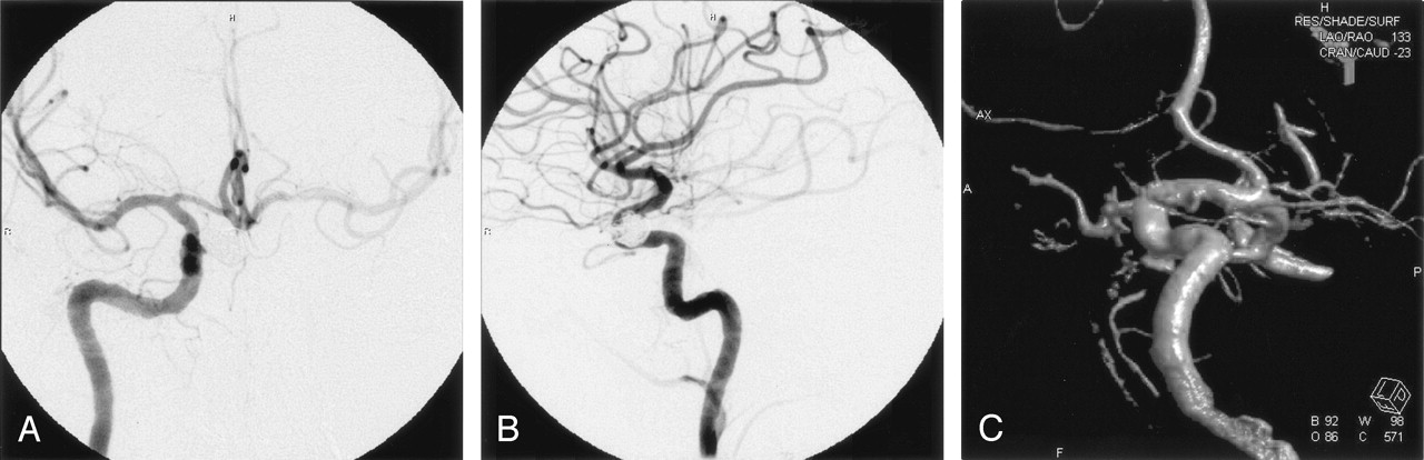
AP (A), lateral (B), and 3D rotational (C) right ICA angiograms at 10 weeks display complete regression of the previously visualized ventral paraclinoid ICA aneurysm.

Fig 4.
  • Download figure
  • Open in new tab
  • Download powerpoint
Fig 4.

AP (A), lateral (B), and 3D rotational (C) right ICA angiograms at 10 months display a stable coil configuration within the superior hypophyseal artery aneurysm and continued absence of the ventral paraclinoid ICA aneurysm.

Discussion

It is well known that mycotic aneurysms, arteriovenous malformation–associated aneurysms, and post-traumatic pseudoaneurysms can regress spontaneously (1–5). The incidence of spontaneous thrombosis of saccular aneurysms is unknown, however, and estimates from various autopsy series have ranged anywhere from 0.01% to 13% (6–8). A serial angiographic review at the Karolinska Institute between 1964 and 1973 revealed only one (1.3%) in 78 patients with a spontaneously disappearing aneurysm (9). These authors believe that the true incidence is probably 1–2%. Because the philosophy for the timing of aneurysm surgery after SAH has changed over the past 20 years, any contemporary series would probably underestimate the true incidence of spontaneous thrombosis.

The first report of a spontaneously disappearing aneurysm has been attributed to Marguth and Schieffer (10) in 1957. Our review of the literature revealed a total of 23 cases of angiographically proven saccular aneurysms that appeared to spontaneously disappear on the basis of follow-up angiography findings (Table 1; 9–28). This does not include reports of aneurysms that reappeared at subsequent angiography, presented with thrombosis (i.e., no angiogram before thrombosis), were found to be thrombosed at surgical exploration (i.e., without a follow-up angiogram), or were partially thrombosed (22, 29–31). To the best of our knowledge, there are no case reports that describe an aneurysm that regressed after treatment of a concurrent aneurysm. Furthermore, our case is unique in that the concurrent aneurysm was treated by endovascular means.

View this table:
  • View inline
  • View popup

Aneurysmal regression series

On closer examination of the literature, there appear to be two separate categories of spontaneously regressing saccular aneurysms—a group that disappeared after SAH and a group that thrombosed without an antecedent rupture. In the SAH group, the two factors that appear to play a role in subsequent thrombosis are the development of vasospasm (with or without parent vessel thrombosis) and treatment with epsilon amino-caproic acid. It is not difficult to believe that significant vasospasm could cause alteration in the hemodynamic forces within the aneurysm to the point at which stasis and thrombosis occur. In fact, some cases have reported thrombosis of the parent vessel along with the aneurysm (13, 17, 19, 22). Similarly, epsilon amino-caproic acid possesses an antifibrinolytic mechanism of action that may predispose the aneurysm to spontaneous thrombosis. Another factor that has been postulated as a contributing factor but has not been well documented in the preceding case reports is systemic hypotension. In fact, there are some early reports of using controlled hypotension to induce spontaneous thrombosis of saccular aneurysms (32, 33).

Although more rare in occurrence, a group of patients exists who had spontaneous regression without antecedent SAH. Within this group, few reports of large or giant aneurysms disappearing after extracranial-intracranial bypass without accompanying parent vessel occlusion can be found (26, 27). In these cases, the plan was to treat the patient with proximal vessel balloon occlusion subsequent to the external carotid artery-middle cerebral artery bypass. Follow-up angiograms, however, showed spontaneous thrombosis of the aneurysms with thrombosis of the parent vessel as well. It was postulated that the high-flow saphenous vein bypass resulted in retrograde filling of the ICA and coupled with the unaltered antegrade flow along the ICA resulted in significant turbulence at the aneurysm site, leading to subsequent thrombosis. The same result, spontaneous aneurysm thrombosis, is usually obtained if the proximal vessel is occluded at the same time the bypass is being performed.

Aneurysm characteristics such as size, neck width, and location have all been considered as potential predictors of spontaneous regression. Stehbens (34) believed that the size of the aneurysmal sac in relation to the caliber of the parent vessel was important in determining the risk for aneurysmal thrombosis. In an experimental model, Black and German (35) demonstrated that the ratio of the aneurysmal volume to the neck width was important with a large volume and a narrow neck predisposing the aneurysm to thrombosis. Most of the regressing aneurysms listed in the Table were medium to large with narrow necks.

On examination of the list of aneurysm locations, there appeared to be few aneurysms found at either the ICA terminus or basilar terminus. It is well established from the endovascular literature that these are the sites where there is significantly increased risk for coil compaction secondary to the pattern of flow into the aneurysm (36). Therefore, it would be logically less likely for an aneurysm in one of these locations to thrombose spontaneously.

After considering all of the above factors, it is apparent that our disappearing aneurysm is different in many respects. Our aneurysm was a small, relatively broad-necked sidewall aneurysm, not a medium to large, narrow-necked aneurysm like most the cases described in the literature. Nor had our patient had an SAH. We postulate, however, that our previous embolization procedure may have somehow altered the hemodynamic flow into the smaller aneurysm, leading to stasis within the aneurysm and subsequent thrombosis. Perhaps the outflow zone of the proximal aneurysm led to the initial formation of the smaller distal aneurysm and, with the obliteration of the proximal aneurysm with coils, the hemodynamic flow was altered significantly enough to result in regression of the distal aneurysm either by changing of the vessel contour or thrombosis. In addition, it was possible that microcatheter or guidewire manipulation within the smaller distal aneurysm may have caused intimal injury promoting thrombosis of the aneurysm. This was less likely, however, because the patient underwent full anticoagulation at the time of the procedure.

Another pertinent issue with regressing aneurysms is the length of follow-up necessary to confirm that the aneurysm does not reappear. In fact, there have been three reports of aneurysms that have angiographically disappeared and subsequently reappeared at follow-up angiography (18, 29, 37). All three cases occurred in the setting of SAH, and in two of the three cases (18, 37) vasospasm appeared to contribute to the nonvisualization, whereas in the remaining case systemic hypotension at the time of the negative angiogram may have played a role (29). In all three cases, the aneurysm reappeared between 1 and 2 weeks after the complicating factors had receded.

Benedetti et al (23) described a similar case in which a ruptured anterior communicating artery aneurysm was demonstrated on an initial angiogram and subsequently re-bled. At follow-up angiography after the re-bleed, no aneurysm opacification was seen despite adequate technical conditions (i.e., no spasm or shifting of vessels secondary to mass effect). On surgical exploration, however, a patent aneurysm was discovered and a clip placed. Another case reported by Atkinson et al (31) involved a fusiform posterior cerebral artery aneurysm that resulted in neurologic deficits related to acute thrombosis of the aneurysm. There was no prethrombosis angiogram documenting the patency of the aneurysm, although a follow-up angiogram obtained 3 months later demonstrated recanalization of the lesion.

Unfortunately, none of these cases can provide any guidance in that they do not match the circumstances of our particular patient, because our patient’s aneurysm was unruptured and was saccular in nature. The 10-month follow-up angiogram obtained in our patient did not show recurrence of the ventral paraclinoid aneurysm. Because our patient had another concurrent aneurysm in which a coil was placed and we characteristically repeat angiography for 2 years after coil placement to monitor for coil compaction and regrowth, we plan to obtain at least one more angiogram. It remains to be seen whether the smaller aneurysm will reappear or whether the cure is durable.

Conclusion

We report on a unique case of an ICA aneurysm that disappeared after endovascular treatment of a concurrent ICA aneurysm. The hemodynamic alterations caused by the embolization procedure may have contributed to the regression.

References

  1. ↵
    Kowada M, Takahashi M, Gito Y. Spontaneous cure of intracranial aneurysm. Acta Neurochir (Wien)1974;31:131–137
  2. Devadiga KV, Mathai KV, Chandy J. Spontaneous cure of intracavernous aneurysm of the internal carotid artery in a 14-month-old child. Case report. J Neurosurg 1969;30:165–168
    PubMed
  3. Koulouris S, Rizzoli HV. Coexisting intracranial aneurysm and arteriovenous malformation: case report. Neurosurgery 1981;8:219–222
    PubMed
  4. Salcman M, Botero E, Bellis E. Giant posttraumatic aneurysm of the intracranial carotid artery: evolution and regression documented by computed tomography. Neurosurgery 1985;16:218–221
    PubMed
  5. ↵
    Mori S, Feliciani M, Guglielmi G, et al. Regression of an internal carotid artery pseudoaneurysm after therapeutic embolization of a post-traumatic carotid-cavernous fistula secondary to gunshot injury. Neuroradiology 1990;32:226–228
    PubMed
  6. ↵
    Schunk H. Spontaneous thrombosis of intracranial aneurysm. AJR Am J Roentgenol 1964;91:1327–1338
  7. Krayenbuhl H. Das hirnaneurysma. Schweiz Arch Neurol Psychiatr 1944;47:155–236
  8. ↵
    Housepian EM, Pool JL. A systematic analysis of intracranial aneurysm from the autopsy file of the Presbyterian Hospital, 1914 to 1956. J Neuropath Exp Neurol 1958;17:409–423
    PubMed
  9. ↵
    Edner G, Forster DM, Steiner L, Bergvall U. Spontaneous healing of intracranial aneurysms after subarachnoid hemorrhage: case report. J Neurosurg 1978;48:450–454
    PubMed
  10. ↵
    Marguth F, Schieffer W. Spontanheilung eines intrakraniellen aneurysms angiographisch nachgewiesen. Acta Neurochir (Wien)1957;5:38–45
    PubMed
  11. Hemmer R, Umbach W. Verlautisformen intrakranieller sackformiger aneurysmen. Arch Psychiatr Nervenkr 1960;200:612–625
  12. Hook O, Norlen G. Aneurysms of the middle cerebral artery: a report of 80 cases. Acta Chir Scand (Suppl) 1958;235:1–39
  13. ↵
    Lindgren SO. Diagnostiska och terapeutiska svarigheter vid vaskulara cerebrala lasioner. Lakartidningen 1958;55:2453
  14. Hollin SA, Gross SW. Changing size of an aneurysm: report of a case. J Neurosurg 1965;24:573–575
  15. Lodin H. Spontaneous thrombosis of cerebral aneurysms. Br J Radiol 1966;39:701–703
    Abstract/FREE Full Text
  16. Bjorkesten G, Troupp H. Changes in the size of intracranial arterial aneurysms. J Neurosurg 1962;19:583–588
    PubMed
  17. ↵
    Scott RM, Ballantine HT Jr. Spontaneous thrombosis in a giant middle cerebral artery aneurysm: case report. J Neurosurg 1972;37:361–363
    PubMed
  18. ↵
    Spetzler RF, Winestock D, Newton HT, Boldrey EB. Disappearance and reappearance of cerebral aneurysm in serial arteriograms: case report. J Neurosurg 1974;41:508–510
    CrossRefPubMed
  19. ↵
    Scott RM, Garrido E. Spontaneous thrombosis of an intracranial aneurysm during treatment with epsilon aminocaproic acid. Surg Neurol 1977;7:21–23
    PubMed
  20. Suzuki J, Onuma T. A giant intracranial aneurysm which disappeared angiographically following pneumoencephalography. Neurol Med Chir (Tokyo)1976;16:105–108
    PubMed
  21. Moritake K, Handa H, Ohtsuka S, Hashimoto N. Vanishing cerebral aneurysm in serial angiography. Surg Neurol 1981;16:36–40
    CrossRefPubMed
  22. ↵
    Fodstad H, Liliequist B. Spontaneous thrombosis of ruptured intracranial aneurysms during treatment with tranexamic acid (AMCA): report of three cases. Acta Neurochir (Wien)1979;49:129–144
    CrossRefPubMed
  23. ↵
    Benedetti A, Curri C, Colombo F. Rebleeding of an angiographically healed aneurysm. Surg Neurol 1983;20:206–208
    PubMed
  24. Spallone A, Peresedov VV, Kandel EI. Spontaneous cure of ruptured intracranial arterial aneurysms. Surg Neurol 1981;16:367–370
    CrossRefPubMed
  25. Davila S, Oliver B, Molet J, Bartumeus F. Spontaneous thrombosis of an intracranial aneurysm. Surg Neurol 1984;22:29–32
    CrossRefPubMed
  26. ↵
    Cantore G, Santoro A, Da Pian R. Spontaneous occlusion of supraclinoid aneurysms after the creation of extra-intracranial bypasses using long grafts: report of two cases. Neurosurgery 1999;44:216–219; discussion 219–220
    PubMed
  27. ↵
    Yeh H, Tomsick TA. Obliteration of a giant carotid aneurysm after extracranial-to-intracranial bypass surgery: case report. Surg Neurol 1997;48:473–476
    PubMed
  28. ↵
    Senn P, Krauss JK, Remonda L, et al. The formation and regression of a flow-related cerebral artery aneurysm. Clin Neurol Neurosurg 2000;102:168–172
    PubMed
  29. ↵
    Bohmfalk GL, Story JL. Intermittent appearance of a ruptured cerebral aneurysm on sequential angiograms: case report. J Neurosurg 1980;52:263–265
    PubMed
  30. Lin JP. Thrombosis of aneurysm of anterior communicating artery: case report. Acta Radiol Diagn (Stockh)1969;8:74–80
    PubMed
  31. ↵
    Atkinson JL, Lane JI, Colbassani HJ, Llewellyn DM. Spontaneous thrombosis of posterior cerebral artery aneurysm with angiographic reappearance: case report. J Neurosurg 1993;79:434–437
    PubMed
  32. ↵
    Mullan S. Experiences with surgical thrombosis of intracranial berry aneurysms and carotid cavernous fistulas. J Neurosurg 1974;41:657–670
    PubMed
  33. ↵
    Khil’ko VA, Shkliarova ED. A method of artificial thrombosis of arterial aneurysms using epsilon-aminocaproic acid and other coagulants in addition to regionally retarded circulation: preliminary report. Vopr Neirokhir 1969;33:6–11
  34. ↵
    Stehbens WE. Hemodynamics and the blood vessel wall. Springfield, IL: Thomas1979 :465–515
  35. ↵
    Black SPW, German WJ. Observations on the relationship between the volume and the size of the orifice of experimental aneurysms. J Neurosurg 1960;17:984–990
    PubMed
  36. ↵
    Mericle RA, Wakhloo AK, Lopes DK, et al. Delayed aneurysm regrowth and recanalization after Guglielmi detachable coil treatment: case report. J Neurosurg 1998;89:142–145
    PubMed
  37. ↵
    Suzuki K, Tone O, Yonemura N, Inaba Y. A case presentation in which cerebral aneurysm disappeared then recurred documented by cerebral angiography. No Shinkei Geka 1981;9:1433–1436
    PubMed
  • Received May 1, 2004.
  • Accepted after revision July 16, 2004.
  • American Society of Neuroradiology
View Abstract
PreviousNext
Back to top

In this issue

American Journal of Neuroradiology: 26 (4)
American Journal of Neuroradiology
Vol. 26, Issue 4
1 Apr 2005
  • Table of Contents
  • Index by author
Advertisement
Print
Download PDF
Email Article

Thank you for your interest in spreading the word on American Journal of Neuroradiology.

NOTE: We only request your email address so that the person you are recommending the page to knows that you wanted them to see it, and that it is not junk mail. We do not capture any email address.

Enter multiple addresses on separate lines or separate them with commas.
Aneurysm Regression after Coil Embolization of a Concurrent Aneurysm
(Your Name) has sent you a message from American Journal of Neuroradiology
(Your Name) thought you would like to see the American Journal of Neuroradiology web site.
CAPTCHA
This question is for testing whether or not you are a human visitor and to prevent automated spam submissions.
Cite this article
Michael M. Chow, William E. Thorell, Peter A. Rasmussen
Aneurysm Regression after Coil Embolization of a Concurrent Aneurysm
American Journal of Neuroradiology Apr 2005, 26 (4) 917-921;

Citation Manager Formats

  • BibTeX
  • Bookends
  • EasyBib
  • EndNote (tagged)
  • EndNote 8 (xml)
  • Medlars
  • Mendeley
  • Papers
  • RefWorks Tagged
  • Ref Manager
  • RIS
  • Zotero
0 Responses
Respond to this article
Share
Bookmark this article
Aneurysm Regression after Coil Embolization of a Concurrent Aneurysm
Michael M. Chow, William E. Thorell, Peter A. Rasmussen
American Journal of Neuroradiology Apr 2005, 26 (4) 917-921;
del.icio.us logo Twitter logo Facebook logo Mendeley logo
  • Tweet Widget
  • Facebook Like
  • Google Plus One

Jump to section

  • Article
    • Abstract
    • Case Report
    • Discussion
    • Conclusion
    • References
  • Figures & Data
  • Info & Metrics
  • Responses
  • References
  • PDF

Related Articles

  • No related articles found.
  • PubMed
  • Google Scholar

Cited By...

  • Republished: Spontaneous regression of intracranial aneurysm following remote ruptured aneurysm treatment with pipeline stent assisted coiling
  • Spontaneous regression of intracranial aneurysm following remote ruptured aneurysm treatment with pipeline stent assisted coiling
  • Crossref
  • Google Scholar

This article has not yet been cited by articles in journals that are participating in Crossref Cited-by Linking.

More in this TOC Section

  • SAVE vs. Solumbra Techniques for Thrombectomy
  • Contrast-Induced Encephalopathy after NeuroIR
  • CT Perfusion&Reperfusion in Acute Ischemic Stroke
Show more Interventional

Similar Articles

Advertisement

Indexed Content

  • Current Issue
  • Accepted Manuscripts
  • Article Preview
  • Past Issues
  • Editorials
  • Editor's Choice
  • Fellows' Journal Club
  • Letters to the Editor
  • Video Articles

Cases

  • Case Collection
  • Archive - Case of the Week
  • Archive - Case of the Month
  • Archive - Classic Case

Special Collections

  • AJNR Awards
  • ASNR Foundation Special Collection
  • Most Impactful AJNR Articles
  • Photon-Counting CT
  • Spinal CSF Leak Articles (Jan 2020-June 2024)

More from AJNR

  • Trainee Corner
  • Imaging Protocols
  • MRI Safety Corner

Multimedia

  • AJNR Podcasts
  • AJNR Scantastics

Resources

  • Turnaround Time
  • Submit a Manuscript
  • Submit a Video Article
  • Submit an eLetter to the Editor/Response
  • Manuscript Submission Guidelines
  • Statistical Tips
  • Fast Publishing of Accepted Manuscripts
  • Graphical Abstract Preparation
  • Imaging Protocol Submission
  • Evidence-Based Medicine Level Guide
  • Publishing Checklists
  • Author Policies
  • Become a Reviewer/Academy of Reviewers
  • News and Updates

About Us

  • About AJNR
  • Editorial Board
  • Editorial Board Alumni
  • Alerts
  • Permissions
  • Not an AJNR Subscriber? Join Now
  • Advertise with Us
  • Librarian Resources
  • Feedback
  • Terms and Conditions
  • AJNR Editorial Board Alumni

American Society of Neuroradiology

  • Not an ASNR Member? Join Now

© 2025 by the American Society of Neuroradiology All rights, including for text and data mining, AI training, and similar technologies, are reserved.
Print ISSN: 0195-6108 Online ISSN: 1936-959X

Powered by HighWire