
We read with great interest the article of Takahashi et al.1 The article points out the use of 3D stereotactic surface projections (3D-SSP) to study the age-effect on regional cerebral blood flow (rCBF). The greatest rCBF reduction observed was in the bilateral anterior cingulate. Although we generally agree with the conclusions, we would like to emphasize some methodologic issues that may have had an impact on the obtained results.
In the study, 31 healthy volunteers between 50 and 79 years were classified in 3 different age classes (50–59, 60–69, and 70–79 years). Statistical analysis was performed 2 by 2 by using unpaired Student t test. Rather than considering age as a discrete variable, the analysis would have been strengthened by performing a multivariate analysis based on the general linear model with regions as intrasubject factor and sex as intersubject factor. The sex should also have been considered as a covariate, because rCBF has been shown to be sex dependent in a second order.
In 2004, we presented a similar analysis by using 3D-SSP2 of a previously published normal data base constituted of 89 healthy volunteers (46 women and 43 men; age range, 21–81 years) acquired on a triple-head camera.3 By using the previously described multivariate analysis, both age and sex had significant interaction with rCBF. Like Takahashi et al, we found a significant age-related decline (P < .001) in the anterior cingulated gyrus and left frontal association cortex, as well as in the left insula and peri-insular cortex. Moreover, we found also a significant relative increased perfusion in the bilateral occipital association and left primary visual cortex. Concerning the sex effect, women showed higher uptake in parietal (P = .001) and right sensorimotor cortex (P = .002) and a lower uptake in the left temporal associative cortex (P = .002). An age-by-sex interaction (P < .01) was found only in the left medial frontal cortex, in line with a known higher vulnerability of the left frontal lobe in men compared with women.
Of major importance, although 3D-SSP seems to be better for the analysis of atrophied brain than other analysis methods, it cannot be stated that the partial volume effect can be totally excluded. It is known that the anterior cingulate shows a marked age-related regional atrophy (eg, based on voxel-based morphometry studies).4 In the latter study, it was shown that in the anterior cingulate and other regions the changes of perfusion with aging fully paralleled underlying atrophy effects. Therefore, in our opinion, it should be acknowledged that atrophy is not fully taken into account by 3D-SSP and that a direct comparison between partial-volume corrected and uncorrected data should be made to assess to what extent the effect of atrophy on a 3D-SSP analysis is less than other voxel-based techniques such as statistical parametric mapping.
Reply:
We thank Pirson et al for their interest in our recent article in which we demonstrated a reduction of the regional cerebral blood flow (rCBF) in the bilateral anterior cingulate gyrus with aging.1 They made 2 important points. One concerns the effect of sex on rCBF with aging. The other is the effect of brain atrophy with aging on rCBF, especially in the anterior cingulate gyri. To address these concerns, we analyzed our data and examined the effect of aging on rCBF in each sex, by using 3D stereotactic surface projection analysis (3D-SSP). There was reduced rCBF in the bilateral anterior cingulate, right inferior frontal, left superior temporal gyrus, and left posterior cingulate gyrus in women in their 70s compared with those in their 50s (Fig 1A), whereas rCBF was reduced in bilateral anterior cingulate, left precentral lobule, and left uncus in men in their 70s compared with those in their 50s (Fig 1B). Thus, although there was no sex difference in the reduction of rCBF in bilateral anterior cingulate gyri with aging, we found differential sex effects on age-related rCBF reduction. We conclude, however, that our dataset was too small to reach a firm conclusion about the sex effect.
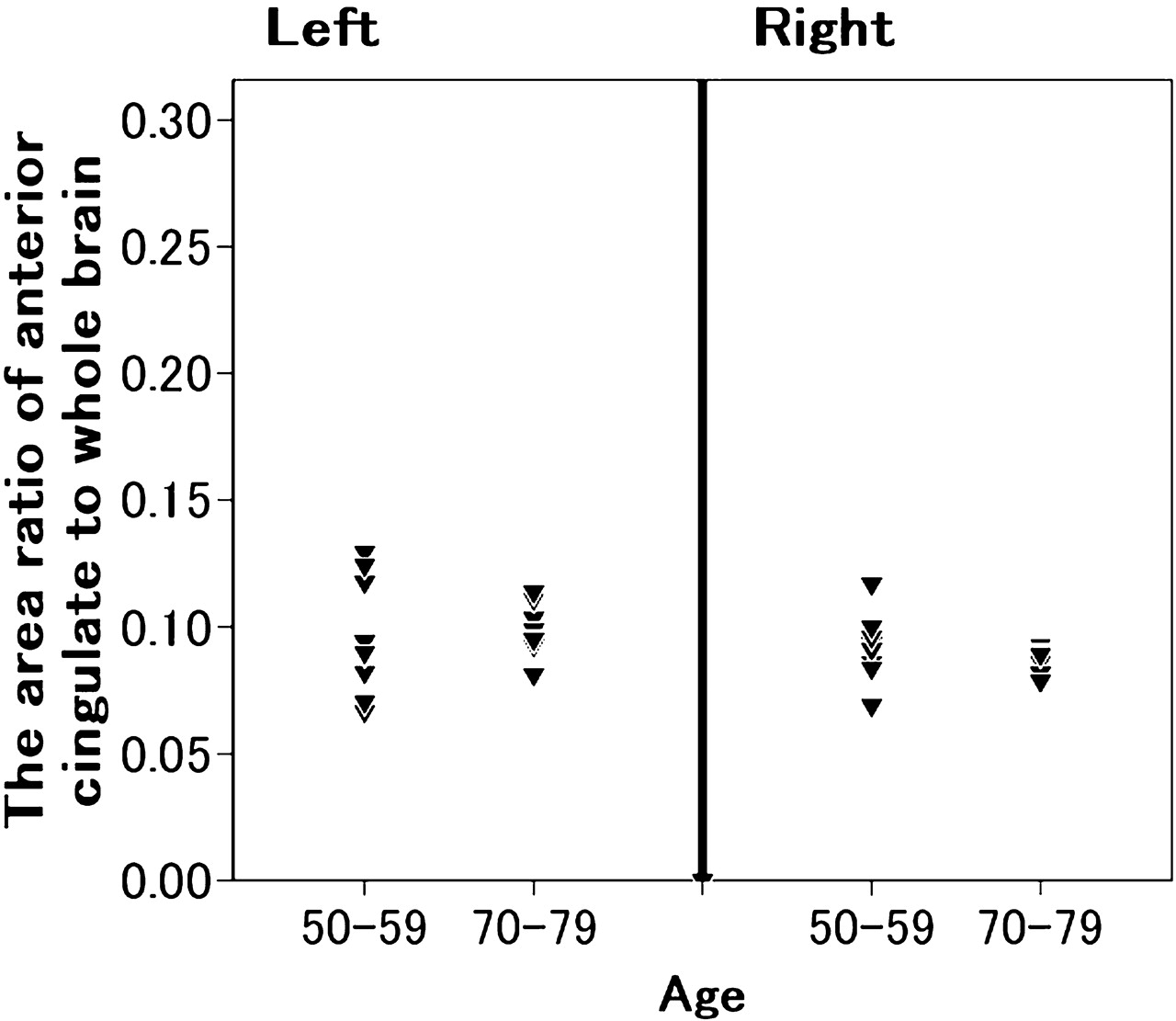
Next, we investigated the effect of brain atrophy. As Pirson et al stated, the anterior cingulate gyrus tends to atrophy with aging. Therefore, we measured the area of the anterior cingulate gyrus in MR images for each subject, by using National Institutes of Health Image (a software for analysis of medical images, free download page: http://rsb.info.nih.gov/nih-image/). Tracing the right and left anterior cingulate gyri and the entire brain of each subject in the sagittal view, we calculated the ratio of the area of the anterior cingulate gyri to that of the entire brain. This measurement yielded no significant difference in the ratio between subjects aged 50–59 years and those aged 70–79 years (Fig 2).
In general, we should always pay attention to the influence of brain atrophy when interpreting 3D-SSP images as suggested by Pirson et al.

Statistical maps analyzed by using 3D-SSP. The color of the outer contour corresponds to a Z score of 7. The relative decrease in rCBF (Z score of ≤2) in subjects 70–79 years of age compared with that in subjects 50–59 years of age (A, women; B, men). A marked reduction of rCBF was observed in the bilateral anterior cingulate gyri in each sex.

The ratio of the area of the right and left anterior cingulate gyri to that of the entire brain.
Reference
- Copyright © American Society of Neuroradiology


