
Abstract
SUMMARY: The presence of edema on MR imaging is a common finding in acute or subacute vertebral body compression fractures. Compression fractures can present in patients with benign osteoporosis, metastases, multiple myeloma, or hemangiomas. We present 2 patients with multiple myeloma who had symptomatic acute and subacute compression fractures documented on imaging studies without associated edema on MR imaging evaluation.
In recent years, the use of percutaneous vertebroplasty for the treatment of vertebral fractures has become very common. Several studies have documented the effectiveness of vertebroplasty in alleviating the pain associated with acute or subacute compression fractures.1–3 Although benign osteoporosis is the most common cause of compression fractures, multiple myeloma is another common contributor. Confirmation of a compression fracture is typically performed with MR imaging or bone scintigraphy. MR imaging generally shows loss of vertebral body height as well as edema in the involved vertebra. This edema is manifested as increased T2 and decreased T1 signal intensity. The visualized edema can be enhanced with the use of fat-saturated T2 techniques. Bone scintigraphy generally reveals increased radiotracer uptake in a linear fashion within the involved vertebral body. We present 2 patients with multiple myeloma in whom no edema was detected on MR imaging, despite clinical and imaging criteria for an acute or subacute vertebral compression fracture.
Case Reports
Patient 1
A 64-year-old man with multiple myeloma presented with severe diffuse back pain. He was evaluated initially for vertebroplasty with an MR imaging of the spine, which showed compression fractures at multiple thoracic and lumbar levels. Because it was difficult to clinically localize the specific levels involved, he elected to delay vertebroplasty and attempt a trial of narcotics. He returned 27 days later for a follow-up evaluation, at which time repeat MR imaging was performed. The previously documented fractures had not changed significantly in the interim. There was, however, a new fracture at the L2 level (Fig 1A, -B). Despite the short imaging interval, no edema was present on the MR imaging examination at the L2 level. Because the patient’s pain was improving on pain medications, he elected to defer vertebroplasty. The patient subsequently underwent bone marrow transplantation and developed multiple severe pulmonary and dermatologic infections. Because of long-term intravenous antibiotic treatments, he has not been considered a candidate for vertebroplasty during the intervening 6 weeks since his initial evaluation.

A, A 64-year-old man with multiple myeloma. Sagittal T2-weighted MR image demonstrates several mild lumbar compression fractures with a normal appearance of L2 (arrow).
B, A follow-up sagittal MR image obtained 4 weeks later demonstrates a new compression fracture of L2 (arrow) without any associated edema.
Patient 2
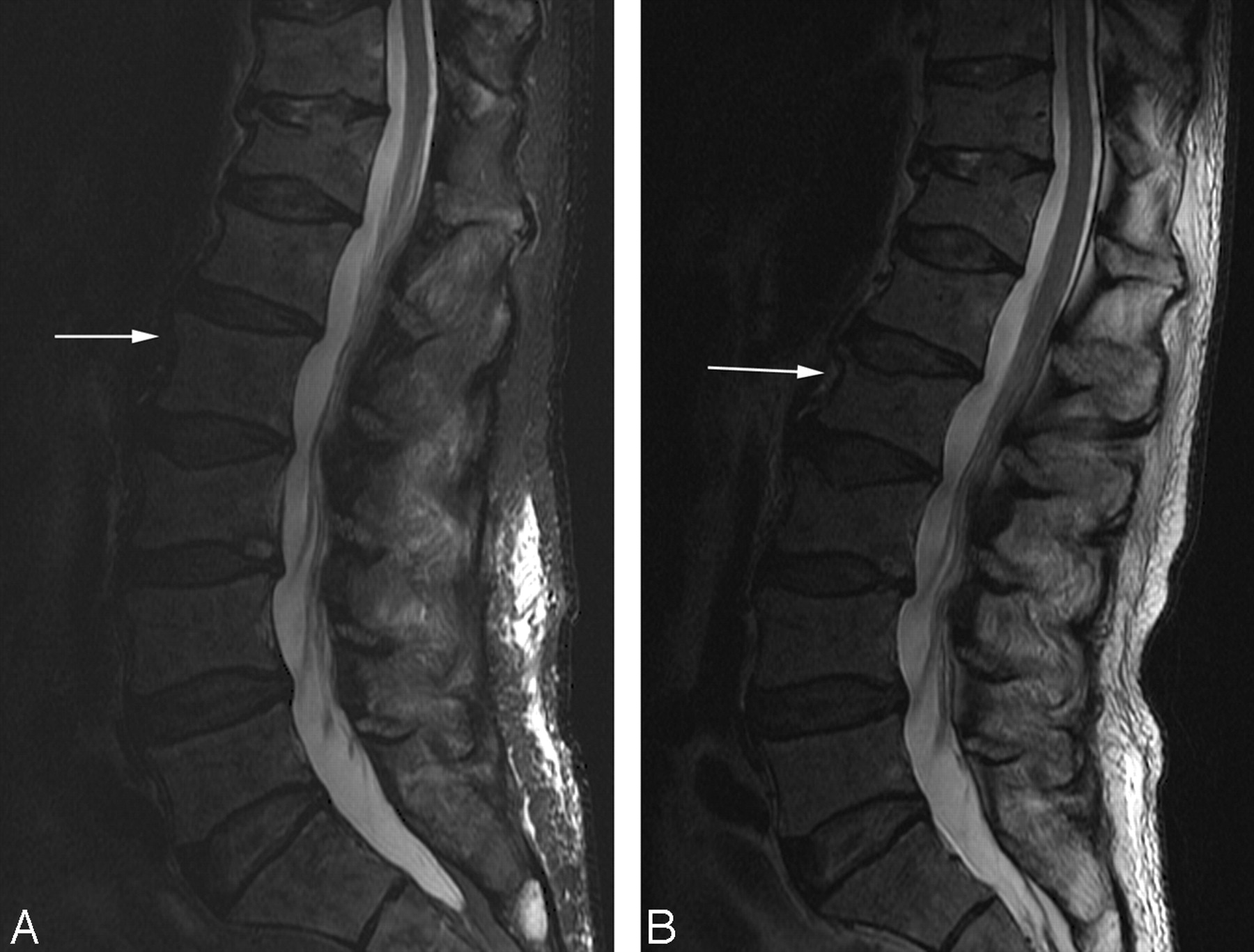
A 47-year-old man with multiple myeloma presented for vertebroplasty evaluation for severe back pain. The patient had fallen from a horse 6 months earlier, resulting in midback pain during this time course, which was rated as 8 of 10, for which he was taking oral narcotics. During the previous week, his pain had become more severe, and he was requiring intravenous narcotics. MR imaging of the thoracic spine demonstrated a compression fracture of the T7 vertebral body with a 20% loss in height but no significant edema (Fig 2A, -B). Manual palpation of the T7 spinous process under fluoroscopy demonstrated severe focal pain at this level. Because the patient was requiring intravenous narcotic administration, he was sent to the department of neuroradiology for a T7 vertebroplasty. With a transpedicular approach bilaterally, a total of 4.3 mL of cement was injected through a 13-gauge needle into the vertebral body. The patient experienced dramatic relief of pain 2 hours following the procedure and was discharged the same day without complication or the need for intravenous narcotics. Four weeks after the procedure, the patient reported continued pain relief, with a visual analog pain score at 3 of 10.

A, A 47-year-old man with multiple myeloma. Sagittal T2-weighted MR image demonstrates a compression fracture of T7 (arrow) without any significant edema.
B, A sagittal T1-weighted MR image reveals no edema in the superior endplate.
Discussion
Patients with acute or subacute compression fractures typically have severe pain that is localized to the fractured level. Inclusion criteria for vertebroplasty in patients with compression fractures have been described.4, 5 Generally, a documented acute or subacute fracture corresponding to the patient’s level of tenderness is required. Clinical correlation with the imaging findings can be confirmed with palpation under fluoroscopy of the spinous process at the involved level. Other causes of pain, such as disk extrusion and central spinal canal or neural foraminal compromise, should also be evaluated because vertebroplasty may not relieve the pain in these patients. Because patients with osteoporosis and multiple myeloma often have chronic vertebral compression fractures, it can be difficult to define precisely which fracture level or levels are the current pain generator. MR imaging is generally well suited to identify the edema associated with acute or subacute compression fractures. Although fat-saturated T2-weighted sequences are recommended to assess vertebral edema, simple T1-weighted images are also very sensitive to the decreased signal intensity of an acute/subacute compression fracture (Fig 3A, -B). In our practice, patients who are unable to undergo MR imaging are evaluated with bone scintigraphy studies (Fig 3C).
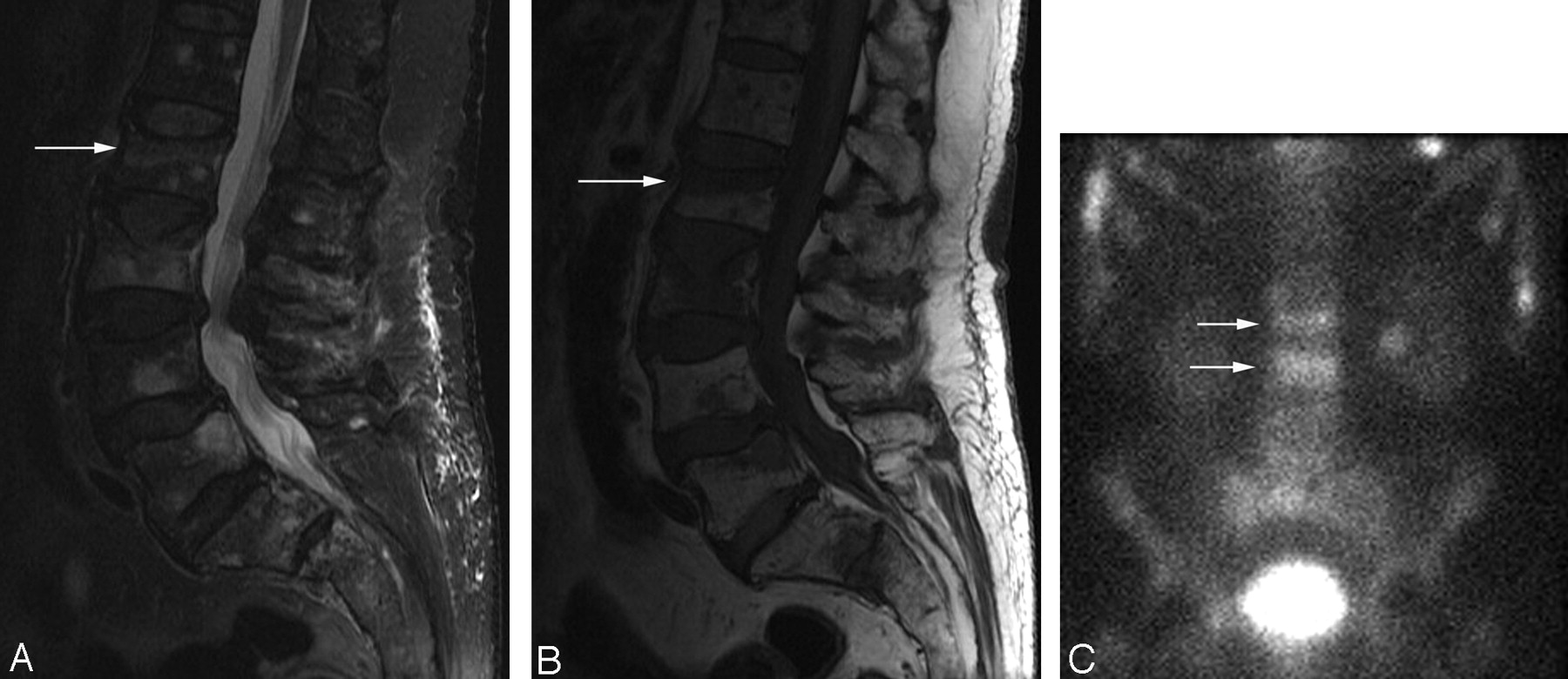
A, A sagittal fat-saturated T2-weighted image demonstrates increased signal intensity (arrow) in the superior endplate from an acute compression fracture in a different patient.
B, A sagittal spin-echo T1-weighted image in this patient nicely demonstrates the decreased signal intensity in the same superior endplate (arrow).
C, An image from a whole-body bone scintigraphy study demonstrates the classic horizontal radiotracer uptake (arrows) in acute vertebral body compression fractures.
The edema in a fractured vertebra is likely related to an inflammatory healing response with increased fluid in the bone. As a compression fracture heals, the edema typically resolves along with the patient’s pain. Previous reports have documented the utility of percutaneous vertebroplasty in chronic compression fractures without associated edema on MR imaging.6, 7 It is important to accurately identify compression fractures that might be amenable to vertebroplasty treatment, given the efficacy of this procedure. Acute or subacute compression fractures in osteoporosis and multiple myeloma generally mount a reparative response with associated edema; however, the edema present in patients with multiple myeloma may be decreased or even absent as demonstrated in the previously mentioned patients (Figs 1B and 2A, -B). Therefore, practitioners who perform percutaneous vertebroplasty procedures should consider this fact when evaluating patients with multiple myeloma and deciding whether to treat a fractured level. In our vertebroplasty experience, in treating patients with multiple myeloma, the edema present in acute or subacute compression fractures is subjectively decreased or absent compared with benign osteoporotic fractures. Because this article includes only 2 patients, further evaluation with a larger series is required to fully define the differences in edema response encountered in patients with multiple myeloma. In these patients with severe back pain, clinical findings along with manual palpation may be more important than edema on MR imaging in determining the potential response to vertebroplasty.
- Received September 21, 2005.
- Accepted after revision September 28, 2005.
- Copyright © American Society of Neuroradiology


