Advertisement

Main menu

  • Home
  • Content
    • Current Issue
    • Publication Preview--Ahead of Print
    • Past Issue Archive
    • Case of the Week Archive
    • Classic Case Archive
    • Case of the Month Archive
    • COVID-19 Content and Resources
  • For Authors
  • About Us
    • About AJNR
    • Editors
    • American Society of Neuroradiology
  • Submit a Manuscript
  • Podcasts
    • Subscribe on iTunes
    • Subscribe on Stitcher
  • More
    • Subscribers
    • Permissions
    • Advertisers
    • Alerts
    • Feedback
  • Other Publications
    • ajnr

User menu

  • Subscribe
  • Alerts
  • Log in

Search

  • Advanced search
American Journal of Neuroradiology

American Journal of Neuroradiology

  • Subscribe
  • Alerts
  • Log in

Advanced Search

  • Home
  • Content
    • Current Issue
    • Publication Preview--Ahead of Print
    • Past Issue Archive
    • Case of the Week Archive
    • Classic Case Archive
    • Case of the Month Archive
    • COVID-19 Content and Resources
  • For Authors
  • About Us
    • About AJNR
    • Editors
    • American Society of Neuroradiology
  • Submit a Manuscript
  • Podcasts
    • Subscribe on iTunes
    • Subscribe on Stitcher
  • More
    • Subscribers
    • Permissions
    • Advertisers
    • Alerts
    • Feedback
  • Follow AJNR on Twitter
  • Visit AJNR on Facebook
  • Follow AJNR on Instagram
  • Join AJNR on LinkedIn
  • RSS Feeds
OtherHEAD & NECK

Optic Tract Hemorrhage after Pituitary Apoplexy

H.J. Kim and W.H. Cho
American Journal of Neuroradiology January 2007, 28 (1) 141-142;
H.J. Kim
  • Find this author on Google Scholar
  • Find this author on PubMed
  • Search for this author on this site
W.H. Cho
  • Find this author on Google Scholar
  • Find this author on PubMed
  • Search for this author on this site
  • Article
  • Figures & Data
  • Info & Metrics
  • References
  • PDF
Loading

Abstract

SUMMARY: Subarachnoid hemorrhage following pituitary apoplexy is rare, and optic tract hemorrhage after the apoplexy is extremely rare. We report a case of optic tract hemorrhage after apoplexy that is not associated with hematologic disorders.

Hemorrhage within pituitary adenomas is relatively common.1 Rarely, hemorrhage following pituitary apoplexy expels into the subarachnoid space.2,3 Optic tract hemorrhage in pituitary apoplexy is extremely rare and has been reported only in a patient with idiopathic thrombocytopenic purpura.4 We report a case of optic pathway hemorrhage after pituitary apoplexy not associated with hematologic disorders.

Case Report

A 50-year-old man was admitted due to acute onset of blindness in the right eye. He had visual disturbance for 2 months and severe headache for 4 days before admission.

A pituitary macroadenoma was incidentally noticed on MR images 6 years before admission (not shown here). He had hypertension and was on medication for this for 7 years. He had no history of hematologic disorders.

On physical examination, the pupil was isocoric and light reflex was fixed in the right eye. The extraocular movement showed a full range. Sensory and motor function was intact. The patient’s right eye revealed no light perception and his left eye demonstrated decreased visual acuity with temporal hemianopia. Pituitary hormone evaluation revealed normal ranges. Platelet count, prothrombin time, and activated partial prothrombin time were within normal limits. The patient had no history of hematologic disorders.

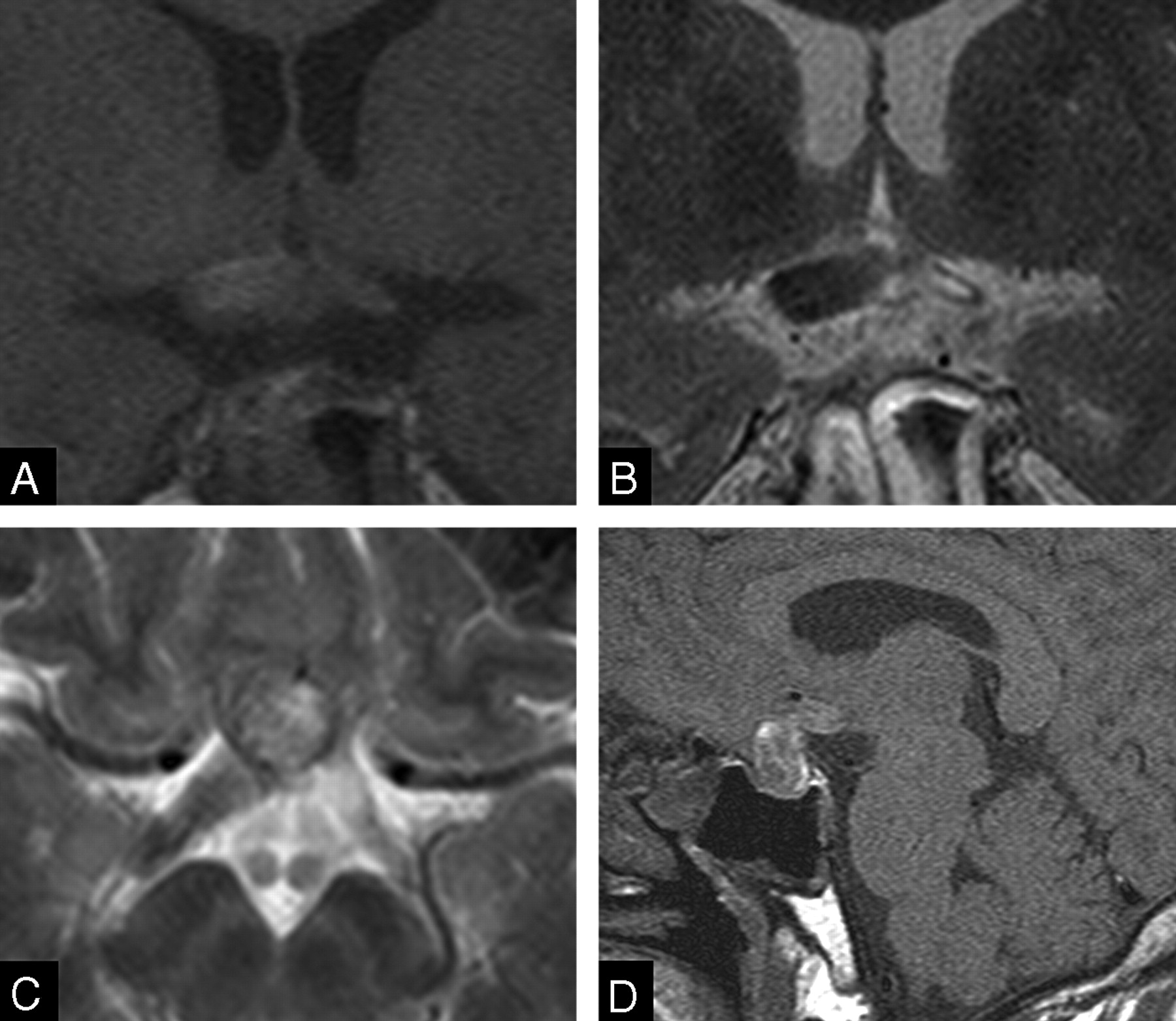
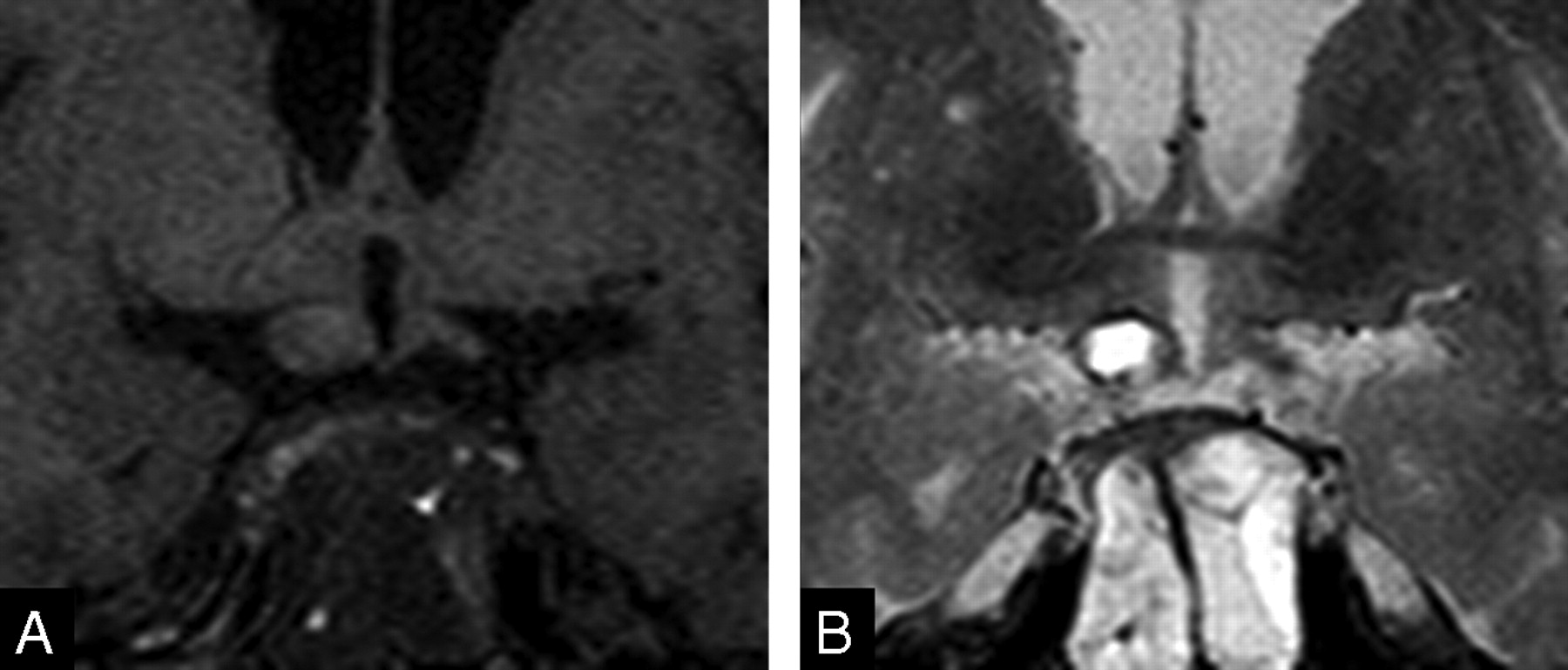
Orbit MR images on admission day 1 demonstrated mixed signal intensity prominence of the pituitary gland on T1- (Fig 1A) and T2-weighted images and mild contrast enhancement. The optic chiasm and optic tract showed no specific change of signal intensity or of size on T1- and T2-weighted images (Fig 1A–C). On admission day 2, T1-weighted coronal images revealed an edematous optic chiasm and right optic tract with hyperintensity on T1-weighted images (Fig 2A, -D). T2-weighted images showed hypointensity of the optic chiasm and right optic tract (Fig 2B, -C). The right optic tract increased in size within 1 day (Figs 1C, 2C). A pituitary mass showed mixed signal intensities on T1- and T2-weighted images. However, pituitary CT scans showed no evidence of subarachnoid hemorrhage.

Fig 1.
  • Download figure
  • Open in new tab
  • Download powerpoint
Fig 1.

MR images of patient on admission. T1-weighted coronal image (A) shows a hyperintense mass in the sellar and the suprasellar regions, suggestive of a subacute bleed. Note that neither the optic chiasm nor the optic tracts reveal evidence of hemorrhage on T1-weighted (A, B) or T2-weighted images (C).

Fig 2.
  • Download figure
  • Open in new tab
  • Download powerpoint
Fig 2.

MR images of patient on admission day 2. Enlargement of the right optic tract with hyperintensity on T1-weighted (A) and hypointensity on T2-weighted images (B, C) is seen, suggestive of acute bleed. Note mild edema in the left optic tract. A sagittal T1-weighted image (D) reveals hyperintensity of the pituitary adenoma and right optic chiasm.

Transsphenoidal aspiration of the mass was performed on admission day 3. Necrotic and hemorrhagic adenoma was confirmed pathologically. On admission day 30, pituitary MR images demonstrated an optic chiasm, and the right optic tract showed isointensity on T1- and T2-weighted images (Fig 3 A, -B). Swelling of both optic tracts was decreased compared with MR images of admission days 1 or 2. Physical examination revealed faint light perception in the patient’s right eye. His left eye revealed improved visual acuity; however, temporal hemianopia was still evident.

Fig 3.
  • Download figure
  • Open in new tab
  • Download powerpoint
Fig 3.

T1-weighted (A) and T2-weighted (B) MR images on admission day 30. Optic tract swelling in the patient has decreased. Both optic tracts are revealed as isointensity on both images.

Discussion

In pituitary apoplexy, blood and necrotic tumor tissue are enclosed and compressed within the confined space of the sella. When the pressure gradient within the sella exceeds the resistance of the surrounding structures, blood is expelled into the subarachnoid space, producing a clinical picture that may be indistinguishable from aneurysmal subarachnoid hemorrhage.3

MR images in the present case showed that hemorrhage occurred acutely not only in the existing pituitary adenoma but also in the optic tract. Severe headache and acute visual loss supported MR imaging findings. Hemorrhage and edema increased on admission day 2. The precise mechanism of hemorrhage extending to the optic tract is unknown. The optic tract hemorrhage may have extended directly from the hemorrhagic adenoma or occurred primarily within an ischemic optic tract.4 In the present case, T2-weighted MR images on admission day 2 demonstrated different signal intensities of the optic tract from those of the hemorrhage of pituitary adenoma, suggesting that the optic tract hematoma occurred more recently than the pituitary hemorrhage. Recent reports have shown connections of Virchow spaces into the optic tracts that become edematous in patients with suprasellar masses.5 The connection of these subarachnoid spaces may be the route of spreading hemorrhage into the tract; alternatively, venous pressure increases may also be responsible for the spread of hemorrhage.

Optic tract thickening is commonly secondary to optic tract tumors such as gliomas, ependymoma, or primary CNS lymphoma,6 and it is also associated with sellar and suprasellar tumors including craniopharyngioma.7,8 Hemorrhage into the optic tract is extremely rare and has been reported only with pituitary apoplexy in the setting of idiopathic thrombocytopenic purpura4 and with a cavernous malformation within the optic tract.6

Transsphenoidal surgery for pituitary macroadenoma results in a progressive recovery of the visual field in 95% of patients.9 Even in apoplectic patients, early decompression of the pituitary mass leads to improvement in the visual deficit.10 However, in the present case, transsphenoidal aspiration was performed because the patient had experienced visual symptoms for 2 months. Delayed decompression may be related to the poor outcome of the vision in the patient.

Abrupt cranial neuropathy is also associated with pituitary apoplexy. Vision is most commonly disturbed; rarely third, fourth, or sixth cranial nerve palsies develop following pituitary apoplexy.11–13 Our patient showed visual disturbance for 2 months before admission and an abrupt decrease of the vision associated with acute hemorrhage of the optic tract.

In conclusion, we report a patient with acute hematoma of the optic tracts associated with pituitary apoplexy from a hemorrhagic pituitary adenoma. While rare, this finding is of interest in that ischemic or hemorrhagic complications of the optic tract with acute expansion of a pituitary adenoma may lead to visual loss that is not soley related to external compression of the optic pathways.

Early decompression of the pituitary adenomas is recommended for preservation or improvement of the vision.

Acknowledgments

The authors are grateful to Bonnie Hami, Department of Radiology, University Hospitals of Cleveland and Haaga Radiology Research, for her assistance in the preparation of this article.

References

  1. ↵
    Mohanty S, Tandon PN, Banerji AK, et al. Haemorrhage into pituitary adenomas. J Neurol Neurosurg Psychiatry 1977;40:987–91
    Abstract/FREE Full Text
  2. ↵
    Wohaibi MA, Russell NA, Ferayan AA. Pituitary apoplexy presenting as massive subarachnoid haemorrhage. J Neurol Neurosurg Psychiatry 2000;69:700–01
    FREE Full Text
  3. ↵
    Chen ST, Chen SD, Ryu SJ, et al. Pituitary apoplexy with intracerebral hemorrhage simulating rupture of an anterior cerebral artery aneurysm. Surg Neurol 1988;29:322–25
    CrossRefPubMed
  4. ↵
    Lenthall R, Gonugunta V, Jaspan T. Pituitary apoplexy with optic tract oedema and haemorrhage in a patient with idiopathic thrombocytopenic purpura. Neuroradiology 2001;43:156–58
    PubMed
  5. ↵
    Saeki N, Nagai Y, Matsuura I, et al. Histologic characteristics of normal perivascular spaces along the optic tract: new pathogenetic mechanism for edema in tumors in the pituitary region. AJNR Am J Neuroradiol 2004;25:1218–22
    Abstract/FREE Full Text
  6. ↵
    Muller-Forell W. Intracranial pathology of the visual pathway. Eur J Radiol 2004;49:143–78
    PubMed
  7. ↵
    Saeki N, Uchino Y, Murai H, et al. MR imaging study of edema-like change along the optic tract in patients with pituitary region tumors. AJNR Am J Neuroradiol 2003;24:336–42
    Abstract/FREE Full Text
  8. ↵
    Sklar EM, Schatz NJ, Glaser JS, et al. Optic tract edema in a meningioma of the tuberculum sellae. AJNR Am J Neuroradiol 2000;21:1661–63
    Abstract/FREE Full Text
  9. ↵
    Gnanalingham KK, Bhattacharjee S, Pennington R, et al. The time course of visual field recovery following transphenoidal surgery for pituitary adenomas: predictive factors for a good outcome. J Neurol Neurosurg Psychiatry 2005;76:415–19
    Abstract/FREE Full Text
  10. ↵
    Bills DC, Meyer FB, Laws ER Jr, et al. A retrospective analysis of pituitary apoplexy. Neurosurgery 1993;33:602–28
    PubMed
  11. ↵
    Rossitch E Jr, Carrazana EJ, Black PM. Isolated oculomotor nerve palsy following apoplexy of a pituitary adenoma. J Neurosurg Sci 1992;36:103–05
    PubMed
  12. Tanioka D, Abe T, Kunii N, et al. A case of a hemorrhagic non-functioning pituitary adenoma presenting with abducens nerve palsy. No Shinkei Geka 2005;33:473–79
    PubMed
  13. ↵
    Riedl M, Clodi M, Kotzmann H, et al. Apoplexy of a pituitary macroadenoma with reversible third, fourth and sixth cranial nerve palsies following administration of hypothalamic releasing hormones: MR features. Eur J Radiol 2000;36:1–4
    PubMed
  • Received October 23, 2005.
  • Accepted after revision December 2, 2005.
  • Copyright © American Society of Neuroradiology
View Abstract
PreviousNext
Back to top

In this issue

American Journal of Neuroradiology: 28 (1)
American Journal of Neuroradiology
Vol. 28, Issue 1
January 2007
  • Table of Contents
  • Index by author
Advertisement
Print
Download PDF
Email Article

Thank you for your interest in spreading the word on American Journal of Neuroradiology.

NOTE: We only request your email address so that the person you are recommending the page to knows that you wanted them to see it, and that it is not junk mail. We do not capture any email address.

Enter multiple addresses on separate lines or separate them with commas.
Optic Tract Hemorrhage after Pituitary Apoplexy
(Your Name) has sent you a message from American Journal of Neuroradiology
(Your Name) thought you would like to see the American Journal of Neuroradiology web site.
CAPTCHA
This question is for testing whether or not you are a human visitor and to prevent automated spam submissions.
Citation Tools
Optic Tract Hemorrhage after Pituitary Apoplexy
H.J. Kim, W.H. Cho
American Journal of Neuroradiology Jan 2007, 28 (1) 141-142;

Citation Manager Formats

  • BibTeX
  • Bookends
  • EasyBib
  • EndNote (tagged)
  • EndNote 8 (xml)
  • Medlars
  • Mendeley
  • Papers
  • RefWorks Tagged
  • Ref Manager
  • RIS
  • Zotero
Share
Optic Tract Hemorrhage after Pituitary Apoplexy
H.J. Kim, W.H. Cho
American Journal of Neuroradiology Jan 2007, 28 (1) 141-142;
Reddit logo Twitter logo Facebook logo Mendeley logo
  • Tweet Widget
  • Facebook Like
  • Google Plus One

Jump to section

  • Article
    • Abstract
    • Case Report
    • Discussion
    • Acknowledgments
    • References
  • Figures & Data
  • Info & Metrics
  • References
  • PDF

Related Articles

  • No related articles found.
  • PubMed
  • Google Scholar

Cited By...

  • No citing articles found.
  • Crossref
  • Google Scholar

This article has not yet been cited by articles in journals that are participating in Crossref Cited-by Linking.

More in this TOC Section

  • Imaging the Tight Orbit: Radiologic Manifestations of Orbital Compartment Syndrome
  • CT and MR Imaging Appearance of the Pedicled Submandibular Gland Flap: A Potential Imaging Pitfall in the Posttreatment Head and Neck
  • Increased Labyrinthine T1 Postgadolinium Signal Intensity Is Associated with the Degree of Ipsilateral Sensorineural Hearing Loss in Patients with Sporadic Vestibular Schwannoma
Show more Head & Neck

Similar Articles

Advertisement

News and Updates

  • Lucien Levy Best Research Article Award
  • Thanks to our 2022 Distinguished Reviewers
  • Press Releases

Resources

  • Evidence-Based Medicine Level Guide
  • How to Participate in a Tweet Chat
  • AJNR Podcast Archive
  • Ideas for Publicizing Your Research
  • Librarian Resources
  • Terms and Conditions

Opportunities

  • Share Your Art in Perspectives
  • Get Peer Review Credit from Publons
  • Moderate a Tweet Chat

American Society of Neuroradiology

  • Neurographics
  • ASNR Annual Meeting
  • Fellowship Portal
  • Position Statements

© 2023 by the American Society of Neuroradiology | Print ISSN: 0195-6108 Online ISSN: 1936-959X

Powered by HighWire