Advertisement

Main menu

  • Home
  • Content
    • Current Issue
    • Publication Preview--Ahead of Print
    • Past Issue Archive
    • Case of the Week Archive
    • Classic Case Archive
    • Case of the Month Archive
    • COVID-19 Content and Resources
  • For Authors
  • About Us
    • About AJNR
    • Editors
    • American Society of Neuroradiology
  • Submit a Manuscript
  • Podcasts
    • Subscribe on iTunes
    • Subscribe on Stitcher
  • More
    • Subscribers
    • Permissions
    • Advertisers
    • Alerts
    • Feedback
  • Other Publications
    • ajnr

User menu

  • Subscribe
  • Alerts
  • Log in

Search

  • Advanced search
American Journal of Neuroradiology

American Journal of Neuroradiology

  • Subscribe
  • Alerts
  • Log in

Advanced Search

  • Home
  • Content
    • Current Issue
    • Publication Preview--Ahead of Print
    • Past Issue Archive
    • Case of the Week Archive
    • Classic Case Archive
    • Case of the Month Archive
    • COVID-19 Content and Resources
  • For Authors
  • About Us
    • About AJNR
    • Editors
    • American Society of Neuroradiology
  • Submit a Manuscript
  • Podcasts
    • Subscribe on iTunes
    • Subscribe on Stitcher
  • More
    • Subscribers
    • Permissions
    • Advertisers
    • Alerts
    • Feedback
  • Follow AJNR on Twitter
  • Visit AJNR on Facebook
  • Follow AJNR on Instagram
  • Join AJNR on LinkedIn
  • RSS Feeds
OtherPEDIATRICS

Lingual Hamartoma in an Infant: CT and MR Imaging

A.L. Goold, B.L. Koch and J.P. Willging
American Journal of Neuroradiology January 2007, 28 (1) 30-31;
A.L. Goold
  • Find this author on Google Scholar
  • Find this author on PubMed
  • Search for this author on this site
B.L. Koch
  • Find this author on Google Scholar
  • Find this author on PubMed
  • Search for this author on this site
J.P. Willging
  • Find this author on Google Scholar
  • Find this author on PubMed
  • Search for this author on this site
  • Article
  • Figures & Data
  • Info & Metrics
  • References
  • PDF
Loading

Abstract

SUMMARY: Lingual hamartoma is a rare tongue mass, primarily diagnosed in childhood. In most cases in the literature, the masses were surgically removed without preoperative imaging. There are only 3 cases reported in the clinical literature that describe preoperative imaging findings. We report the clinical and imaging findings in an infant with lingual hamartoma and review the literature.

Lingual hamartoma is a rare benign tumorlike malformation of the tongue composed of tissue indigenous to the area in which it is found, but abnormally arranged. These malformations may occur as isolated lesions or in association with syndromes such as oral-facial-digital syndrome. Although one might expect that they would occur in Cowden syndrome (multiple hamartoma syndrome), the classic lesion of the tongue in these patients is a scrotal tongue rather than a focal lingual hamartoma.1 In the clinical literature, the imaging characteristics (in 2 children and 1 adult) have been reported as hypointense on T1-weighted images, hyperintense on T2-weighted images, and intermediate attenuation on CT. We report the clinical, CT, and MR imaging findings in an infant with an asymptomatic lingual hamartoma.

Case Report

A 5-month-old male infant presented with a posterior tongue mass visible initially by his mother, only when he laughed with his mouth wide open (Fig 1). Subsequent evaluation by the otolaryngologist demonstrated a smooth yellowish mass arising near the foramen cecum, thought to be potentially a lingual thyroid. The mass did not interfere with deglutition or respiration. The family history was unremarkable and the child was otherwise healthy.

Fig 1.
  • Download figure
  • Open in new tab
  • Download powerpoint
Fig 1.

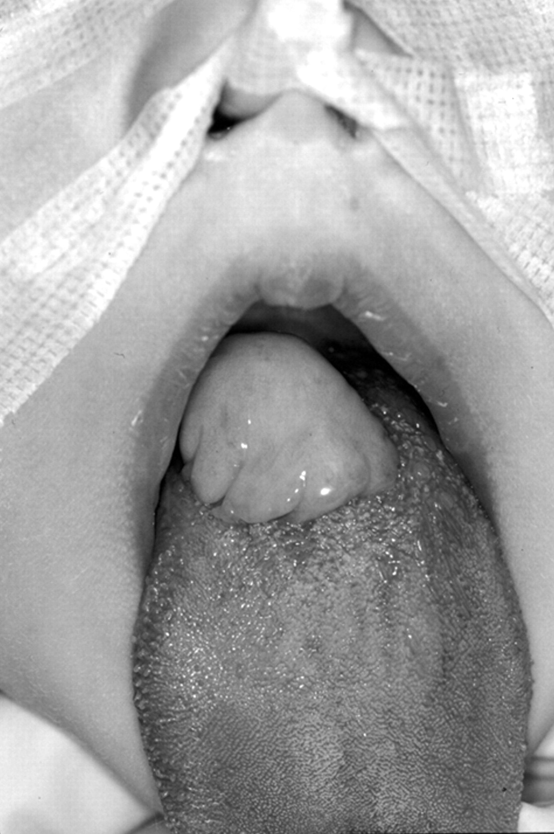
Preoperative photograph of the infant in the operating room after anesthesia induction and nasotracheal intubation, with manual traction applied to the patient’s tongue, shows a lobulated midline tongue base mass.

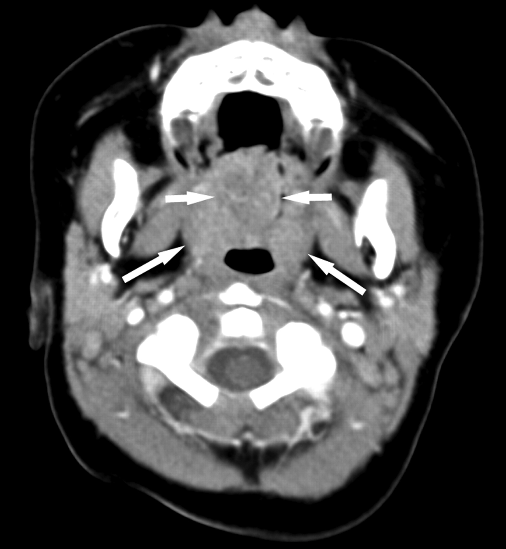
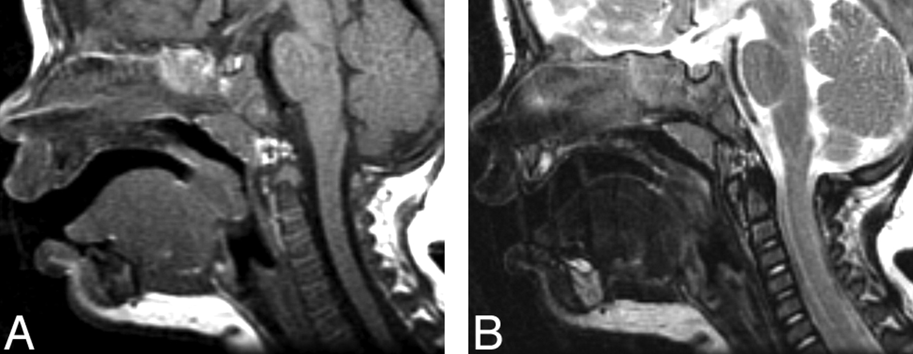
Imaging was requested to characterize the lesion better, evaluate the extent of deep tongue involvement, and aid in preoperative planning to determine whether surgery would be via an intraoral approach or an open procedure through the neck. CT scans revealed a round intermediate-attenuation soft-tissue mass arising from the midline posterior tongue. There was only minimal heterogeneous contrast enhancement (Fig 2). A homogeneously enhancing bilobed normal-appearing thyroid gland was present in the lower neck (image not shown). MR imaging showed a well-defined mass at the surface of the posterior tongue, mildly hypointense on T1-weighted images and having mixed signal intensity on T2-weighted images, with a hyperintense irregular center and a hypointense periphery (Fig 3).

Fig 2.
  • Download figure
  • Open in new tab
  • Download powerpoint
Fig 2.

Axial postcontrast CT scan shows a lobulated soft-tissue midline posterior tongue mass with minimal heterogeneous enhancement (short arrows). On this axial image, the mass is at the same level as the normal-appearing palatine tonsils (long arrows).

Fig 3.
  • Download figure
  • Open in new tab
  • Download powerpoint
Fig 3.

A, Sagittal T1-weighted image demonstrates a mildly hypointense well-defined mass arising from the posterior aspect of the tongue abutting the inferior surface of the uvula. B, T2-weighted image demonstrates a central area of hyperintensity with irregular margins and hypointense periphery.

At surgery, the smooth and slightly lobulated mass measured 2.3 × 2 × 1.2 cm and arose near the foramen cecum. It was noted to have a short 5-mm-thick stalk, which was amputated by using electrocautery. Surrounding mucosa and a thin layer of tongue musculature were removed without complication. Histology confirmed a hamartoma composed mainly of smooth muscle with components of squamous epithelium and mucous glands.

Discussion

Hamartoma is a benign tumorlike overgrowth in which normal tissue components of the organ of origin are abnormally arranged, most commonly located in the liver, spleen, kidney, lung, skin, and pancreas. Lingual hamartoma was first described in 1945 by Stamm and Tauber2 and may occur in isolation or in association with syndromes such as oral-facial-digital syndrome (originally described by Gorlin and Psaume as orodigitofacial dysostosis).3 Lingual hamartomas presenting as an isolated anomaly are extremely rare, with only 15 previously reported patients, 13 of whom were children.2,4-16 Isolated lingual hamartomas occur as solitary lesions on the dorsum of the tongue, usually posterior and midline in the area of the foramen cecum. Most lingual hamartomas are polypoid and more common in females, with a female-to-male ratio of almost 2:1. Multiple hamartomas in the tongue have been reported, but such occurrence has been limited to an association with other syndromes, such as oral-facial-digital syndrome,17 incomplete cleft palate,18 and tuberous sclerosis.15 Cowden syndrome (multiple hamartoma syndrome) is known to feature multiple hamartomas throughout the body, including the mucous membrane of the oral cavity. However, the classic lesion of the tongue in Cowden syndrome is a scrotal tongue, not a lingual hamartoma.1

Differential diagnosis of congenital tongue lesions in children includes lingual thyroid, thyroglossal duct cyst, vascular malformation, and duplication cyst. Pediatric tongue neoplasms include teratoma and sarcoma.19 Of these lesions, lingual thyroid and thyroglossal duct cyst (TGDC) occur in the region of the foramen cecum, as do lingual hamartomas. The lesion in this report was not cystic on imaging or clinical appearance; therefore, TGDC and duplication cyst were unlikely possibilities. The presence of a normal-appearing thyroid in the lower neck did not exclude lingual thyroid. However, the lesion in this report was not as hyperattenuated on CT as lingual thyroid usually is. The lesion did not contain fat or calcium, which would have increased the likelihood of a diagnosis of teratoma.

To our knowledge, imaging findings are discussed in only 3 clinical reports in the English literature.5,6,15 Halfpenny et al5 reported a 4-year-old girl with a left posterior tongue mass, with MR imaging showing a predominately cystic nonenhancing lesion, hypointense on T1-weighted images and hyperintense T2-weighted images. Takimoto et al6 reported a 6-year-old girl with a left-of-midline posterior tongue mass, hyperintense on T1-weighted images and moderately hyperintense on T2-weighted images. Wallace et al15 reported a 41-year-old woman with tuberous sclerosis and a large base-of-tongue mass, intermediate in attenuation on postcontrast CT. In our patient, the lesion was mildly hypointense on T1-weighted images and heterogeneous on T2-weighted images, with only minimal postcontrast enhancement on CT.

Conclusion

Lingual hamartoma is a rare cause of tongue base mass, with only 15 previously reported cases occurring without associated syndromes. To our knowledge, there are only 3 reports in the clinical literature that describe the imaging findings of lingual hamartomas. Imaging features are nonspecific. However, the differential diagnosis of a posterior midline tongue mass, in the region of the foramen cecum in a child, should include lingual hamartoma.

Footnotes

  • Paper previously presented at: Annual Meeting of the American Society of Head and Neck Radiology, October 1–4, 2003; Rancho Mirage, Calif.

References

  1. ↵
    Lloyd KM 2nd, Dennis M. Cowden’s disease: a possible new symptom complex with multiple system involvement. Ann Intern Med 1963;58:136–42
    CrossRefPubMed
  2. ↵
    Stamm C, Tauber R. Hamartoma of tongue. Laryngoscope 1945;140–46
  3. ↵
    Gorlin RJ, Psaume J. Orodigitofacial dysostosis: a new syndrome—a study of 22 cases. J Pediatr 1962;61:520–30
    CrossRefPubMed
  4. ↵
    Owen G, Berry J, Bicknell P. Hamartoma of the tongue. J Laryngol Otol 1993;107:363–67
    PubMed
  5. ↵
    Halfpenny W, Odell EW, Robinson PD. Cystic and glial mixed hamartoma of the tongue. J Oral Pathol Med 2001;30:368–71
    PubMed
  6. ↵
    Takimoto T, Yoshizaki T, Umeda R. Hamartoma of the tongue. Int J Pediatr Otorhinolaryngol 1989;18:157–61
    PubMed
  7. Herzog S, Bressman J, Giglio JA. Case 61: tongue mass in an infant. J Oral Maxillofac Surg 1986;44:463–66
    PubMed
  8. Takato T, Fukuda O, Ohhara Y, et al. Hamartoma of the tongue: two case reports. Plast Reconstr Surg 1985;75:258–62
    PubMed
  9. Cutchavaree A, Rungruxsirivorn S, Suphanakorn S. Hamartoma of the tongue. J Med Assoc Thai 1985;68:216–19
    PubMed
  10. Becker GD, Ridolfi R, Ingber C. Lingual hamartoma in a newborn. Otolaryngol Head Neck Surg 1984;92:357–59
  11. Kobayashi A, Amagasa T, Okada N. Leiomyomatous hamartoma of the tongue: case report. J Oral Maxillofac Surg 2001;59:337–40
    PubMed
  12. Horn C, Thaker HM, Tampakopoulou DA, et al. Tongue lesions in the pediatric population. Otolaryngol Head Neck Surg 2001;124:164–69
    PubMed
  13. Goldsmith P, Soames JV, Meikle D. Leiomyomatous hamartoma of the posterior tongue: a case report. J Laryngol Otol 1995;109:1190–91
    PubMed
  14. Hinshaw CT Jr. Unusual lesions of the tongue: hamartoma. J Kans Med Soc 1963;64:154–57
    PubMed
  15. ↵
    Wallace H, Davis A, Spedding A. Tongue-base hamartoma in tuberous sclerosis. J Laryngol Otol 2001;115:149–50
    PubMed
  16. ↵
    Perri FA. Myoepithelial hamartoma of tongue. AMA Arch Otolaryngol 1956;64:289–90
    CrossRefPubMed
  17. ↵
    Doege TC, Thuline HC, Priest JH, et al. Studies of a family with the oral-facial-digital syndrome. N Engl J Med 1964;271:1073–78
    PubMed
  18. ↵
    Ishii T, Takemori S, Suzuki JI. Hamartoma of the tongue. Arch Otolaryngol 1968;88:79–81
  19. ↵
    Donnelly LF, Jones BV, Strife JL. Imaging of pediatric tongue abnormalities. AJR Am J Roentgenol 2000;175:489–93
    PubMed
  • Received December 7, 2005.
  • Accepted after revision December 22, 2005.
  • Copyright © American Society of Neuroradiology
View Abstract
PreviousNext
Back to top

In this issue

American Journal of Neuroradiology: 28 (1)
American Journal of Neuroradiology
Vol. 28, Issue 1
January 2007
  • Table of Contents
  • Index by author
Advertisement
Print
Download PDF
Email Article

Thank you for your interest in spreading the word on American Journal of Neuroradiology.

NOTE: We only request your email address so that the person you are recommending the page to knows that you wanted them to see it, and that it is not junk mail. We do not capture any email address.

Enter multiple addresses on separate lines or separate them with commas.
Lingual Hamartoma in an Infant: CT and MR Imaging
(Your Name) has sent you a message from American Journal of Neuroradiology
(Your Name) thought you would like to see the American Journal of Neuroradiology web site.
CAPTCHA
This question is for testing whether or not you are a human visitor and to prevent automated spam submissions.
Citation Tools
Lingual Hamartoma in an Infant: CT and MR Imaging
A.L. Goold, B.L. Koch, J.P. Willging
American Journal of Neuroradiology Jan 2007, 28 (1) 30-31;

Citation Manager Formats

  • BibTeX
  • Bookends
  • EasyBib
  • EndNote (tagged)
  • EndNote 8 (xml)
  • Medlars
  • Mendeley
  • Papers
  • RefWorks Tagged
  • Ref Manager
  • RIS
  • Zotero
Share
Lingual Hamartoma in an Infant: CT and MR Imaging
A.L. Goold, B.L. Koch, J.P. Willging
American Journal of Neuroradiology Jan 2007, 28 (1) 30-31;
Reddit logo Twitter logo Facebook logo Mendeley logo
  • Tweet Widget
  • Facebook Like
  • Google Plus One

Jump to section

  • Article
    • Abstract
    • Case Report
    • Discussion
    • Conclusion
    • Footnotes
    • References
  • Figures & Data
  • Info & Metrics
  • References
  • PDF

Related Articles

  • No related articles found.
  • PubMed
  • Google Scholar

Cited By...

  • No citing articles found.
  • Crossref
  • Google Scholar

This article has not yet been cited by articles in journals that are participating in Crossref Cited-by Linking.

More in this TOC Section

  • Neuroimaging Features of Biotinidase Deficiency
  • Medullary Tegmental Cap Dysplasia: Fetal and Postnatal Presentations of a Unique Brainstem Malformation
  • Diagnostic Utility of 3D Gradient-Echo MR Imaging Sequences through the Filum Compared with Spin-Echo T1 in Children with Concern for Tethered Cord
Show more PEDIATRICS

Similar Articles

Advertisement

News and Updates

  • Lucien Levy Best Research Article Award
  • Thanks to our 2022 Distinguished Reviewers
  • Press Releases

Resources

  • Evidence-Based Medicine Level Guide
  • How to Participate in a Tweet Chat
  • AJNR Podcast Archive
  • Ideas for Publicizing Your Research
  • Librarian Resources
  • Terms and Conditions

Opportunities

  • Share Your Art in Perspectives
  • Get Peer Review Credit from Publons
  • Moderate a Tweet Chat

American Society of Neuroradiology

  • Neurographics
  • ASNR Annual Meeting
  • Fellowship Portal
  • Position Statements

© 2023 by the American Society of Neuroradiology | Print ISSN: 0195-6108 Online ISSN: 1936-959X

Powered by HighWire