
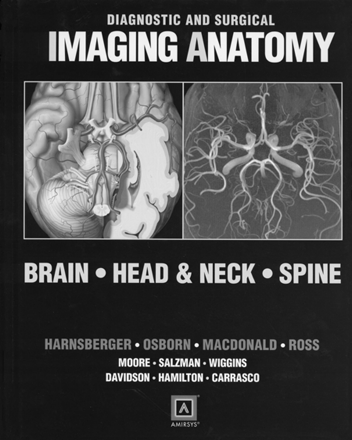
H.R. Harnsberger, A.G. Osborn, A. MacDonald, J. Ross, K.R. Moore, K.L. Salzman, R.H. Wiggins, H.C. Davidson, B.E. Hamilton, and C.R. Carrasco, eds. Salt Lake City: Amirsys; 2006. 1000 pages, 2870 illustrations, $249.
In a single, beautifully illustrated book, the neuroradiology group from the University of Utah has presented the neuroradiology and neuroscience community with detailed structural anatomy of the brain, head and neck, spine, and peripheral nerves by using anatomic drawings, CT, MR imaging, and angiography. Nine authors have contributed to this 1000-page book, which is structured as follows: The brain section is divided into a) scalp, skull, meninges, b) supratentorial brain, c) infratentorial brain, d) CSF spaces, e) cranial nerves, f) extracranial arteries, g) intracranial arteries, and h) veins and dural sinuses. The head and neck section is divided into a) temporal bone and skull base, b) orbit, nose, sinuses, c) supra and infrahyoid neck, and d) oral cavity. The spine section is divided into a) vertebral column/disks/paraspinal muscles, b) spinal cord/meninges/spinal spaces, c) spinal vasculature, d) plexuses, and e) peripheral nerves. Many of the illustrations are drawn from the authors’ previous 3-volume set of books on diagnostic imaging.
The labeling of structures is precise, and the anatomy is not obscured by overly abundant labels. There are so many positive aspects of this large and information-packed book that they all cannot be mentioned in this review; however, like the authors’ bulletlike presentation, just a few highlights (in this reviewer's opinion) will be similarly described.
There are outstanding color-plate drawings (called “graphics”) of key anatomic features, which drive home those imaging correlations often needed when reviewing a topic or reviewing high-resolution images. Outstanding examples are (just to name a few): the limbic system; internal structure of the brain stem (medulla/pons/mesencephalon); detailed courses, origins, interconnections, and terminations of the cranial nerves (each dealt with individually); the temporal bone with exquisite details of the inner ear including the cochlea, membranous labyrinth, and facial nerve; the spaces of the neck (if only the clinical MR and CT images came out colorized to show the fascial layers and the various compartments!) laryngeal/hypolaryngeal area; lymph node anatomy; oral cavity/sublingual space; the brachial, lumbar, and sacral plexuses; and peripheral nerves.
High-quality MR images and CT scans demonstrate the key findings in all areas. This is particularly useful in the skull base and head/neck areas.
Descriptive material for every major area includes the following subheadings–“Terminology,” “Imaging Anatomy,” “Embryology” (where appropriate), “Anatomy-Based Imaging Issues,” and “Clinical Implications.” The latter 2 categories (subheadings) are particularly well done throughout the book. The amount of anatomy-based imaging issues varies in length from section to section and includes key concepts, imaging recommendations, the approach to imaging, and pitfalls. Under the subheading of clinical implications, clinical importance and functional anatomy are described. Take the pterygopalatine fossa (6 pages) as just 1 small isolated example. This area, composed of different named spaces containing a complex mixture of vascular structures and nerves and irregularly shaped bony borders, is illustrated and described so that these anatomic points stick. One can easily flip back and forth from the drawings to the CT images to gain an appreciation of the exact relationship between the vidian nerve, the pterygopalatine (sphenopalatine) ganglia, and the foramen rotundum and how (where) the nerves interconnect. The critical role this fossa plays in the interconnection of numerous spaces and the subsequent spread of disease is emphasized. Although it may be added work for the authors, this reviewer suggests that a future edition contain 2 images at each level: 1 as currently shown and the 2nd directly adjacent to it showing in color the critical structures running through that space. This could be done in many other sections of the book also. The information under “Function-Dysfunction” briefly describes the innervation of structures in this area. This excellent section is detailed in this review to give the reader a flavor of what type of information to expect in every area of the book.
Some pathology (limited) is included to emphasize the anatomy under review, such as a submucosal/mucosal mass in the parapharyngeal space or an abscess in the prevertebral/retropharyngeal space.
Both the text material (written in phrases, not sentences) and the figure legends (written in sentences, not phrases) contain important information. Take the 8-page description and corresponding illustration of the limbic system—one quickly gains an appreciation of the components of the limbic system and limbic lobe, the “arches” of which the limbic system is composed; its MR imaging details; and the details of the inner zones of the hippocampus. The T2- and T1-weighted MR images of this area are excellent and allow an appreciation of the key imaging features of the limbic system. This segment exemplifies again the persistent high quality throughout the book.
It is easy to forget much of the detailed anatomy buried in images, which a neuroradiologist or a neurologist/neurosurgeon views every day. There is no better way or a more effective publication to accomplish a review of the anatomy than to use this book with its drawings and images side by side, a value amplified by the accompanying figure legends, which, incidentally, are amply robust. Few will read this book cover to cover, but the vexing questions that often arise in reviewing films or in consulting with clinicians are painlessly answered. While it is true that the drawings show far more details than any of our images are currently capable of depicting, lesion localization/significance is more greatly appreciated with the details afforded by this text. The illustrative material (drawings in particular) would have made a nice CD add-on in this book; perhaps the authors would consider marketing that, along with pathology drawings from other texts in their series, in a single CD. These would rapidly take their place alongside the legendary illustrations by Frank Netter.
This book should find its way onto the shelves of every department and neuroradiology section library, and for those who treasure the fine details of neuroanatomy, this book is a highly recommended purchase. Standing alongside the other neuroradiology volumes in this series (see review AJNR 2005;26:1876–78), this book would provide one with as complete an illustrative and descriptive neuroimaging collection as is possible.
- Copyright © American Society of Neuroradiology
In this issue


Jump to section
Related Articles
Cited By...
- No citing articles found.