Advertisement

Main menu

  • Home
  • Content
    • Current Issue
    • Publication Preview--Ahead of Print
    • Past Issue Archive
    • Case of the Week Archive
    • Classic Case Archive
    • Case of the Month Archive
    • COVID-19 Content and Resources
  • For Authors
  • About Us
    • About AJNR
    • Editors
    • American Society of Neuroradiology
  • Submit a Manuscript
  • Podcasts
    • Subscribe on iTunes
    • Subscribe on Stitcher
  • More
    • Subscribers
    • Permissions
    • Advertisers
    • Alerts
    • Feedback
  • Other Publications
    • ajnr

User menu

  • Subscribe
  • Alerts
  • Log in

Search

  • Advanced search
American Journal of Neuroradiology

American Journal of Neuroradiology

  • Subscribe
  • Alerts
  • Log in

Advanced Search

  • Home
  • Content
    • Current Issue
    • Publication Preview--Ahead of Print
    • Past Issue Archive
    • Case of the Week Archive
    • Classic Case Archive
    • Case of the Month Archive
    • COVID-19 Content and Resources
  • For Authors
  • About Us
    • About AJNR
    • Editors
    • American Society of Neuroradiology
  • Submit a Manuscript
  • Podcasts
    • Subscribe on iTunes
    • Subscribe on Stitcher
  • More
    • Subscribers
    • Permissions
    • Advertisers
    • Alerts
    • Feedback
  • Follow AJNR on Twitter
  • Visit AJNR on Facebook
  • Follow AJNR on Instagram
  • Join AJNR on LinkedIn
  • RSS Feeds
Research ArticleHEAD & NECK

Diagnostic Value of High-Resolution MR Imaging in Giant Cell Arteritis

T.A. Bley, M. Uhl, J. Carew, M. Markl, D. Schmidt, H.-H. Peter, M. Langer and O. Wieben
American Journal of Neuroradiology October 2007, 28 (9) 1722-1727; DOI: https://doi.org/10.3174/ajnr.A0638
T.A. Bley
  • Find this author on Google Scholar
  • Find this author on PubMed
  • Search for this author on this site
M. Uhl
  • Find this author on Google Scholar
  • Find this author on PubMed
  • Search for this author on this site
J. Carew
  • Find this author on Google Scholar
  • Find this author on PubMed
  • Search for this author on this site
M. Markl
  • Find this author on Google Scholar
  • Find this author on PubMed
  • Search for this author on this site
D. Schmidt
  • Find this author on Google Scholar
  • Find this author on PubMed
  • Search for this author on this site
H.-H. Peter
  • Find this author on Google Scholar
  • Find this author on PubMed
  • Search for this author on this site
M. Langer
  • Find this author on Google Scholar
  • Find this author on PubMed
  • Search for this author on this site
O. Wieben
  • Find this author on Google Scholar
  • Find this author on PubMed
  • Search for this author on this site
  • Article
  • Figures & Data
  • Info & Metrics
  • References
  • PDF
Loading

Article Figures & Data

Figures

  • Tables
  • Fig 1.
    • Download figure
    • Open in new tab
    • Download powerpoint
    Fig 1.

    Enlargements of 3T transversal postcontrast fat-suppressed T1-weighted SE image of the superficial temporal arteries of 4 different patients representing typical images of each grade of the 4-point ranking scale. Temporal artery biopsy is negative in cases A and B, and suspected diagnosis of giant cell arteritis is validated by histology in cases C and D. The concomitant veins (arrowheads in A and C) display homogeneous signal intensity increase because of low venous flow. A, Mural thickness <0.5 mm and no mural enhancement; rating “0.” Note the intraluminal signal intensity void (light arrow) because of arterial flow. B, Mural thickness <0.5 mm with only slight contrast enhancement (light arrow), probably because of enhancing vasa vasorum; rating “1.” C, Mural thickening >0.6 mm and prominent mural enhancement (arrow); rating “2.” D, Strong mural thickening >0.7 mm and strong mural enhancement (arrow); rating “3.” The arterial lumen is still patent, as signal intensity void consistent with flow can be seen.
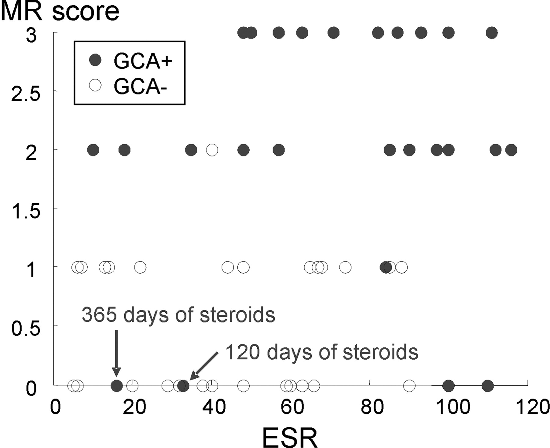
  • Fig 2.
    • Download figure
    • Open in new tab
    • Download powerpoint
    Fig 2.

    Feature plot MR score of mural inflammation versus ESR. Patients with an elevated ESR and a high MR score are all diagnosed GCA positive according to the ACR criteria. Patients with a low ESR and a low MR score are mostly diagnosed GCA negative. Please note that 2 of the false-negative MR findings with a very low MR score are imaged after long treatment with corticosteroids. Single points in the plot may represent >1 patient in case of identical values.

  • Fig 3.
    • Download figure
    • Open in new tab
    • Download powerpoint
    Fig 3.

    3T transversal contrast fat-suppressed T1-weighted SE image acquired with the large FOV that covers the entire cranial circumference. Enlargements of the temporal branch of the superficial temporal arteries (A and B) and of the superficial occipital arteries (C and D) demonstrate the cranial involvement pattern. Mural thickening and inflammatory changes are depicted in the left temporal artery (enlargement B, 0.7-mm mural thickness, rated as “3”) and occipital artery (enlargement D, 0.7-mm mural thickness, rated as “3”), whereas the right-sided arteries display no signs of mural inflammation (enlargements A and C, 0.2-mm mural thickness, both rated as “0”). Temporal artery biopsy validates GCA in this patient.

Tables

  • Figures
    • View popup
    Table 1:

    MR imaging vs. clinical critera according to the American College of Rheumatology

    ComparisonnTPTNFPFNSensSpecPPVNPV
    MR vs ACR (all patients)6425321680.697.096.284.2
    MR vs ACR (<10 days of steroids)5024211485.795.596.084.0
    MR vs ACR (>10 days)141110233.310010084.6
    Histo vs ACR322150677.810010045.5
    MR vs ACR (patients with histo)322250581.510010050.0
    MR vs histo321983290.572.786.480.0
    Wall thickness vs ACR (all patients)6422267971.078.875.974.3
    • Note:—Results of the MR evaluation and temporal artery biopsy in the diagnosis of giant cell arteritis (GCA). The number of subjects (n), true-positive (TP), true-negative (TN), false-positive (FP), and false-negative (FN) cases, as well as the values for sensitivity (Sens), specificity (Spec), positive predictive value (PPV), and negative predictive value (NPV) are reported for all of the patients in the study and various subgroups. Histo indicates histology; ACR, American College of Rheumatology.

    • View popup
    Table 2:

    Biochemical characteristics and MR imaging measurements with performance ranking

    GCASubjects, nCRP, mg/dLESRWall, mmLumen, mmLumen/wallMR score
    Positive3111.2 ± 7.176.1 ± 30.90.74 ± 0.320.65 ± 0.381.23 ± 1.12.03 ± 1.05
    Negative337.05 ± 7.348.8 ± 32.30.39 ± 0.180.84 ± 0.292.63 ± 1.430.45 ± 0.56
    P value—0.0330.00240.000140.0360.0006760.000009
    P value ranking—542631
    • Note:—Various parameters for the GCA-positive and GCA-negative patient collectives. GCA indicates giant cell arteritis. The C-reactive protein (CRP), erythrocyte sedimentation rate (ESR), wall thickness, lumen diameter, ratio of lumen and wall, and the MR mural inflammation score are presented as mean ± SD. The bottom row shows the ranking of their P values when used as a single predictor for the ACR-based diagnosis, where 1 represents the lowest P value and 6 the highest.

PreviousNext
Back to top

In this issue

American Journal of Neuroradiology: 28 (9)
American Journal of Neuroradiology
Vol. 28, Issue 9
October 2007
  • Table of Contents
  • Index by author
Advertisement
Print
Download PDF
Email Article

Thank you for your interest in spreading the word on American Journal of Neuroradiology.

NOTE: We only request your email address so that the person you are recommending the page to knows that you wanted them to see it, and that it is not junk mail. We do not capture any email address.

Enter multiple addresses on separate lines or separate them with commas.
Diagnostic Value of High-Resolution MR Imaging in Giant Cell Arteritis
(Your Name) has sent you a message from American Journal of Neuroradiology
(Your Name) thought you would like to see the American Journal of Neuroradiology web site.
CAPTCHA
This question is for testing whether or not you are a human visitor and to prevent automated spam submissions.
Citation Tools
Diagnostic Value of High-Resolution MR Imaging in Giant Cell Arteritis
T.A. Bley, M. Uhl, J. Carew, M. Markl, D. Schmidt, H.-H. Peter, M. Langer, O. Wieben
American Journal of Neuroradiology Oct 2007, 28 (9) 1722-1727; DOI: 10.3174/ajnr.A0638

Citation Manager Formats

  • BibTeX
  • Bookends
  • EasyBib
  • EndNote (tagged)
  • EndNote 8 (xml)
  • Medlars
  • Mendeley
  • Papers
  • RefWorks Tagged
  • Ref Manager
  • RIS
  • Zotero
Share
Diagnostic Value of High-Resolution MR Imaging in Giant Cell Arteritis
T.A. Bley, M. Uhl, J. Carew, M. Markl, D. Schmidt, H.-H. Peter, M. Langer, O. Wieben
American Journal of Neuroradiology Oct 2007, 28 (9) 1722-1727; DOI: 10.3174/ajnr.A0638
Reddit logo Twitter logo Facebook logo Mendeley logo
  • Tweet Widget
  • Facebook Like
  • Google Plus One
Purchase

Jump to section

  • Article
    • Abstract
    • Materials and Methods
    • Results
    • Discussion
    • Conclusion
    • Acknowledgments
    • Footnotes
    • References
  • Figures & Data
  • Info & Metrics
  • References
  • PDF

Related Articles

  • No related articles found.
  • PubMed
  • Google Scholar

Cited By...

  • Imaging in diagnosis, outcome prediction and monitoring of large vessel vasculitis: a systematic literature review and meta-analysis informing the EULAR recommendations
  • Comparison of High-Resolution MR Imaging and Digital Subtraction Angiography for the Characterization and Diagnosis of Intracranial Artery Disease
  • 3T MRI Reveals Extra- and Intracranial Involvement in Giant Cell Arteritis
  • Imaging of Inflammation by PET, Conventional Scintigraphy, and Other Imaging Techniques
  • Giant cell arteritis
  • Imaging of Inflammation by PET, Conventional Scintigraphy, and Other Imaging Techniques
  • Steroid-Responsive Large Vessel Vasculitis: Application of Whole-Brain 320-Detector Row Dynamic Volume CT Angiography and Perfusion
  • High-resolution MRI for assessment of middle meningeal artery involvement in giant cell arteritis
  • Crossref
  • Google Scholar

This article has not yet been cited by articles in journals that are participating in Crossref Cited-by Linking.

More in this TOC Section

  • CT and MR Imaging Appearance of the Pedicled Submandibular Gland Flap: A Potential Imaging Pitfall in the Posttreatment Head and Neck
  • Increased Labyrinthine T1 Postgadolinium Signal Intensity Is Associated with the Degree of Ipsilateral Sensorineural Hearing Loss in Patients with Sporadic Vestibular Schwannoma
  • MRI for Cushing Disease: A Systematic Review
Show more HEAD & NECK

Similar Articles

Advertisement

News and Updates

  • Lucien Levy Best Research Article Award
  • Thanks to our 2022 Distinguished Reviewers
  • Press Releases

Resources

  • Evidence-Based Medicine Level Guide
  • How to Participate in a Tweet Chat
  • AJNR Podcast Archive
  • Ideas for Publicizing Your Research
  • Librarian Resources
  • Terms and Conditions

Opportunities

  • Share Your Art in Perspectives
  • Get Peer Review Credit from Publons
  • Moderate a Tweet Chat

American Society of Neuroradiology

  • Neurographics
  • ASNR Annual Meeting
  • Fellowship Portal
  • Position Statements

© 2023 by the American Society of Neuroradiology | Print ISSN: 0195-6108 Online ISSN: 1936-959X

Powered by HighWire